Cytotoxicity and antimicrobial activity of mouthwash obtained from the extract
of Libidibia ferrea Mart
Citotoxicidade e atividade antimicrobiana de enxaguatório bucal obtido do
extrato de Libidibia ferrea Mart
DOI:10.34117/bjdv6n9-433
Recebimento dos originais: 15/08/2020 Aceitação para publicação: 18/09/2020
Gisely Naura Venâncio
Doutora em Clínicas Odontológicas pela Faculdade São Leopoldo Mandic – SP Instituição: Secretaria Municipal de Saúde de Manaus – SEMSA
Endereço: Av. Ministro Waldemar Pedrosa, 1539 – Praça 14 de Janeiro, Manaus – AM, Brasil E-mail: [email protected]
Wanderleia Monteiro de Souza
Mestranda em Ciências Odontológicas pela Faculdade de Odontologia/UFAM Instituição: Faculdade de Odontologia da Universidade Federal do Amazonas – UFAM Endereço: Av. Ministro Waldemar Pedrosa, 1539 – Praça 14 de Janeiro, Manaus – AM, Brasil
E-mail: [email protected]
Fábio Correia Sampaio
Pós-Doutor em Odontologia pela Universidade de São Paulo
Instituição: Faculdade de Odontologia da Universidade Federal da Paraíba – UFPB Endereço: Campus I - Cidade Universitária - João Pessoa - PB - Brasil
E-mail: [email protected]
Maria Fulgência Costa Lima Bandeira
Doutora em Dentística pela Universidade Estadual Júlio Mesquita Filho/ Faculdade de Odontologia de Araraquara-SP
Instituição: Faculdade de Odontologia da Universidade Federal do Amazonas – UFAM Endereço: Av. Ministro Waldemar Pedrosa, 1539 – Praça 14 de Janeiro, Manaus – AM, Brasil
E-mail: [email protected]
Marne Carvalho de Vasconcellos
Doutora em Farmacologia pela Universidade Federal do Ceará - UFCE
Instituição: Faculdade de Ciências Farmacêuticas da Universidade Federal do Amazonas – UFAM Endereço: Av. General Rodrigo Octávio Jordão Ramos, 1200, Campus da UFAM, setor Sul –
prédio da Faculdade de Ciências Farmacêuticas – Coroado I, Manaus – AM, Brasil E-mail: [email protected]
Tatiane Pereira de Souza
Doutora em Ciências Farmacêuticas pela Universidade Federal do Rio Grande do Sul Instituição: Faculdade de Ciências Farmacêuticas da Universidade Federal do Amazonas - UFAM
Endereço: Av. General Rodrigo Octávio Jordão Ramos, 1200, Campus da UFAM, setor Sul – prédio da Faculdade de Ciências Farmacêuticas – Coroado I, Manaus – AM, Brasil
Carina Toda
Doutora em Reabilitação Oral pela Universidade Estadual Júlio Mesquita Filho/ Faculdade de Odontologia de Araraquara-SP
Instituição: Faculdade de Odontologia da Universidade Federal do Amazonas – UFAM Endereço: Av. Ministro Waldemar Pedrosa, 1539 – Praça 14 de Janeiro, Manaus – AM, Brasil
E-mail: [email protected]
Nikeila Chacon de Oliveira Conde
Doutora em Estomatologia pela Universidade Federal da Paraíba - UFPB Instituição: Faculdade de Odontologia da Universidade Federal do Amazonas – UFAM Endereço: Av. Ministro Waldemar Pedrosa, 1539 – Praça 14 de Janeiro, Manaus – AM, Brasil
E-mail: [email protected]
ABSTRACT
The aim of this research was to evaluate the in vitro cytotoxicity and antimicrobial activity of a new mouthwash containing the extract of juca (Libidibia ferrea Mart.) against oral microorganisms. The dry plant extract was obtained by decoction of juca dried fruits in a hydroalcoholic solution (1:1, v/v) and submitted to a spray dryer appliance. The mouthwash formulations containing 1.0 % (w/v) of the dry extract were prepared with vehicle composed for: distilled water, sodium benzoate, saccharine, glycerin, Tween, mint essence and 10% sodium hydroxide. Chlorhexidine (0.12 %) and the vehicle were used as positive and negative control, respectively. The oral rinsing formulations from juca were tested at baseline (day 0), 30 and 60 days after the preparation. The cytotoxicity of these formulations was evaluated by hemolysis (2 % (w/w) erythrocytes from Mus musculus mice) and cell culture (MRC-5) tests. The antimicrobial activity was evaluated by microdilution against Streptococcus mutans (ATCC 25175), Streptococcus salivarius (ATCC 7073), Lactobacillus casei (ATCC 7469) and Candida albicans (DPUA 1706). The mouthwash from juca extract did not promote cytotoxic effect in human fibroblasts. This oral rinsing formulation presented antimicrobial activity against bacteria and fungi common from oral cavity. The mouthwash was considered efficient and safe for using as product of oral health under the conditions studied.
Keywords: Juca, Oral health, Phytotherapy, Biological activity. RESUMO
O objetivo deste trabalho foi avaliar a citotoxicidade in vitro e a atividade antimicrobiana de um enxaguatório bucal contendo o extrato de jucá (Libidibia ferrea Mart.) contra micro-organismos orais. O extrato seco da planta foi obtido por decocção de frutos secos de jucá em solução hidroalcoólica (1: 1, v / v) e submetido a um aparelho de secagem por atomização. As formulações bucais contendo 1,0% (p / v) do extrato seco foram preparadas com água, conservante, surfactante e solução tampão. O enxaguatório bucal de clorexidina (0,12%) e o veículo foram utilizados como controle positivo e negativo, respectivamente. As formulações de enxaguante de jucá foram testadas no início (dia 0), 30 e 60 dias após a preparação. A citotoxicidade dessas formulações foi avaliada por hemólise (eritrócitos a 2% (p / p) de camundongos Mus musculus) e cultura de células (MRC-5). A atividade antimicrobiana foi avaliada por microdiluição contra Streptococcus mutans (ATCC 25175), Streptococcus salivarius (ATCC 7073), Lactobacillus casei (ATCC 7469) e Candida albicans (DPUA 1706). O enxaguatório bucal do extrato de jucá não promoveu efeito citotóxico em fibroblastos humanos. Esta formulação de enxaguante bucal apresentou atividade antimicrobiana contra bactérias e fungos comuns da cavidade oral. O enxaguatório bucal foi considerado eficiente e seguro para uso como produto de saúde bucal nas condições estudadas.
1 INTRODUCTION
The main species of Libidibia (Caesalpinaceae family) genus are native from Brazil, and they can be found in different regions of the country. In Brazilian Amazon region, Libidibia (Caesalpinia) ferrea var. parvifolia (Mart. ex Tul.) L. P. Queiroz (L. ferrea), is known as iron wood or juca. This plant is widely used by folk medicine for many purposes (Ferreira and Soares, 2015). Different parts of the plant have been already evaluated for several therapeutic properties such as: anti-inflammatory, analgesic, antimicrobial, antipyretic and wound healing (Sampaio et al., 2009; De Araujo et al., 2014). Fruits of L. ferrea are important sources of polyphenols. The total phenol content in hydroalcoholic extracts of fruits ranges from 7.3 up to 28.0 %, reaching values of 460 mg/g of gallic acid2. Condensed and hydrolysable tannins were frequently identified1 and ellagic acid, gallic acid and methyl gallate were already isolated supporting that a variety of phenolic compounds can be found in L. ferrea fruits (Ueda et al., 2001; Pedrosa et al., 2016).
The antimicrobial activity of polyphenols from medicinal plants against oral pathogens has particularly interest (Jeonet al., 2011). If a direct destructive effect does not occur it is expected that polyphenols interfere on many bacterial regulatory mechanisms (e.g. quorum sensing) with or without a measurable effect on bacterial growth(Silva et al., 2016). Thus, there is a good chance to obtain pharmaceutical formulations based on polyphenols from plants for future clinical trials.
In the study with a mouthwash formulation containing plant extract to control dental biofilms was tested its stability under several parameters (pH, density, sedimentation, and time stability up to 60 days) (Marreiro et al., 2014a). The organoleptic properties were also monitored and the minimum inhibitory concentration (MIC) values against Streptococcus mutans (S. mutans) and Streptococcus oralis (S. oralis) were 4.3 and 3.7 μg/mL, respectively(Venâncio et al., 2015).
The purpose of this research was to evaluate the cytotoxicity and antimicrobial activity of a new mouthwash containing extract of L. ferrea against oral microorganisms.
2 MATERIAL AND METHODS
Samples of L. ferrea dry fruit were collected and voucher specimens (228.022) were deposited at the National Institute of Amazon Research (INPA). The fruits were processed at the School of Pharmaceutical Sciences, Federal University of Amazonas (UFAM), Brazil.
The extraction process was carried out according to the methodology described by Venâncio et al. (2015). A sample of 7.5 g of the plant was submitted to decoction in a hydroalcoholic solution (1:1, v/v) for 15 minutes. The plant extract was cooled, filtered and dried in a spray dryer appliance (MSD 1.0, Labmaq, Ribeirão Preto, São Paulo, Brazil).
The characterization of L. ferrea extract by liquid chromatography coupled to tandem mass spectrometry (LC–MS/MS) was previously performed and published by Pedrosa et al. (2016).
2.1 MOUTHWASH FORMULATION
The components of the product were: distilled water, L. ferrea extract, saccharin, glycerin, mint essence, sodium benzoate, surfactants and sodium hydroxide. The plant extract ranged a final concentration of 1 % in the mouthwash.
The mouthwash was subjected to a natural aging process under controlled environmental conditions (room temperature of 24°C - 26°C) for 30 and 60 days. A standard mouthwash solution was subjected to immediate evaluation starting at day 0 (baseline).
A solution of 0.12 % chlorhexidine digluconate (Sigma-Aldrich, St. Louis-MO, USA) and a mouthwash solution containing only the vehicle components were used as positive and negative control, respectively. The plant extract without any components of the mouthwash was also tested. Finally, the six testing groups were: chlorhexidine mouthwash as positive control (PC), mouthwash components as negative control (NC), L. ferrea hydroalcoholic extract (Lf-E), L. ferrea mouthwash day 0 (Lf-0), L. ferrea mouthwash day 30 (Lf-30) and L. ferrea mouthwash day 60 (Lf-60).
2.2 CYTOTOXICITY ASSAYS
2.2.1 Hemolysis Test
The hemolysis test was performed as described by Fischer et al. (2003). A solution of erythrocytes (2.0 %, v/v) was prepared using blood samples (1 mL) collected from Mus musculus mice (EAEC-UFAM 116/2012). Then, 1 mL of blood was added to 10 mL of saline solution (0.85 % sodium chloride and 10 mM calcium chloride) and centrifuged (700 g / 5 min) (5804R Centrifuge / Eppendorf / AG / Germany). The supernatant was removed, resuspended in 10 mL of saline solution and centrifuged at every cycle. At the end of 3 cycles, the plasma (supernatant) was removed and 2 mL of the red blood cells were transferred (infranatant) to 98 mL of phosphate buffer solution.
The assays were performed in a 96-wells plate. In each well were added 100 μL of the extracts test and 100 μL of the erythrocyte solution. For this assay, 0.2 % of TritonTM X-100 (PBST, Sigma-Aldrich, St. Louis-MO, USA) was used as positive control and DMSO (Dimethyl Sulfoxide) was used as negative control. All the substances were tested in triplicate. The plate was incubated (1 h / 25°C) under constant agitation and centrifuged (900 g / 10 min). After the visual reading, according to the intensity of color in the supernatant, it was transferred to another plate and subjected to a spectrophotometer at 540 nm (Multimode Detector DTX 800 - Beckman Coulter). Less than 10 % of hemolysis was considered as non-toxic effect.
2.2.2 Fibroblast Cells Assay
The fibroblasts MRC-5 cells were grown in DMEM medium supplemented with 10 % of fetal bovine serum, 2 mM glutamine, 100 mg/mL streptomycin and 100 U/mL of penicillin. They were incubated at 37 οC with a 5 % CO2 atmosphere.
The Alamar Blue assay for measuring the cytotoxicity was performed for all testing groups according to Ahmed et al. (1994). The MRC-5 cells were seeded at a density of 1 x 10⁴ in 100 μL of
DMEM per well in a 96-well microplate. After 24 hours of seeding, the cells were treated with 100 μL of the extracts (250-1.95 μg/mL) during 24 hours. For this test, doxorubicin was used as positive control (25-0.19 μg/mL) and PBS (phosphate buffer solution) was used as negative control. After the treatment period, 10 μL of Alamar Blue (0.02 %) was added to each well for 3 h. Then, the fluorescence was measured in an ELISA reader (Thermofisher, Synergy HT. Detector, Biotek, São Paulo, Brazil).
2.3 ANTIMICROBIAL ACTIVITY
The following microorganisms were used in the microbiological assay: S. mutans (ATCC 25175), S. salivarius (ATCC 7073), Lactobacillus casei (L. casei) (ATCC 7469) and Candida albicans (C. albicans) (DPUA 1706).
The bacteria strains were provided by the National Institute of Health Quality Control of the Oswaldo Cruz Foundation (Rio de Janeiro, Brazil) and the yeast culture from the DPUA Culture Collection, UFAM. The bacteria were cultivated in brain heart infusion medium (BHI) (DifcoTM, MI, USA) and C. albicans in Sabouraud-dextrose (DifcoTM, MI, USA). All microorganisms were incubated under aerobic conditions at 37 oC. They were reactivated in nutrient broth BHI (37 °C / 24 h) under aerophilic conditions for S. salivarius and L. casei, and microaerophilic conditions for S. mutans. For the yeast, the reactivation was done on Sabouraud-dextrose broth (37 °C / 48 h) in aerophilic conditions.
The inoculum of the microorganisms was prepared under aseptic conditions: for bacteria, culture aliquots from BHI broth were inoculated into 5 mL of sterile saline solution. For C. albicans, the inoculum was prepared from young colonies with the aid of a sterile platinum loop (5 mm diameter) in a test tube containing sterile saline solution. The suspensions were vigorously mixed in a vortex mixer until they became homogeneous.
The standardization of the inoculum was performed according to the 0.5 McFarland scale to provide a pattern of 108 CFU.mL-1 for bacteria and 106 cells.mL-1 for the yeast.
2.4 MINIMUM INHIBITORY CONCENTRATION (MIC)
The MIC was determined using the resazurin microdilution broth assay in microplates (Sampaio et al., 2009; CLSI, 2011; Andrews 2001).
Sterile 96-well microplates (U-bottom shape) were used for dilution, totalizing 100 μL of final volume in each plate. A standard volume of inoculum (20 μL) was added in the wells, except in controls for sterility of the testing groups (plant extract or mouthwash + medium without inoculum). A solution of 0.12 % chlorhexidine digluconate (Sigma-Aldrich, St. Louis-MO, USA) was used as positive control (PC). A test for the viability of microorganisms (medium + inoculum without any test product) was also prepared.
The microplates were sealed and incubated (37 °C / 24 h) in aerophilic conditions for S. salivarius, L. casei and C. albicans, and microaerophilic conditions for S. mutans. After the incubation period, 30 μL of 0.01 % resazurin aqueous solution was placed. Readings were carried out after 1 h of incubation (Andrews 2001). The L. ferrea mouthwash was distributed in log dilution according to extract concentration (mg/mL).
The color change (blue to pink) represented the presence of viable cells after exposition to the tested product. For confirmation of the MIC aliquots of each well were taken and incubated in BHI agar distributed in Petri dishes (S. mutans, S. salivarius and L. casei), and in Sabouraud agar (C. albicans). The bacteria and yeast cultures were incubated as described to confirm the growth of microorganisms.
2.5 STATISTICAL ANALYSIS
The results of hemolysis tests and antimicrobial activity were tabulated and described by descriptive statistics and for the cell culture test results were obtained by non-linear regression and IC50 values with 95 % confidence intervals (95 % CI).
3 RESULTS
L. ferrea dry extract composition was analyzed by LC–MS/MS. This method evidenced the presence of phenolic acids and flavonoids as the substances of higher amount and, in minor proportions, were determined the presence of organic acids, secoridoids and phytohormones. The main components were kaempferol, gallic acid and quinic acids, respectively (Pedrosa et al., 2016). The results of hemolysis test are shown on Table 1.
Table 1: Hemolysis Test results after statistical analysis of EC50 (µg / mL)
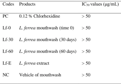
The test in fibroblasts cells by Alamar BlueTM method showed that none of the samples tested was able to induce cytotoxic effect with IC50 values (Inhibitory Concentration) above 50 µg / mL
(Table 2).
Table 2: Alamar Blue Cytotoxicity Test results
IC50 – Inhibitory Concentration
Statistical analysis and IC50 values obtained by a nonlinear regression/
GraphPad Prism 5.0 program.
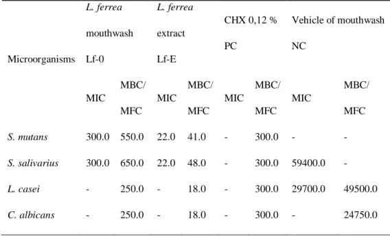
Table 3 shows the MIC and Minimal Bactericidal Concentration (MBC) values of the mouthwash from L. ferrea extract demonstrating its antimicrobial activity against all the microorganisms tested. The results of MIC values were 300.0 μg/mL against S. mutans and S. salivarius, corresponding to 22.0 μg/mL of the extract used. The Minimal Fungicidal Concentration (MFC) of the L. ferrea mouthwash was 250.0 μg/mL against C. albicans.
Codes Products EC50 values(μg/ mL)
PC 0,12 % Chlorhexidine 75.14 (69.83 - 80.86) Lf-0 L. ferrea mouthwash (time 0) > 2000
Lf-30 L. ferrea mouthwash (30 days) > 2000 Lf-60 L. ferrea mouthwash (60 days) > 2000 Lf-E L. ferrea extract > 2000
NC Vehicle of mouthwash 300.5 (259.1 - 348.6)
Codes Products IC50 values (μg/mL)
PC 0.12 % Chlorhexidine > 50 Lf-0 L. ferrea mouthwash (time 0) > 50 Lf-30 L. ferrea mouthwash (30 days) > 50 Lf-60 L. ferrea mouthwash (60 days) > 50 Lf-E L. ferrea extract > 50 NC Vehicle of mouthwash > 50
Table 3. Minimal Inhibitory Concentration (MIC), Minimal Bactericidal Concentration (MBC) and Minimal Fungicidal Concentration (MFC)
The other components tested did not show any antimicrobial activity.
4 DISCUSSION
The plant extract mouthwash was effective in reducing plaque and gingivitis. This natural product may be considered as a good alternative for using like oral rising due the chlorhexidine mouthwash promotes many side effects (staining of the teeth and restorations, mucosa desquamation, and taste changes), which limit its acceptability and long-term use (De Araújo et al., 2018).
A possible limitation of this new and natural product is the color. It can decrease the prolonged use of this mouthwash. This is what already happens with the use of chlorhexidine. However, empirical observations as the use of infusion and decoction by the local population show that the use of the plant does not result in significant tooth staining.
However, in order to improve the use of L. ferrea mouthwash in Dentistry and to confirm its efficacy, it is necessary to subject the product to diverse tests. One of them is the performance when used in the oral cavity and then use the product in daily clinical routine (Venâncio et al., 2015; Oliveira et al., 2011).
In this research, the use of a new herbal mouthwash from extracts of L. ferrea, is considered ecological correct, because it does not compromise the nature, since the L. ferrea plant is not destroyed and only the fruits are used. This plant has been utilized by folk medicine and is composed of phenolic compounds as condensed tannins, catechins and hydrolysable tannins such as gallic acid and ellagic acid (Ferreira and Soares, 2015; Vasconcelos et al, 2011).
Microorganisms L. ferrea mouthwash Lf-0 L. ferrea extract Lf-E CHX 0,12 % PC Vehicle of mouthwash NC MIC MBC/ MFC MIC MBC/ MFC MIC MBC/ MFC MIC MBC/ MFC S. mutans 300.0 550.0 22.0 41.0 - 300.0 - - S. salivarius 300.0 650.0 22.0 48.0 - 300.0 59400.0 - L. casei - 250.0 - 18.0 - 300.0 29700.0 49500.0 C. albicans - 250.0 - 18.0 - 300.0 - 24750.0
The isolation of active components from L. ferrea extract by successive high performance liquid chromatography was performed. The results showed the presence of two compounds that were identified as gallic acid and epicatechin by retention time and spectral comparisons of respective standards (Pedrosa et al., 2016).
The crude extract contains anthraquinones, alkaloids, depsides, depsidones, flavonoids, lactones, saponins, sugars, sesquiterpenes and triterpenes, but tannins are regarded as the major component (Souza et al., 2006). The aqueous and acetone water extracts are active against all Gram-positive bacteria and against some Gram-negative bacteria (De Araújo et al., 2014).
The mouthwash was evaluated according to pharmacological stability and quality conditions, showing to be absent from microorganisms and no changes were observed in the organoleptics and sedimentation characteristics. The average pH values were 6.21, 6.15 and 5.85 at 0, 30 and 60 days, respectively, and density values of 1.029, 1.033 and 1.035 g/ mL, respectively, without interfering in the final characteristics of the formulation (Venâncio et al., 2015). Souza et al. (2020) analyzed the aforementioned formulation with the objective of evaluating its stability, in three storage conditions and in six time intervals, demonstrating favorable conditions of stability and absence of microorganisms.
In the process of evaluation and validation of a product, it is necessary to prove the benefits of its use, its effectiveness as drug and its risks to human health, animals and the environment. Therefore, it is essential to conduct toxicological tests to evaluate the safety and the risk / benefit of possible therapeutic use of this new product (Evans, 2009).
Thus, this study is justified as a way to use a medicinal herb extract in dentistry therapeutic practice. The analysis of the mouthwash formulation based on L. ferrea extract using cytotoxicity tests is important because herbal solutions are more affordable and have wide drug benefits.
In the hemolysis test it was found that the L. ferrea mouthwash showed no hemolytic activity at the times studied. However, the vehicle and the 0.12 % chlorhexidine mouthwashes caused hemolysis with EC50 of 75.14 and 300.5 µg / mL, respectively. This result is considered satisfactory
since the concentration of vehicle in the mouthwash formulation is lower than the effective concentration of hemolysis.
According to the Alamar BlueTM method for cell culture test it was found that no tested sample
was able to induce a cytotoxic effect in contact with the cells. The values of IC50 above 50 µg / mL
demonstrated the absence of cytotoxicity under the tested conditions.
The results obtained by Marreiro et al. (2014b), using the same cytotoxicity assay methodology of this study, showed that both formulations (mouthwash and vehicle of L. ferrea) were cytotoxic causing the fibroblasts death. The mouthwash presented feasibility cell of 8 % and the
vehicle of 7 % when compared to the positive control. However, when the study was carried out in vivo, the toxicity of the product showed no inflammatory changes.
The antimicrobial activity of L. ferrea fruit extract in the research of Sampaio et al. (2009) was carried out against the same oral pathogens of this study, using the microdilution method for planktonic cells (MIC) and a multispecies biofilm model. The results showed in MIC values of 40.0 μg/mL, 100.0 μg/mL, 66.0 μg/mL, 66.0 μg/mL and 25.0 μg/mL for S. mutans, S. oralis, S. salivarius, L. casei and C. albicans, respectively. For the biofilm assay, chlorhexidine and plant extract showed no growth at 10−4 and 10−5 microbial dilution, respectively. The study concluded that L. ferrea fruit extract can inhibit in vitro growth of oral pathogens in planktonic and biofilm models supporting its use against oral infections.
Although this study did not tested multispecies biofilms, it has great perspective to be efficient in vivo, since the extract in vitro had antimicrobial activity against oral microorganisms. About this topic, Moreira et al. (2013) affirmed that in vitro studies have limitations conditions, no matter how much they approximate to real clinical situations they do not reproduce the real oral conditions, which are known to be extremely complex. However, in vitro researches have their values as initial screening tests, which may suggest valuable future clinical trials or observational studies.
Jeon et al. (2011) and Moreira et al. (2013) affirmed that bacteria in classical MIC/MBC assays are in suspension, whereas the oral bacteria associated with caries are enmeshed in the plaque biofilm matrix. There is a plethora of evidence showing that biofilm cells are more resistant to antimicrobials than cells in suspension. Thus, describing antibacterial activity against planktonic oral bacteria with constant exposure to an agent at a high concentration over a period of 24 hour is relevant to know how oral bacteria would respond in the mouth.
Marreiro et al. (2014a) evaluated the in vitro antimicrobial activity of a mouthwash containing L. ferrea extract to control oral biofilm using the same methodology of this study. The results showed MIC values against S. mutans, S. oralis and L. casei in concentrations of 4.37 μg/mL, 3.75 μg/mL and 4.37 μg/mL, respectively.
Candida albicans was the most susceptible microorganism in this study since all tested compounds had fungicidal action forward to this yeast in all tested concentrations. This result is important because Canela et al. (2018) showed that Candida spp. is responsible for 80 % of all systemic fungal infections and is associated with high mortality rates. C. albicans was the predominant specie (44 %) involved.
According to Alavarce et al. (2015) with the increasing development of antifungal resistance, there is a persistent requirement in research for newer effective agents. Thus, our results suggest the
potential use of L. ferrea extract in the treatment of infectious conditions such as candidiasis and denture stomatitis.
The mouthwashes used to control plaque biofilms may be very important since these agents can remove these biofilms and/or kill disease-associated with bacteria; they can also even inhibit at sub-lethal levels the expression of pathogenic traits (Marsh, 2010). In this way, the beneficial activities of the resident oral microbiota will be retained and the risk of dysbiosis occurring will be reduced (Marsh; Head; Devine, 2014).
Studies of antimicrobial activity have been carried out with several medicinal plants indicating great potential for combating several diseases (Antunes et al., 2020; Martins and Casali, 2019).
Jeon et al. (2011) and Jain et al. (2015) affirmed that natural products remain a largely unexplored source of effective antibiofilm molecules with potentially low toxicity that could be used in alternative or adjunctive anticaries therapies.
This study suggests that L. ferrea mouthwash would be a future alternative choice for prevention of caries and oral candidiasis. For that reason, further investigation must be conduct to allow the formulation of this product as an adjuvant to oral health maintenance.
5 CONCLUSION
L. ferrea mouthwash (1%, v/v) is not cytotoxic in human fibroblasts and has antimicrobial activity against some oral pathogenic microorganisms. According to the in vitro tests, the results support the use of L. ferrea extract for oral health due its biofilm inhibation and related diseases as dental caries and candidiasis. Clinical trials with this antimicrobial product are warranted to the determination of its efficacy and safety parameters in vivo.
ACKNOWLEDGMENTS
This work was support by Amazonas State Foundation for Research Support (FAPEAM - 450.002.001.002.001/019.451-4). We are also grateful for the very important help and support in the routine function at the laboratory of Raquel de Oliveira Marreiro, Patrícia Sâmea Lêdo Lima Milério, Glauber Oliveira Palmaand Taciana de Amorim Silva.
REFERENCES
Ahmed SA, Gogal Jr RM, Walsh JE (1994). A new rapid and simple nonradioactive assay to monitor and determine the proliferation of lymphocytes: an alternative to [3H] thymidine incorporation assay. J Immunol Methods, 170: 211-24.
Alavarce RA, Saldanha LL, Almeida NL, Porto VC, Dokkedal AL, Lara VS (2015). The beneficial effect of Equisetum giganteum L. against Candida biofilm formation: New Approaches to Denture Stomatitis. Evid Based Complement Alternat Med, 2015: 939625. doi: 10.1155/2015/939625. Andrews JM (2001). Determination of minimum inhibitory concentrations. J Antimicrob Chemother, 48(1): 5-16.
Antunes BF, Otero DM, Oliveira FM, Jacques AC, Gandra EA, Zambiazi RC (2020). Antioxidant and antimicrobial activity of olive trees cultivated in the Campanha Gaúcha region. Braz. J. of Develop, 6(4): 21791-21805. DOI:10.34117/bjdv6n4-374.
Canela HMS, Cardoso B, Vitali LH, Coelho HC, Martinez R, Ferreira MEDS (2018). Prevalence, virulence factors and antifungal susceptibility of Candida spp. isolated from bloodstream infections in a tertiary care hospital in Brazil. Mycoses, 61(1): 11-21. doi: 10.1111/myc.12695.
CLSI - Clinical and Laboratory Standards Institute. (2011). Performance Standards for Antimicrobial Susceptibility Testing, 21st Informational Supplement. CLSI Document M100-S21. Clinical and
Laboratory Standards Institute, Wayne.
http://vchmedical.ajums.ac.ir/_vchmedical/documents/CLSI%202011.pdf
De Araújo AA, Soares LA, Assunção Ferreira MR, de Souza Neto MA, da Silva GR, de Araújo RF Jr, Guerra GC, de Melo MC (2014). Quantification of polyphenols and evaluation of antimicrobial, analgesic and anti-inflammatory activities of aqueous and acetone-water extracts of Libidibia ferrea, Parapiptadenia rigida and Psidium guajava. J Ethnopharmacol, 156: 88-96. doi:10.1016/j.jep.2014.07.031.
De Araújo JSC, de Castilho ARF, Lira AB, Pereira AV, de Azevêdo TKB, de Brito EMMC, Pereira MDSV, Pessôa HLF, Pereira JV (2018). Antibacterial activity against cariogenic bacteria and cytotoxic and genotoxic potential of Anacardium occidentale L. and Anadenanthera macrocarpa (Benth.) Brenan extracts. Arch Oral Biol, 85: 113-119. doi: 10.1016/j.archoralbio.2017.10.008. Evans GO (2009). Animal Hematotoxicology: a partial guide for toxicologists and biomedical researchers. [S.I.]: CRC. 224p.
Ferreira MRA, Soares LAL (2015). Libidibia ferrea (Mart. ex Tul.) L. P. Queiroz: A review of the biological activities and phytochemical composition. J Med Plants Res, 9: 140-50.
Fischer D, Youxin L, Ahlemeyer B, Krieglstein J, Kissel T (2003). In vitro cytotoxicity testing of polycations: influence of polymer structure on cell viability and hemolysis. Biomaterials, 24: 1121-31.
Jain I, Jain P, Bisht D, Sharma A, Srivastava B, Gupta N (2015). Use of traditional Indian plants in the inhibition of caries-causing bacteria--Streptococcus mutans. Braz Dent J, 26(2): 110-5. doi: 10.1590/0103-6440201300102.
Jeon JG, Rosalen PL, Falsetta ML, Koo H (2011). Natural products in caries research: current (limited) knowledge, challenges and future perspective. Caries Res, 45(3): 243-63. doi: 10.1159/000327250.
Marreiro RO, Bandeira MF, de Souza TP, de Almeida MC, Bendaham K, Venâncio GN et al (2014a). Evaluation of the stability and antimicrobial activity of an ethanolic extract of Libidibia ferrea. Clin Cosmet Investig Dent, 28(6): 9-13. doi: 10.2147/CCIDE.S54319.
Marreiro RO, Bandeira MFCL, de Almeida MC, Coellho CN, Venâncio GN, Conde NCO (2014b). Cytotoxicity evaluation of a mouthwash containing extract of Libidibia ferrea. BRPDIC, 3: 34-42. doi: 10.4034/PBOCI.2014.14s3.04.
Marsh PD (2010). Controlling the oral biofilm with antimicrobials. J Dent, 1: 11-5.
Marsh PD, Head DA, Devine DA (2014). Prospects of oral disease control in the future - an opinion. J Oral Microbiol, 27(6): 26176. doi: 10.3402/jom.v6.26176.
Martins FWP, Casali AK (2019). In vitro antimicrobial activity of ethanolic extracts of Pomegranate (Punica granatum, L.) on the bacteria Escherichia coli and Staphylococcus aureus. Braz. J. of Develop, 5(11): 22970-22980. DOI:10.34117/bjdv5n11-027.
Moreira AD, Mattos CT, de Araújo MV, Ruellas AC, Sant'anna EF (2013). Chromatic analysis of teeth exposed to different mouthrinses. J Dent, 41(5): e24-7. doi: 10.1016/j.jdent.2012.12.002. Oliveira LA, Souza-Moreira TM, Cefali LC, Chiari BG, Corrêa MA, Isaac VLB, Salgado HRN, Pietro RCLR (2011). Design of antiseptic formulations containing extract of Plinia cauliflora. Braz J Pharmac Sci, 47(3): 525-33.
Pedrosa TD, Barros AO, Nogueira JR, Fruet AC, Rodrigues IC, Calcagno DQ et al (2016). Anti-wrinkle and anti-whitening effects of jucá (Libidibia ferrea Mart.) extracts. Arch Dermatol Res, 308(9): 643-54.
Sampaio FC, Pereira MS, Dias CS, Costa VC, Conde NC, Buzalaf MA (2009). In vitro antimicrobial activity of Caesalpinia ferrea Martius fruits against oral pathogens. J Ethnopharmacol, 124(2): 289-94. doi: 10.1016/j.jep.2009.04.034.
Silva LN, Zimmer KR, Macedo AJ, Trentin DS (2016). Plant Natural Products Targeting Bacterial Virulence Factors. Chem Rev, 116(16): 9162-236. doi: 10.1021/acs.chemrev.6b00184.
Souza AB, Souza LMS, Carvalho JCT, Maistro EL (2006). No clastogenic activity of Caesalpinia ferrea Mart. (Leguminosae) extract on bone marrow cells of Wistar rats. Genet Mol Biol, 29: 380-3. doi: 10.1590/S1415-47572006000200028.
Souza LAL, Melo KS, Gomes LSS, Souza TP, Bandeira MFCL, Toda C (2020). Quality control of a formulation mouthwash based on Libidibia Ferrea L. Braz. J. of Develop, 2020; 6(7): 47236-47246. Ueda H, Tachibana Y, Moriyasu M, Kawanishi K, Alves SM (2001). Aldose reductase inhibitors from the fruits of Caesalpinia ferrea Mart. Phytomed, 8(5): 377-81. doi: 10.1078/0944-7113-00043. Vasconcelos CFB, Maranhão HML, Batista TM, Carneiro EM, Ferreira F, Costa J et al (2011). Hypoglycaemic activity and molecular mechanisms of Caesalpinia ferrea Martius bark extract on streptozotocin-induced diabetes in Wistar rats. J Ethnopharmocol, 137: 1533-41. doi: 10.1016/j.jep.2011.08.059.
Venâncio GN, Rodrigues IC, Souza TP, Marreiro RO, Bandeira MFCL, Conde NCO (2015). Herbal mouthwash based on Libidibia ferrea: microbiological control, sensory characteristics, sedimentation, pH and density. Rev Odontol UNESP, 44(2): 118-24. doi: 10.1590/1807-2577.1064.