Literature Review
Master’s Degree in Dental Medicine
Erbium Lasers Effects on the Dentin Organic Matrix
André Alves Vieira
Literature Review
Master’s Degree in Dental Medicine
Erbium Lasers Effects on the Dentin Organic Matrix
André Alves Vieira
Supervisor: Ana Catarina Nogueira da Silva
Invited Assistant, Faculty of Dental Medicine, Porto University
ACKNOWLEDGMENTS
Este trabalho representa o fim de uma boa e gratificante jornada. Deste modo, não poderia deixar de fazer alguns agradecimentos:
À minha orientadora, Ana Catarina Nogueira Silva, pela disponibilidade e exigência demonstradas na realização desta dissertação e por me ter ajudado a ultrapassar várias dificuldades, num ano bastante atípico.
À minha família, em especial aos meus Pais, por todo o apoio demonstrado, de forma incondicional. Só eles saberão todos os sacrifícios que fizeram, prejudicando, muitas vezes, as suas vidas e os seus dias, para me darem as melhores condições possíveis, tornando todo este caminho mais fácil e menos tumultuoso.
À Rute, a minha namorada, pela paciência, o carinho, força e ambição que sempre me conseguiu transmitir. Foi um pilar que surgiu a meio deste percurso, mas que se revelou fundamental para que o seu desfecho fosse o melhor possível.
A todos os meus amigos, que foram, muitas vezes, a alegria dos meus piores dias e com os quais vivi momentos únicos e que recordarei para sempre.
“Recomeça... se puderes, sem angústia e sem pressa e os passos que deres, nesse caminho duro do futuro, dá-os em liberdade, enquanto
não alcances não descanses, de nenhum fruto queiras só metade.” Miguel Torga
INDEX
INTRODUCTION ... 1
MATERIALS AND METHODS ... 4
RESULTS ... 6
RESULTSOFER:YAGANDER,CR:YSGGLASERS ... 13
DISCUSSION ... 18
CONCLUSION ... 22
REFERENCES ... 23
ABBREVIATIONS INDEX
Er,Cr:YSGG. – erbium/chromium-doped yttrium aluminum garnet Er:YAG - erbium-doped yttrium aluminum garnet
GAGs – glycosaminoglycans G - group HL – hybrid layer h - hours Hz – hertz LP – long pulse MPa – megapascals min - minutes mJ – milijoules ml – mililiters n – number nm - nanometers pgs – proteoglycans pps – pulses per second s – second
sbs – shear bond strength
SEM – scanning electron microscopy SiC – silicon carbide
SP – short pulse
tbs – tensile bond strength
TEM – transmission electron microscopy
VLP – very long pulse VSP – very short pulse W – watts ws – water spray wt – weight 3D – three-dimension µm – micrometers V
TABLES INDEX
TABLE 1 – INCLUSION AND EXCLUSION CRITERIA ... 4
TABLE 2 - FLOWCHART: ARTICLES SELECTION AFTER ELECTRONIC SEARCH ... 5
TABLE 3 – ER:YAG LASER PARAMETERS ... 15
TABLE 4 – ER,CR:YSGG LASER PARAMETERS... 15
RESUMO
Introdução: Preparar cavidades dentárias, utilizando lasers erbium, é um método cada
vez mais utilizado na Medicina Dentária, com a utilização dos lasers Er:YAG e o Er,Cr:YSGG. No entanto, a adesão dos materiais restauradores nas superfícies preparadas com laser tem originado resultados controversos, com a literatura disponível a mostrar maior eficácia quando utilizados métodos convencionais. Na origem destes resultados, poderão estar os efeitos que estes lasers têm na matriz orgânica da dentina. Este trabalho pretende recolher os estudos existentes sobre a ação dos lasers Er:YAG e Er,Cr:YSGG na matriz orgânica da dentina e como isso pode influenciar os processos adesivos.
Objetivos: Estudar e descrever os efeitos que os lasers Erbium têm sobre a matriz orgânica da dentina, e o seu efeito na adesão. Materiais e métodos: A pesquisa foi realizada através do recurso a bases de dados como a PubMed®, SciELO e Cochrane
Library. Foram incluídos artigos do tipo revisão bibliográfica, revisão sistemática, meta-análises, estudos comparativos e estudos in vitro com um limite temporal de 20 anos.
Resultados: Depois de aplicar os critérios de inclusão e exclusão, 10 artigos foram
selecionados para esta revisão. Discussão: Alguns pormenores interessantes são assinaláveis. Irrigação com água é fundamental para não ocorrer um maior aumento da temperatura na dentina. Pulsos longos mostraram piores resultados, pois a energia transforma-se em calor no tecido dentinário, provocando alterações significativas na sua matriz orgânica. Também a energia e a frequência usadas influenciam as características da dentina sendo necessário uma boa gestão destes dois parâmetros de forma a não provocar destruição nos seus componentes orgânicos. Conclusão: Dependendo dos parâmetros usados, a utilização de lasers de erbium na dentina pode afetar negativamente a sua matriz orgânica e, consequentemente, afetar a adesão de biomateriais na dentina. A falta de estudos e a heterogeneidade entre os estudos selecionados foram limitações desta revisão.
Palavras-chave: “adhesion”, “erbium lasers”, “Er:YAG”, “Er,Cr:YSGG”, “collagen”,
“dentin organic matrix”.
ABSTRACT
Introduction: Cavity preparation and conditioning with lasers is becoming widely used
in Dentistry, over the last years. In this aspect, Er:YAG and Er,Cr:YSGG lasers are emerging as the most safe and effective. Nevertheless, adhesion to dentin prepared with these methods is showing controversy results, and most of the literature concludes that preparation with conventional procedures has better adhesion. Probably, one of the main reasons for this to happen is the effects produced by irradiation on the dentin organic matrix. This review will gather the information on the action of Er:YAG and Er,Cr:YSGG lasers in this dentin zone and try to understand how it can influence adhesive procedures.
Aim: The aim of this study is to describe the effects of erbium lasers on the dentin organic
matrix, and its effect on dentin adhesion. Materials and Methods: In this review, electronic search was performed on PubMed, SciELO and Cochrane Library. Scientific magazines and “Repositório aberto da Universidade do Porto” were also consulted. The search was limited to studies between 2000 and 2020. Results: After applying the inclusion and exclusion criteria, 10 articles were included in this review. Discussion: From the analysed articles, some aspects could be referred. Laser irradiation under water irrigation is fundamental to avoid uncontrolled increasing in the dentin temperature. Longer pulses showed worst results, because energy is transformed in conductive heat, leading to significant alterations on the dentin organic components. Output energy and frequency used also influence the dentin characteristics and a good management of these two parameters is needed to not provoke greater damage on the dentin organic matrix.
Conclusion: Depending on the parameters used. erbium lasers irradiation can negatively
affect the dentin organic matrix and, consequently, affect the adhesion to dentin. The lack of studies and the heterogeneity between the studies selected were limitations of this review.
Keywords: “adhesion”, “erbium lasers”, “Er:YAG”, “Er,Cr:YSGG”, “collagen”, “dentin
organic matrix”.
INTRODUCTION
Through the years, dentists have been using traditional mechanical cutting and drilling systems to remove carious dental hard tissues and to prepare cavities for restorations (1). Laser technology appearance offers a new alternative. Unlike the common systems, laser irradiation involves selective ablation of dental tissue without vibration, diminishing the requirement of local anaesthesia and increasing the patient’s comfort, resulting in an attractive option (2). In dentistry, lasers can be applied in different ways: for ablation of dental hard tissues, oral surgery, periodontal therapy, endodontics and implantology, for example (3). The first lasers used for dental ablation, generated an increase of the dental pulp temperature as well as microcracks and carbonization (4).
Later (1990s), investigators found that erbium-doped yttrium aluminum garnet (Er:YAG) and erbium/chromium-doped yttrium aluminum garnet (Er, Cr:YSGG) lasers with the use of water cooling could promote effective caries and hard tissue removal (5).
Er:YAG (2.94 µm) and Er,Cr:YSGG (2.78 µm) are both highly absorbed by water and hydroxyapatite (6). When the light encounters the tooth, it is absorbed by water molecules present in the dental hard tissues. As consequence, the water temperature rises, rapidly, and it vaporizes. The reaction creates a high localized pressure and a micro-explosion, which results in ablation of dental hard tissue (7). This affinity to water and the difference in water content between peri and intertubular dentin results in a selective ablation of intertubular dentin, leaving protruding dentinal tubules with a cuff-like appearance (8). The use of erbium lasers to irradiate dentin also results in a distinctive rough surface, without smear layer and micro-irregularities (9).
These lasers allow a minimally invasive cavity preparation and the possibility of condition the dental substrate (depending on the parameters used), before the application of the adhesive materials (10).
All these aspects propose that the resulting dentin surface is open to resin bonding. However, the erbium lasers can also have repercussions on the dentin subsurface and can cause thermal denaturation of the dentin organic matrix, which is thought to be
responsible for the decrease in bond strength of adhesive systems on irradiated dentin, according to Lopes, R.M. et. al. (8).
So, bonding to irradiated dentin remains a contradictory issue in the literature. A great number of current restorative procedures use dentin as a substrate and the success of restorative dental materials invariably depends on a good understanding of the structural and mechanical properties that characterize this hard tissue organic matrix and the changes that occur in its structure during adhesive procedures (11)(12).
Dentin consists of 70wt% mineral component, 20wt% organic matrix, and 10wt% fluids (13). Its composition can differ in different areas of the tooth, depending on the distance to the pulp tissue, as well as whether the matrix is demineralized or caries affected/infected (12). The dentin structure and nature as well as its morphological variations make it a complex substrate to which a good bond may be achieved (2). Its micro-morphology is arranged of tubules (∼1–2 μm diameter) surrounded by a hypermineralized layer (∼1 μm), called peritubular dentin, and a softer intertubular matrix, the “house” of the organic material (14).
The organic matrix consists of 90% fibrillar collagen type I, and the remaining 10% is noncollagen proteins, proteoglycans, in particular phosphoproteins (13).
Type I collagen fibrils represent the main structure of the collagen network. They are perpendicularly connected by non-collagenous proteins (12). Collagen plays a fundamental role in the tensile strength, in the elastic modulus and biochemical properties of dentin (15). The most prevalent of the non-collagenous proteins are the proteoglycans (PGs), which consist of a core protein, glycosaminoglycans (GAGs) and linkage proteins (12).
PGs drag water molecules into the interfibrillar spaces of the dentin matrix regulating a hydraulic mechanical support system to the type I collagen fibrils. The intricate interactions between PGs and collagen suggest that the first ones interact with type I collagen by protein core binding to four or more collagen microfibrils via hydrogen bonds, assuming a helical configuration (16).
Simplifying, proteoglycans may be responsible for the 3D appearance of the dentin organic matrix due to their ability to fill space, bind and organize water molecules (17). Regarding the bonding process to dentin, it is a form of tissue engineering, in which mineral is replaced with resin monomers to form a biocomposite, composed by dentin collagen and cured resin (18). So, to keep the stability of bonding to dentin, this biocomposite must be homogenous and compact (19). Adhesive monomers infiltrate and invade dentinal tubules and the collagen network, forming two well defined structures, known as resin tags and hybrid layer, respectively (20). It involves a series of topical treatments to dentin that completely changes its physical and chemical properties turning it into a more hydrophobic, organic, highly permeable and acid-resistant surface (21). However, the considerable complexity of the supramolecular structures that form the dentin matrix on the nanometer scale is many times underestimated (11). When collagen is not fully enveloped by adhesive or porosity are present in the hybrid layer, micro- and nano- pathways can speed up resin/dentin interface degradation (22). For example, the so-called nanoleakage phenomenon, described as the main degradation mechanism of tooth-biomaterial interfaces, has been shown to result from the poor ability of monomers to impregnate the demineralized dentin organic matrix. Consequently, this leads to water sorption and hydrolytic degradation, even when there is no visible marginal gap at the tooth–biomaterial interface (11).
Nowadays literature has revealed lower bond strengths of laser-irradiated dentin compared with dentin prepared by conventional means and, although, some studies show that laser is capable of creating good conditions to bond, others say that these conditions are not enough or can present a substrate less receptive to adhesive procedures (8)(9). The objective of this literature review is to describe how the dentin organic matrix and collagen network, functions and reacts to erbium laser irradiation in order to understand the reasons why some studies refer that the resin/irradiated dentin interface bond strength is lower when compared to the traditional methods, which can lead to future investigation to improve the bond to lased dentin.
MATERIALS AND METHODS
This dissertation will take the form of a literature review. PubMed, SciELO and Cochrane Library searches were carried out. A combination of the following terms was used: “adhesion”, “erbium lasers”, “Er:YAG”, “Er,Cr:YSGG”, “collagen”, “dentin organic matrix”. Clinical cases, systematic reviews, meta-analysis, comparative and in vitro studies from the last 20 years (2000-2020) with full text available were included. Articles on non-human teeth were excluded and only those written in English, Portuguese or Spanish were selected. Tests in root dentin were not considered, as well as the ones in primary dentition only (Table 1).
INCLUSION CRITERIA EXCLUSION CRITERIA
Articles from the last 20 years (2000-2020)
Articles in languages other than English, Spanish or Portuguese
Meta-analysis, systematic reviews, in vitro studies, comparative studies
Articles with access restriction
Articles in English, Spanish or Portuguese Non-human teeth
Access to full text Tests in root dentin
Human teeth Tests in primary dentition only
Studies using Er:YAG and Er,Cr:YSGG lasers
Table 1 – Inclusion and Exclusion Criteria
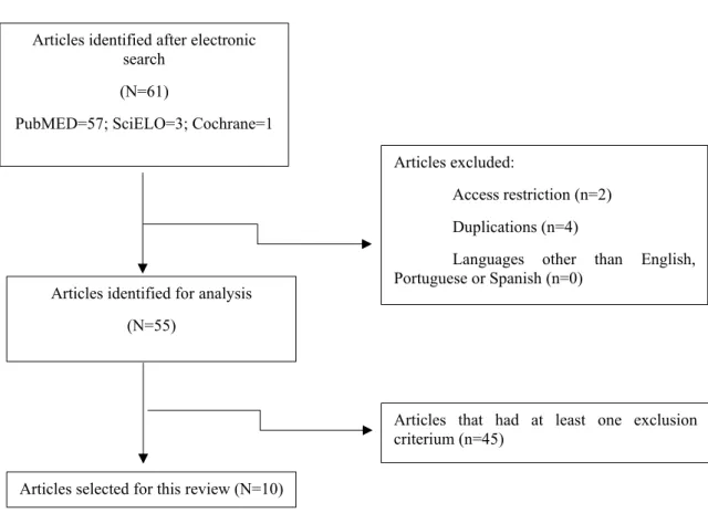
Initially, 61 articles were found through electronic search, based on the keywords. After that, articles with restriction access, duplicated and written in languages other than English were excluded, which left 55 articles for analysis. After analysis 46 articles were
excluded because they had at least one exclusion criterium. In the end, 10 articles were selected for this review.
Table 2 - Flowchart: Articles selection after electronic search Articles identified after electronic
search (N=61)
PubMED=57; SciELO=3; Cochrane=1
Articles identified for analysis (N=55)
Articles selected for this review (N=10)
Articles excluded:
Access restriction (n=2) Duplications (n=4)
Languages other than English, Portuguese or Spanish (n=0)
Articles that had at least one exclusion criterium (n=45)
RESULTS
Author (year)
Title Aim of the study Sample and Laser
Parameters
Results Bond tests (if present)
Trevelin et. al. (2019)
Influence of Er:YAG laser pulse duration
on the long-term stability of organic
matrix and
resin-dentin interface
Explore the influence of Er:YAG laser irradiation
with different pulse durations on the organic
matrix, micromorphology of the
hybrid layer (HL), and bond strength over time
N=60 G1 – Control Er:YAG G2 – 80mJ; 50µs; 21ml/min WS; 40% air; 2Hz G3 – 80mJ; 300µs; 21ml/min WS; 40% air 2Hz G4 - 80mJ; 600µs; 21ml/min WS; 40% air; 2Hz
Curiously, long resin tags were observed only in specimens irradiated with 50 μs pulse duration, but it did
not have no effect on the immediate bond strength. Group 1 exhibited immediate higher values of μSBS with statistical significance difference, comparing to the
laser groups.
The Er:YAG laser irradiation altered the organic matrix and denatured collagen fibers below the irradiated area.
For short pulse duration, a narrow zone of altered organic matrix was observed, but for long pulses this
zone was thicker, exhibiting black voids.
µSBS (MPa) G1 24h - 26.17 12 months – 24.97 G2 24h – 22.14 12 months – 22.85 G3 24h – 21.25 12 months – 22.13 G4 24h – 20.62 12 months – 23.01 6
Nahas et. al. (2018) Effect of Er:YAG laser energy densities on thermally affected dentin layer: Morphological study
Evaluate the effect of Er:YAG laser radiation
at different levels of energy on the morphology of thermally
affected dentin layer
N=48 G0 – Control
Er:YAG G1 – 40mJ; 50µs;4 mL/min WS; 6 mL/min air;
10Hz G2 - 60mJ j; 50µs;4 mL/min WS;6 mL/min air;
10Hz
G3 - 80mJ; 50µs;4mL/min WS; 6 mL/min air; 10Hz
G4 - 100mJ; 50µs;4 mL/min WS; 6 mL/min air;
10Hz G5 - 120mJ; 50µs;4 mL/min WS; 6 mL/min air;
10Hz
A superficial black layer, representing dentinal affected collagen fibers, was present in all groups except for control group. Thickness of the black layer was 28.17 µm at 40 mJ, then decreased to 15.19 µm at 60 mJ and then increased again for 80 mJ to 19.93 µm, 100 mJ to
22.87 µm and 120 mJ to 28.53 µm.
Group 2 (60 mJ) presented the lowest values of collagen damaged layer and significant differences comparing to
the other irradiated groups
He, Z. et. al. (2017) Mechanical properties and molecular structure analysis of subsurface dentin after Er:YAG laser
irradiation
Evaluate the chemical and mechanical modifications in subsurface dentin layer
after Er: YAG laser irradiation
N=15 All specimens were irradiated with Er:YAG (100mJ ; 300µs; under WS)
Comparing to the control groups that had no treatment, significant differences on the dentin organic components were found at the depths of 5µm and 10µm, as well as in the Young’s Modulus of this lased layers. From 15µm to
50µm, no significant differences were mentioned.
Moretto, S.G. et. al. (2011) Effects of ultramorphological changes on adhesion to lased dentin-Scanning electron microscopy and transmission electron microscopy analysis
Evaluate the effect of erbium laser irradiation
on dentin morphology and microtensile bond strength (µTBS) of an adhesive to dentin N=72 G1 – Control Er:YAG G2 – 250mJ; 5ml/min WS; 5 mL/min air; 4Hz G3 - 200mJ; 5ml/min WS; 5 mL/min air; 4Hz G4 - 180mJ j; 5ml/min WS; 5 mL/min air; 10Hz G5 - 160mJ; 5ml/min WS; 5 mL/min air; 10Hz
SEM and TEM analysis revealed different morphologic patterns of the organic structures when compared to the control group, with partially denatured collagen fibrils
after irradiation.
The parameters used for erbium lasers in this study showed that irradiation interacts with the dental hard tissue resulting in a specific morphological pattern of dentin and collagen fibrils that negatively affected the
bond strength to composite resin
µTBS (MPa) G1 – 39.92 Er:YAG G2 – 17.33 G3 – 15.26 G4 – 15.12 G5 – 14.97 Er,Cr:YSGG G6 – 17.56 G7 – 17.54 G8 – 14.97 G9 – 16.16 8
Er,Cr:YSGG G6 – 2.0W; 55% WS; 65% air; 20Hz G7 - 2.5W; 55% WS; 65% air; 20Hz G8 - 3.0W; 55% WS; 65% air; 20Hz G9 - 4.0W; 55% WS; 65% air; 20Hz Aljdaimi, A. et. al. (2017) Effect of 2.94 µm Er: YAG laser on the
chemical composition of hard
tissues
Investigate the effect of the Er-YAG laser radiation on morphology
and chemical composition of enamel,
dentin, and bone
N=20 Er:YAG (100mJ;
750-900ms; 15Hz)
Laser radiation caused a significant decrease in the organic content of all tissues.
The morphological alterations expressed signs of fusion in all the samples.
Soares, L. E. S. et. al.
(2006)
Combined
FT-Raman and SEM studies of the effects of Er:YAG laser irradiation on dentin
Evaluate the mineral and organic components of human dentin before and after laser irradiation and
acid etching N=6 G0 – Control Er:YAG G1 – 80mJ; 2ml/min WS; 3Hz G2 - 120mJ; 2ml/min WS; 3Hz G3 - 180mJ; 2ml/min WS; 3Hz
Organic content was less affected in the control group and group 1 (80mJ).
In group 3 (180mJ), organic (p<0.01) content was more affected.
Ramos et. al. (2010) Adhesives bonded to erbium: yttrium-aluminum-garnet laser-irradiated dentin: Transmission electron microscopy, scanning electron microscopy and tensile bond strength
analyses
Investigate the effect of erbium:yttrium– aluminum–garnet
(Er:YAG) laser irradiation on dentinal collagen by transmission electron microscopy and
to analyse the resin– dentin interface by scanning electron microscopy and test the
tensile bond strength. The samples were divided into three groups, one control and two irradiated, and into several sub-groups. For TEM analysis they were acid etched and unetched and for SEM and TBS
two adhesive systems were applied.
N= 138 specimens (12 for TEM analysis; 54 for SEM;
72 for TBS) Er:YAG G1 – 250mJ;150-250 µs; 1,5ml/min WS; 2Hz G2 – 400mJ; 150-250 µs; 1,5ml/min WS; 4Hz
Unetched samples showed a diffuse layer of granular appearance
This layer was 1µmm thick for the 250mJ-2Hz group and 4x thicker for 400mJ-4hz
250mj-2hz group subjacent layer contained well-preserved collagen fibrils, after acid etching In Specimens irradiated with 400 mJ energy/pulse, the underlying layer appeared to be damaged and remained
so, even when etching had been performed.
TBS (MPa) Control Group Etched – 18.38 Single bond Non etched – 5.59 Clearfil SE – 17.51 Group 1 Etched – 18.03 Single bond Non etched – 7.62 Clearfil SE – 17.5 Group 2 Etched – 18.19 Single bond Non etched – 7.63 Clearfil SE – 16.32 11
Bakry, A.S. et. al. (2007)
Analysis of Er:YAG lased dentin using
attenuated total reflectance fourier transform infrared and X-ray diffraction
techniques
Investigate dentin chemical alterations after
Er:YAG irradiation using different output energies, with or without
water irrigation N=10 Er:YAG G1 - 100mJ, 300µs; WS; 1-25 pps G2 - 200mJ, 300µs; WS; 1-25 pps G3 - 250mJ, 300µs; WS; 1-25 pps G4 - 100mJ, 300µs; NO WS; 1-25 pps G5 - 200mJ, 300µs; NO WS; 1-25 pps G6 - 250mJ, 300µs; NO WS; 1-25 pps
No detectable changes were observed after laser irradiation with 100mJ output energy combined with
water irrigation.
Groups using higher energy output or with no water irrigation affected the organic portion of dentin
Camerlingo, C. et. al. (2004) Er:YAG laser treatments on dentine surface: micro-Raman spectroscopy and SEM analysis
Study the chemical and structural modifications that occur in laser treated
dentin surface N=50 Er:YAG VSP:350mJ; 10Hz; 100µs; 10mL/min WS SP:350mJ; 10Hz; 300µs 10mL/min WS LP:350mJ; 10Hz; 750µs 10mL/min WS VLP:350mJ; 10Hz; 1000µs 10mL/min WS
Cavity surface prepared with laser using VSP mode is irregular, without smear layer and with the orifices of dentinal tubules exposed, which was the same for VLP
mode.
No evidence of thermal damages such as surface cracking or carbonisation was found.
A larger fragmentation of dentin is seen in VSP samples, probably due to the presence of residual parts of collagen
components originated by the ablation process. Short pulse laser treatment seemed to increase the defects in the collagen component of dentine comparing
to those occurring in long pulse laser and mechanical treated surfaces.
RESULTS OF Er:YAG AND Er,Cr:YSGG LASERS
In the article by Trevelin et al., they tested the influence of laser pulse duration on long-term stability of organic matrix, using Er:YAG for surface pre-treatment with the following parameters for every groups (besides control): 80mJ, 40% air 60% water and 20Hz. The first group was control and the lased ones differed on the pulse width, respectively 50µs, 300µs and 600µs. After that, a universal adhesive system was applied. The specimens were evaluated in 24h and 12 months after saliva storage. In every irradiated groups, alterations on the collagen structure were visible, showing irregular hybrid layers and denatured collagen fibers, specially below the irradiated area. The ones that showed the worst results were the groups with bigger pulse width (G3 and G4), with strong signs of organic matrix degradation. The adhesion results showed that the control group exhibited statistically significant higher values of µSBS at 24h storage. Between laser groups, no significant differences were found. For all groups, 12 months saliva storage did not reduce the bond strength values (23).
Another study that used different pulse widths with Er:YAG laser, was the one performed by Camerlingo, C. et. al. that studied the modifications evidenced in the chemical and morphological aspects of the irradiated surface. The pulse widths ranged from VSP (100µs) to VLP (1000µs), and they found that short pulse laser treatment seemed to increase the defects in the collagen component of dentine comparing to those occurring in long pulse laser and mechanical treated surfaces. In this study, no evidence of thermal damages such as surface cracking or carbonisation was found. Cavity surface prepared with laser using VSP mode is irregular, without smear layer and with the orifices of dentinal tubules exposed, which was the same for VLP mode (24).
The in vitro study done by Nahas et. al. evaluated the effect of Er:YAG laser radiation at different levels of energy on the morphology of thermally affected dentin layer. All the lased groups shared these parameters: 50µs of pulse width, 10hz of frequency, 6 ml/min of air spray and 4ml/min of water spray. Energy levels varied: 40, 60, 80, 100, 120mj, respectively. The group using Er:YAG laser at 60mJ, showed the least alterations on the dentin organic matrix, presenting shorter thickness of affected collagen layer (15.19 µm), when compared to the other irradiated groups. The 40mJ and 120mJ had the worst results (28.17µm and 28.53 µm, respectively) (25).
The authors He Z. et. al. performed a study with the aim to evaluate alterations on the molecular and mechanical properties of the subsurface dentin after irradiation. Er:YAG laser was used with 300µs pulse duration and 100mJ set at the control panel, under water spray. These laser effects were evaluated in different dentin depths (5µm, 10µm, 15µm, 20µm, 25µm and 50µm). Comparing to the control groups that had no treatment, significant differences on the dentin organic components were found at the depths of 5µm and 10µm, as well as in the Young’s Modulus of this lased layers. From 15µm to 50µm, no significant differences were mentioned (26).
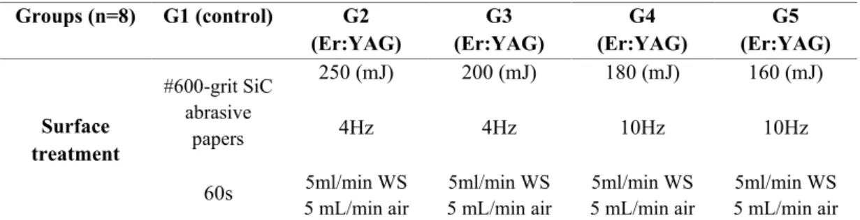
Moretto, S. G. et. al. compared the effects of Er:YAG and Er,Cr:YSGG lasers on dentin surface after irradiation and, consequently, tested the bonding to irradiated dentin using an etch and rinse adhesive system. The laser parameters are described on tables 3 and 4:
Table 3 – Er:YAG laser parameters
Groups (n=8) G1 (control) G6 (Er,Cr:YSGG) G7 (Er,Cr:YSGG) G8 (Er,Cr:YSGG) G9 (Er,Cr:YSGG) Surface treatment #600-grit SiC abrasive papers 60s 2.0W 20Hz 55% WS 65% air 2.5W 20Hz 55% WS 65% air; 3.0W 20Hz 55% WS 65% air 4.0W 20Hz 55% WS 65% air Table 4 – Er,Cr:YSGG laser parameters
The results showed alteration of the dentin superficial layer. SEM and TEM analysis revealed different morphologic patterns of the organic structures when compared to the control group, with partially denatured collagen fibrils after irradiation. It also showed the presence of horizontal microcracks on the peritubular dentin, that were more evident in the Er,Cr:YSGG groups. Control group had organized collagen fibrils, without alterations. Comparing lasers, groups 2, 6 and 7 had the best bond strength results,
Groups (n=8) G1 (control) G2 (Er:YAG) G3 (Er:YAG) G4 (Er:YAG) G5 (Er:YAG) Surface treatment #600-grit SiC abrasive papers 60s 250 (mJ) 4Hz 5ml/min WS 5 mL/min air 200 (mJ) 4Hz 5ml/min WS 5 mL/min air 180 (mJ) 10Hz 5ml/min WS 5 mL/min air 160 (mJ) 10Hz 5ml/min WS 5 mL/min air 15
although the µTBS test showed no statistically significant difference between all the laser groups. This was different from when these groups were compared to the control group, that exhibited the higher bond strength values, with statistically significant difference. The parameters used for erbium lasers in this study showed that the laser irradiation negatively affected the bond strength values (27).
Another in vitro study, this one by Aljdaimi, A. et. al. tried to investigate the effect of Er:YAG laser on the composition of hard tissues using low energy and a high repetition rate. The authors used 100mJ of pulse energy, 15Hz of repetition rate with very long pulse mode (750-950 ms). Concerning the dentin, this tissue expressed a significant reduction in all the examined organic contents (10).
Soares, L.E.S. et. al. performed two similar in vitro studies where they compared the morphological and molecular effects of laser etch on dentin, using the frequency of 3Hz for all groups and 3 levels of output energy with Er:YAG laser , in both studies: 80, 120 and 180mJ. The organic content was greatly affected by the 180mJ group and the lowest laser energy (80mJ) did not affect collagen content with statistical significance. Both experiments shared the same conclusions (28)(29).
Ramos, A.C.B. et. al. performed an in vitro study, in which one of the aims was to evaluate the effects of Er:YAG laser irradiation on dentinal collagen fibrils. All groups had two subgroups, with etched and unetched samples. One was the control group, that was just polished. Er:YAG groups consisted in one with energy/pulse of 250mJ and a repetition rate of 2Hz and the other delivered 400mJ of energy/pulse and had a repetition rate of 4Hz to see if, increasing the ablation speed, would result in different bond strengths and dentin ultrastructural characteristics. The pulse duration ranged from 150µs and 250µs and irradiation was made under water spray (1.5ml/min). The 400mJ group left a three times thicker superficial granular layer, comparing to the 250mJ group in the unetched samples. When acid etch was applied to the specimens of both, the granular layer disappeared on the lower energy group and left a subjacent layer that contained well-preserved collagen fibrils. The other lased group still had a superficial granular layer and the subsurface appeared to be damaged. Bond strength testes showed no significant differences between all the groups when etch and rinse adhesive system plus acid etching and the self-etch adhesive system were compared. On the etch and rinse system,
statistically significant differences were seen between the acid etched and the unetched samples on the bond strength, with the etched samples showing higher TBS values (30). A study performed by Bakry, A.S. et. al. tried to evaluate what would be dentin chemical characteristics after Er:YAG laser irradiation using different output energies, with or without water irrigation. All of them had a pulse duration of 300µs. Two groups used 100mJ/pulse, another two used 200mJ/pulse and the rest used 250mJ/pulse. Half of them had water irrigation and the other half did not. Groups that were not combined with irrigation have shown signs of thermal damage and carbonization. The group that had 100mJ/pulse output energy and water irrigation did not cause any detectable chemical changes and thermal damage in irradiated dentin (31).
DISCUSSION
The comfort and acceptance by patients, regarding erbium lasers, along with the advances in restorative techniques leads to numerous investigations and studies so that a standard
protocol withconsistent good bond results emerges (8). Er:YAG and Er,Cr:YSGG emit
wavelengths that are highly absorbed by water and hydroxyapatite and the preferential removal of the intertubular dentin (more water content) creates an irregular and rough surface with open dentinal tubules and no smear layer, a pattern that should be helpful for bonding to dentin. However, other factors should be put into consideration when the adhesion is to this dental hard tissue and the preservation of the collagen matrix integrity is probably the key to improve dentin bonding durability (32)(23)(18).
Research has shown that dentin collagen has a typical diameter of approximately 80–100 nm and collagen fibrils in dentin are essentially comprised of an array of laterally organized microfibrils (11). The structure of these fibrils makes it very stable and resistant to degradation but, unlike insoluble collagen in other bodily systems, dentinal collagen does not suffer turn over, which means it cannot be replaced (12)(15).
The spaces between collagen fibrils serve as diffusion paths for solvated adhesive monomers to infiltrate and impregnate around them (22). Some studies have indicated that the thermomechanical effects of laser could extend to the dentin subsurface and denature these fibrils (27). The thermal damage can lead to fusion of the organic matrix resulting in a lack of inter-fibrillary space that would limit bonding system diffusion (25). Since the main mechanism of bonding to dentin surface depends directly upon the infiltration of this monomers to the exposed dentin collagen web (33) and the lack of resin penetration in laser-ablated dentin is the most likely explanation for lower bond strengths (34), it comes of great importance to disclose the impact of Er,Cr:YSGG and Er:YAG lasers on the dentin organic matrix.
An article by Lee, B.S. et. al. demonstrated that water cooling is important to reduce thermal effects induced by laser irradiation (35). This is in accordance with the in vitro study performed by Bakry et. al. in which all the irradiated specimens without water irrigation have shown signs of thermal damage and carbonisation (31).
From the analysed articles, few conclusions can be extracted, since there is not much consistency on the parameters used between the different studies. However, some promising details were demonstrated, concerning the use of Er:YAG laser.
When pulse width was compared (50µs, 300µs and 600µs), the group with shorter pulse duration showed less thermal damage to the irradiated subsurface dentin comparing to the other lased groups, which showed black voids beneath the irradiation zone, although all pulse widths provoked similar structural modifications. The chemical analyses showed an absence of protein intensity, which can indicate damage or removal of collagen fibrils (23). Laser irradiation can develop micro rupture of collagen fibers and the literature has mentioned that if the collagen structure collapses or is altered, the penetration of primers and monomers is incomplete (33). However, bond strengths were not statistically significant different between the laser groups, not even after 12 months saliva storage, which was concluded to not be sufficient time to see the consequences of the organic matrix degradation. Even the specimens of the 50µs of pulse width group that presented long resin tags showed no significant differences in bond strength, which corroborates the idea that micromechanical retention is not the factor responsible for bonding to dentin, but the entire resin/dentin interface (23). In the Camerlingo, C. et. al paper no signs of carbonisation or surface cracking were mentioned. However, they found that short pulse laser treatment seemed to increase the defects in the collagen component of the dentin surface, comparing to those occurring in long pulse laser and mechanical treated surfaces. The reduction of organic substances is related to a clearer cavity with the absence of smear layer and debris, with minimal thermal induced changes in the surrounding tissues (24). When long pulses were tested, significant differences were seen on dentin organic components in the study by Aljdaimi, A. et. al. and signs of vitrification and fusion on the irradiated dentin were pointed out (10). This is probably explained by what some authors state in their papers that is when pulse duration is shorter, the emitted energy is completely transferred to the tissue during ablation, but when it is longer, the ablative efficiency is reduced due to the conductive heat loss through the interaction with the tissue (36). So, decreasing the pulse time length might be helpful to reduce the risk of thermal damage to surrounding tissues (26).
Another parameter usually tested is different output energies and frequencies.
Some papers have shown that when laser is used at a high level of energy, the superficial layer of dentin undergoes melting and cracking condition of its organic components. Using Er:YAG laser with 10Hz of frequency with super short pulse duration (50µs), Nahas, P. et. al. found that the group of the lower level of energy (40mJ) produced the smaller crater in dentin (depth of ablated dentin) but originated one of the thickest
thermally affected dentinal layer (black layer). The black affected layer represented altered collagen fibers. This happened because 40mJ were just enough to ablate the minimum of dentin and great part of this energy was transformed into heat around the tissue. The group that showed the least alterations on the organic matrix was the 60mJ group, leaving a 15.19 µm of thermally affected dentinal layer thickness, followed by the 80mJ group. The group that presented the worst results regarding the effects on the organic matrix was the 120mJ group (25). Contrary to this, Ramos, A.C.B. et. al. used two groups with higher levels of energy (250mJ and 400mJ, respectively), but the repetition rate was lower (2Hz and 4Hz). The 250mJ group left a 1µm of thermally affected layer, which disappeared after acid etching, leaving a subsurface with organized collagen fibrils, that could be capable of interacting with resin monomers. In the 400mJ group, this subsurface remained affected, even after acid etching (30). These results with such different output energies can be, probably, explained by the lower repetition rates used in the study by Ramos, A.C.B. et. al. comparing to the one by Nahas et. al., since decreasing the frequency of laser irradiation might help reducing the risk of thermal damage (26). These organic matrix alterations did not affect the immediate bond strength and no statistically significant differences were found on the tensile bond strength values between the groups (laser and control), except when etch and rinse adhesive system plus no etch was used, which showed lower values (30).
The only study selected for this review regarding Er,Cr:YSGG laser was the one by Moretto, S.G. et. al. and used different laser parameters for Er,Cr:YSGG and Er:YAG lasers. The irradiated groups revealed a diffuse superficial layer with granular aspect, with denatured collagen fibrils. The in vitro study by Ramos, A.C.B. et. al. showed that phosphoric acid attack can remove this diffuse superficial layer and leave an organized collagen network on the subsurface, depending on the parameters used. These alterations were transformed in a transition zone with more organized fibrils until it reached an unaltered zone that had the collagen network preserved. This was different from the pattern observed on the control group, that had an organized collagen web from the most superficial region. Comparing the bond strength tests between control and laser groups, they showed a statistically significant difference, with the control group showing higher µTBS values. Between the two types of erbium lasers used, no statistically significant differences were found, comparing all the groups. The parameters used altered the dentin organic matrix and its collagen fibrils, affecting the bond strength to composite resin (27)
Determining the lowest quantity of residual heat during irradiation with erbium lasers will be important to determine the best parameters for irradiating dentin and to preserve its organic matrix (27).
It is fundamental to cross the studies that evaluate the dentin organic matrix characteristics after erbium laser irradiation with the studies that test the bond strength to dentin to form a good protocol. There are still few studies that investigate the first part and the laser parameters used are completely different from a study to another which makes it hard to take conclusions.
Even with these differences, the analysed articles showed that shorter pulse durations, the use of water irrigation and a balance between frequency and energy delivered should be considered to minimize and control damages on the dentin organic matrix
This is a technique that can be very promising in a clinical context, from the patient’s point of view and from the clinicians’, but concerning the effects on dentin organic matrix, the limitations evidenced on this review show that more studies are needed, specially using Er,Cr:YSGG laser.
A limitation of this review was also the lack of studies comparing the organic matrix immediately after irradiation and after some time of storage and the respective bond strengths, which could be interesting, given the importance of this structure on the bond stability and durability.
CONCLUSION
The use of erbium lasers on the dentin, can interfere with the dentin organic matrix. Depending on the parameters used, can negatively affect the dentin organic matrix and cause thermal damage and fusion of collagen fibrils, diminishing the interfibrillar space and reducing the possibility of monomer diffusion through the collagen network to form a stable and homogenous hybrid layer. Laser irradiation can also denature these fibrils, making it lose its 3D conformation, which can lead to a decrease in its important properties.
The alterations produced by erbium lasers on the dentin organic matrix can affect negatively the bond strength values, also depending on the parameters used.
More studies combining bond strength tests and organic matrix alterations (immediately and after some storage time) are needed.
Although some interesting aspects could be used as reference (such as the use of shorter pulse widths, water irrigation and a good management of frequency and output energy) for future studies, the heterogeneity of the laser parameters, differences in storage of the specimens and the lack of studies on this matter were limitations of this review.
Testing the effects of Er,Cr:YSGG laser on the organic components of dentin is needed.
REFERENCES
1. Lee BS, Lin PY, Chen MH, Hsieh TT, Lin CP, Lai JY, et al. Tensile bond strength of Er,Cr:YSGG laser-irradiated human dentin and analysis of dentin-resin interface. Dent Mater [Internet]. Abril de 2007 [citado 29 de Março de
2020];23(5):570–8. Disponível em:
http://www.ncbi.nlm.nih.gov/pubmed/16820200
2. Ramos TM, Ramos-Oliveira TM, Moretto SG, De Freitas PM, Esteves-Oliveira M, De Paula Eduardo C. Microtensile bond strength analysis of adhesive systems to Er:YAG and Er,Cr:YSGG laser-treated dentin. Lasers Med Sci [Internet]. Março de 2014 [citado 29 de Março de 2020];29(2):565–73. Disponível em: http://www.ncbi.nlm.nih.gov/pubmed/23354743
3. Najeeb S, Khurshid Z, Zafar MS, Ajlal S. Applications of Light Amplification by Stimulated Emission of Radiation (Lasers) for Restorative Dentistry. Vol. 25, Medical Principles and Practice. S. Karger AG; 2016. p. 201–11.
4. Montedori A, Abraha I, Orso M, D’Errico PG, Pagano S, Lombardo G. Lasers for caries removal in deciduous and permanent teeth. Vol. 2016, Cochrane Database of Systematic Reviews. John Wiley and Sons Ltd; 2016.
5. Bishnoi A, Adyanthaya R, Singh S, Kapasi A, Jain K. Effect of Er:YAG laser cavity preparation on the bond strength of 2-hydroxyethyl methacrylate-free and 2-hydroxyethyl methacrylate-rich self-etch adhesive systems: An in vitro study. Dent Res J (Isfahan). 1 de Novembro de 2019;16(6):389–97.
6. Van As G. Erbium lasers in dentistry [Internet]. Vol. 48, Dental Clinics of North America. 2004 [citado 12 de Abril de 2020]. p. 1017–59. Disponível em: http://www.ncbi.nlm.nih.gov/pubmed/15464563
7. Jacobsen T, Norlund A, Englund GS, Tranæus S. Application of laser technology for removal of caries: A systematic review of controlled clinical trials [Internet]. Vol. 69, Acta Odontologica Scandinavica. 2011 [citado 12 de Abril de 2020]. p. 65–74. Disponível em: http://www.ncbi.nlm.nih.gov/pubmed/21319941
8. Lopes RM, Trevelin LT, Da Cunha SRB, De Oliveira RF, De Andrade Salgado
DMR, De Freitas PM, et al. Dental Adhesion to Erbium-Lased Tooth Structure: A Review of the Literature [Internet]. Vol. 33, Photomedicine and Laser Surgery. Mary Ann Liebert Inc.; 2015 [citado 29 de Fevereiro de 2020]. p. 393–403. Disponível em: http://www.ncbi.nlm.nih.gov/pubmed/26226169
9. Ayar MK, Erdermir F. Bonding strength of universal adhesives to Er,Cr:YSGG Laser-Irradiated Dentin. Niger J Clin Pract [Internet]. 1 de Janeiro de 2018 [citado 30 de Março de 2020];21(1):93–8. Disponível em: http://www.ncbi.nlm.nih.gov/pubmed/29411731
10. Aljdaimi A, Devlin H, Dickinson M, Alfutimie A. Effect of 2.94 µm Er: YAG laser on the chemical composition of hard tissues. Microsc Res Tech. 1 de Agosto de 2018;81(8):887–96.
11. Bertassoni LE. Dentin on the nanoscale: Hierarchical organization, mechanical behavior and bioinspired engineering. Vol. 33, Dental Materials. Elsevier Inc.; 2017. p. 637–49.
12. Breschi L, Maravic T, Cunha SR, Comba A, Cadenaro M, Tjäderhane L, et al. Dentin bonding systems: From dentin collagen structure to bond preservation and clinical applications. Vol. 34, Dental Materials. Elsevier Inc.; 2018. p. 78–96. 13. Silva A, Melo P, Ferreira J, Oliveira T, Gutknecht N. Adhesion in dentin prepared
with Er,Cr:YSGG laser: Systematic review. Contemp Clin Dent. 2019;10(1):129– 34.
14. Bertassoni LE, Orgel JPR, Antipova O, Swain M V. The dentin organic matrix - Limitations of restorative dentistry hidden on the nanometer scale. Vol. 8, Acta Biomaterialia. Elsevier; 2012. p. 2419–33.
15. Betancourt DE, Baldion PA, Castellanos JE. Resin-Dentin Bonding Interface: Mechanisms of Degradation and Strategies for Stabilization of the Hybrid Layer. Int J Biomater. 2019;2019.
16. de Mattos Pimenta Vidal C, Leme-Kraus AA, Rahman M, Farina AP, Bedran-Russo AK. Role of proteoglycans on the biochemical and biomechanical properties of dentin organic matrix. Arch Oral Biol. 1 de Outubro de 2017;82:203–8. 17. Breschi L, Mazzoni A, Ruggeri A, Cadenaro M, Di Lenarda R, De Stefano Dorigo
E. Dental adhesion review: Aging and stability of the bonded interface [Internet]. Vol. 24, Dental Materials. 2008 [citado 23 de Abril de 2020]. p. 90–101. Disponível em: http://www.ncbi.nlm.nih.gov/pubmed/17442386
18. Tjäderhane L. Dentin bonding: Can we make it last? Oper Dent [Internet]. 1 de Janeiro de 2015 [citado 30 de Março de 2020];40(1):4–18. Disponível em: http://www.ncbi.nlm.nih.gov/pubmed/25615637
19. Karadas M, Çağlar İ. The effect of Er:YAG laser irradiation on the bond stability of self-etch adhesives at different dentin depths. Lasers Med Sci. 1 de Julho de 2017;32(5):967–74.
20. Matos AB, Trevelin LT, da Silva BTF, Francisconi-Dos-Rios LF, Siriani LK, Cardoso MV. Bonding efficiency and durability: Current possibilities. Vol. 31, Brazilian Oral Research. Sociedade Brasileira de Hematologia e Hemoterapia; 2017. p. 3–22.
21. Frassetto A, Breschi L, Turco G, Marchesi G, Di Lenarda R, Tay FR, et al. Mechanisms of degradation of the hybrid layer in adhesive dentistry and therapeutic agents to improve bond durability - A literature review. Dent Mater. 1 de Fevereiro de 2016;32(2):e41–53.
22. Dos Santos PH, Karol S, Bedran-Russo AKB. Nanomechanical properties of biochemically modified dentin bonded interfaces. J Oral Rehabil. 2011;38(7):541– 6.
23. Trevelin LT, da Silva BTF, de Freitas PM, Matos AB. Influence of Er:YAG laser pulse duration on the long-term stability of organic matrix and resin-dentin interface. Lasers Med Sci [Internet]. 1 de Setembro de 2019 [citado 27 de Fevereiro
de 2020];34(7):1391–9. Disponível em:
http://www.ncbi.nlm.nih.gov/pubmed/30762196
24. Camerlingo C, Lepore M, Gaeta GM, Riccio R, Riccio C, De Rosa A, et al. Er:YAG laser treatments on dentine surface: Micro-Raman spectroscopy and SEM analysis. J Dent. Julho de 2004;32(5):399–405.
25. Nahas P, Zeinoun T, Namour M, Ayach T, Nammour S. Effect of Er:YAG laser energy densities on thermally affected dentin layer: Morphological study. LASER
Ther. 2018;27(2):90–90.
26. He Z, Chen L, Hu X, Shimada Y, Otsuki M, Tagami J, et al. Mechanical properties and molecular structure analysis of subsurface dentin after Er:YAG laser irradiation. J Mech Behav Biomed Mater. 1 de Outubro de 2017;74:274–82. 27. Moretto SG, Azambuja N, Arana-Chavez VE, Reis AF, Giannini M, Eduardo C de
P, et al. Effects of ultramorphological changes on adhesion to lased dentin-Scanning electron microscopy and transmission electron microscopy analysis. Microsc Res Tech. Agosto de 2011;74(8):720–6.
28. Soares LES, Resende EBPS, Brugnera A, Zanin FAA, Martin AA. Combined FT-Raman and SEM studies of the effects of Er:YAG laser irradiation on dentin. Photomed Laser Surg. 2007;25(4):239–44.
29. Soares LES, Brugnera A, Zanin F, Pacheco MTT, Martin AA. Molecular analysis of Er:YAG laser irradiation on dentin. Braz Dent J. 2006;17(1):15–9.
30. Ramos ACB, Esteves-Oliveira M, Arana-Chavez VE, De Paula Eduardo C. Adhesives bonded to erbium: yttrium-aluminum-garnet laser-irradiated dentin: Transmission electron microscopy, scanning electron microscopy and tensile bond strength analyses. Lasers Med Sci. Março de 2010;25(2):181–9.
31. Bakry AS, Sadr A, Takahashi H, Otsuki M, Tagami J. Analysis of Er:YAG lased dentin using attenuated total reflectance fourier transform infrared and X-ray diffraction techniques. Dent Mater J. 2007;26(3):422–8.
32. Ramos TM, Ramos-Oliveira TM, Moretto SG, De Freitas PM, Esteves-Oliveira M, De Paula Eduardo C. Microtensile bond strength analysis of adhesive systems to Er:YAG and Er,Cr:YSGG laser-treated dentin. Lasers Med Sci. 26 de Janeiro de 2014;29(2):565–73.
33. Aranha ACC, Eduardo CDP, Gutknecht N, Marques MM, Ramalho KM, Apel C. Analysis of the interfacial micromorphology of adhesive systems in cavities prepared with Er,Cr:YSGG, Er:YAG laser and bur. Microsc Res Tech. 2007;70(8):745–51.
34. Koliniotou-Koumpia E, Kouros P, Zafiriadis L, Koumpia E, Dionysopoulos P, Karagiannis V. Bonding of adhesives to Er:YAG laser-treated dentin. Eur J Dent.
Janeiro de 2012;6(1):16–23.
35. Lee BS, Lin CP, Hung YL, Lan WH. Structural changes of Er:YAG laser-irradiated human dentin. Photomed Laser Surg. Agosto de 2004;22(4):330–4. 36. Trevelin LT, Marques MM, Aranha ACC, Arana-Chavez VE, Matos AB. Effect
of super short pulse Er: YAG laser on human dentin-Scanning electron microscopy analysis. Microsc Res Tech. 1 de Junho de 2015;78(6):472–8.
Parecer
(Entrega do trabalho final da monografia)
Informo que o Trabalho de Monografia desenvolvida pelo estudante André Alves Vieira com o título “Erbium Lasers effects on the dentin organic matrix”, está de acordo com as regras estipuladas pela FMDUP, foi por mim conferido e encontra-se em condições de ser apresentado em provas públicas
A orientadora