Rev Odontol UNESP. 2017 Sept-Oct; 46(5): 273-277 © 2017 - ISSN 1807-2577
ORIGINAL ARTICLE
Doi: http://dx.doi.org/10.1590/1807-2577.03917
Efect of caries-afected dentin on one-step universal and
multi-step etch-and-rinse adhesives’ bond strength
Efeito da dentina cariada na resistência adesiva de um sistema adesivo universal e um de
condicionamento ácido total
Clecila MÜLLER
a, Gabriela Cauduro da ROSA
a, Gabriela Simões TEIXEIRA
a*, Ivo KREJCI
b,
Tissiana BORTOLOTTO
b, Alexandre Henrique SUSIN
aaUFSM – Universidade Federal de Santa Maria, Santa Maria, RS, Brasil bSchool of Dentistry, University of Geneva, Geneva, Switzerland
Resumo
Objetivo: Avaliar a influência da dentina afetada na resistência de união de um sistema adesivo universal e de um adesivo de condicionamento ácido total acetonado. Material e método: As faces oclusais de 60 terceiros molares humanos, hígidos e cariados, foram removidas a fim de expor o substrato dentinário. Os dentes foram divididos aleatoriamente em 6 grupos: Single Bond Universal (3M Dental Products, Seefeld, Germany), nos protocolos de condicionamento total e autocondicionante, e Prime&BondNT (DentsplyDeTrey, Konstanz, Germany), em dentina hígida e afetada. A smearlayer de 30 espécimes de dentina hígida foi padronizada com lixas de granulação 600. A dentina residual infectada de 30 espécimes foi removida com uma broca carbide número 4 até que nada fosse detectável por uma inspeção clínica. Pinos de resina composta (Filtek Z350 XT, 3M ESPE, St. Paul, MN) foram construídos usando tubos de amido como matriz. O teste de microcisalhamento foi realizado em uma máquina universal de testes até que houvesse a fratura. Resultado: Diferenças significantes foram observadas na resistência de união apenas para os espécimes de dentina afetada. A resistência de união do Single Bond Universal não foi influenciada pelo protocolo de aplicação nos espécimes de dentina hígida, mas diminuiu para os espécimes de dentina afetada, enquanto que o desempenho da adesão do Prime&BondNT não foi influenciada pela condição dentinária. Conclusão: A dentina afetada reduziu a resistência de união do Single Bond Universal, em comparação à dentina hígida. A resistência de união do Prime & Bond NT não foi alterada pela condição dos substratos.
Descritores: Dentina afetada por cárie; substrato dentinário; sistema adesivo multimodo.
Abstract
Objective: To evaluate the influence of caries-affected dentin on bond strength of a universal one-step and a multi-step etch-and-rinse adhesive system. Material and method: Enamel of 60 third human molars with and without caries was removed to expose dentin. The teeth were randomly assigned to six groups: Single Bond Universal (3M ESPE, St. Paul, MN, USA) in etch-and-rinse and in self-etch mode and Prime & Bond NT (Dentsply Co, Konstanz, Germany), all on sound and caries-affected dentin. Smear layer of the 30 sound dentin specimens was standardized by polishing with 600-grit SiC paper under water cooling. Residual infected dentin of the 30 caries-affected specimens was removed with a number 4 CA carbide bur until no caries smooth tissue was detectable by tactile-visual inspection. Cylinders of a light cured composite resin (Filtek Z350 XT, 3M ESPE) were built up using starch tubes and microshear test was performed until failure. The data was analyzed by one-way ANOVA and Tukey’s post hoc test. Result: Significant differences in microshear bond strength (μSBS) were observed for the caries-affected groups, but not for sound dentin. The μSBS of Single Bond Universal were not influenced by the application protocol on sound dentin, however they were lower in the caries-affected group with both application protocols. The μSBS for Prime & Bond NT was not influenced by the dentin conditions. Conclusion: Caries-affected dentin decrease in bond strength of Single Bond Universal in comparison to sound dentin. The bond strength of Prime & Bond NT was not altered by substrate conditions.
INTRODUCTION
Interfacial integrity inluences the eicacy and longevity of composite resin restorations1. he researchers and manufacturers
have dedicated eforts to obtain adequate hybridization of enamel and dentin introducing modiications on the adhesive systems and protocols through the last years. Some of these alterations resulted in better bond performances concerning to hybrid layer formation and mechanical properties2,3.
Due to the non-uniform permeability and moisture of the dentin, adhesion in this substrate is still considered a challenge in clinical practice. Based on this, in order to facilitate clinical procedure, the new generation of simpliied adhesive systems, also known as universal or multimode, can be used as self-etching or etch-and-rise. In addition, they would also be efective in both wet and dry demineralized dentin, thus overturning the critical wash and drying phase of the acid conditioning step4.
Studies involving bonding to normal dentin are still predominant in restorative dentistry5-7. Although major eforts concerning
caries-afected dentin have been observed lately, since selective caries removal has been widely advocated to preserve tooth structure and avoid unnecessary pulp tissue exposure. hereby, resin composite is bonded into a prepared cavity ater removal of infected dentin, in which the cavity loor frequently consists of caries afected dentin8.
Comparisons between normal and caries-afected dentin have shown the inluence of the dentinal condition on adhesive systems in mechanical tests9. Studies indicate that bonding to
caries-afected dentin results in lower bond strengths as related to sound dentin. his is due to the weaker structure of the demineralized caries-afected dentin that limits adhesive iniltration because of the tubules illed with acid-resistant mineral deposits and to the unusual conformation of the hybrid layer, which is commonly thicker. Costa et al.10, showed low resin tags formation with short
penetration into demineralized dentin regions and the increasing water content and the more permeable condition of caries-afected dentin are concerns that may compromise bonding quality/stability over time when using hydrophilic adhesives systems10-12.
he aim of this study was to evaluate the inluence of caries-afected dentin on bond strengths of a multimode and an acetone-based etch-and-rinse adhesive systems by a microshear test. he null hypothesis tested was that caries-afected dentin negatively inluences the microshear bond strength of the multimode adhesive system.
MATERIAL AND METHOD
he in vitro study was approved by the local Committee of Ethics in Research (protocol n. 556.453). hirty caries-afected and thirty caries-free extracted human third molars were selected. Before the experiments, they were disinfected in 0.5% chloramine T and stored in saline solution at 5°C, until the use in this study.
he enamel of all teeth was transversally sectioned at the mid coronal third by using a 22mm-diameter thin KG 7010 double-surface diamond saw (Medical Burs, Ind. & Com. Ltda., Cotia, Brazil) under copious water irrigation. In the caries-free dentin teeth, 600-grit silicon carbide paper was used for 60s under running water to produce a standardized smear layer. he caries-afected teeth had the infected dentin removed with a number 4 CA carbide bur (Medical Burs, Ind. & Com. Ltda.) until no caries smooth tissue was detectable by tactile-visual inspection.
A one-step universal adhesive system (Single Bond Universal, 3M ESPE, St. Paul – MN, USA) and a multi-step, acetone-based etch-and-rinse adhesive system (Prime&BondNT, DentsplyDeTrey, Konstanz, Germany) were used according to the manufacturer instructions. he prepared teeth were randomly assigned to six equal groups (n = 10) according to dentin conditions and adhesive systems:
- Group 1 - sound dentin and Single Bond Universal applied in etch-and-rinse mode;
- Group 2: sound dentin and Single Bond Universal applied in self-etch mode;
- Group 3: sound dentin and Prime & Bond NT;
- Group 4: caries-afected dentin and Single Bond Universal in etch-and-rinse mode;
- Group 5: caries-afected dentin and Single Bond Universal applied in self-etch mode and;
- Group 6: caries-afected dentin and Prime & Bond NT.
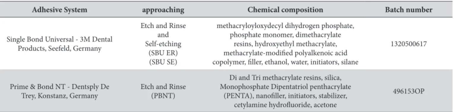
Application mode, chemical composition and batch number of the adhesive systems tested are shown in Table 1.
Ater the adhesive procedures, as recommended for manufacturer, cylindrical restorations with a light-cured composite resin (Filtek Z350 XT shade A2, 3M ESPE, St. Paul, MN, USA) were built up on dentin using starch tubes of 0.96 mm inner diameter and 2.0 mm
Table 1. Adhesive Systems with approaching, chemical composition and batch number
Adhesive System approaching Chemical composition Batch number
Single Bond Universal - 3M Dental Products, Seefeld, Germany
Etch and Rinse and Self-etching
(SBU ER) (SBU SE)
methacryloyloxydecyl dihydrogen phosphate, phosphate monomer, dimethacrylate
resins, hydroxyethyl methacrylate, methacrylate-modiied polyalkenoic acid copolymer, iller, ethanol, water, initiators, silane
1320500617
Prime & Bond NT - Dentsply De Trey, Konstanz, Germany
Etch and Rinse (PBNT)
Di and Tri methacrylate resins, silica, Monophosphate Dipentatriol penthacrylate
(PENTA), nanoiller, initiators, stabilizer, cetylamine hydroluoride, acetone
height13. One tube per sample was positioned on the caries-afected
dentin and another one on the sound dentin surface. Ater light curing of adhesive for 20 s - light-emitting diode (LED Schuster Ind. Ltda, Santa Maria, Brazil, at 800 mW/cm2 - the tubes were
illed in with two increments of composite resin and light-cured for 40 s. All the procedures were performed by a previously trained operator at room temperature of 20°C.
he specimens were stored in distilled water at 37°C for 24 h. he starch tubes and residues were removed with air–water spray. hey were examined under 10× magniication by using a stereomicroscope (Discovery V20, Carl Zeiss AG, Oberkochen, Germany) to identify bonding defects.
he specimens were placed in a jig attached to a universal testing machine (EMIC DL 1000, Instron Brazil, São José dos Pinhais, Brazil). A 0.5mm diameter stainless steel wire loop was adapted precisely to the dentin–adhesive interface and shear load was applied at a crosshead speed of 0.5 mm/min until failure. he fracture pattern was examined at 70× magniication under a stereomicroscope and classiied as adhesive, mixed and cohesive.
Means of microshear bond strengths were compared by two-way ANOVA followed by Tukey’s test at a signiicance level of 5%.
RESULT
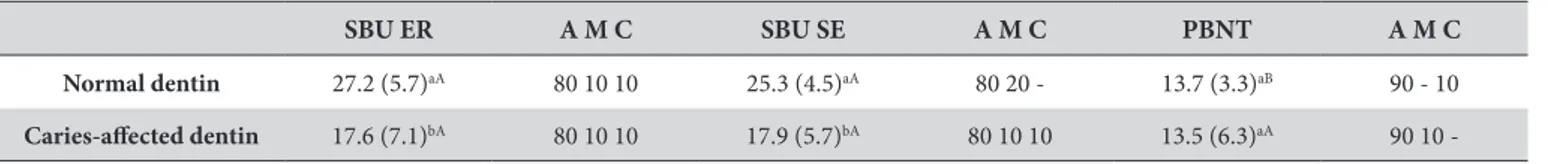
he dentin condition signiicantly afected microshear bond strength values of Single Bond Universal (p < 0.001). he adhesive system signiicantly afected the microshear bond strength values as well (p < 0.001) (Tables 2 and 3).
Figure 1 (Boxplot) shows that the bond strength values of Single Bond Universal were not inluenced by the application mode, (i.e. etch-and-rinse or self-etch), but in both application modes it decreased in the caries-afected dentin group. he bond performance of Prime & Bond NT was not signiicantly inluenced by the dentinal condition.
Mainly adhesive failures occurred in all groups. he dentinal condition did not inluence the failure pattern. Mixed and cohesive failures were well distributed among the groups.
DISCUSSION
Mechanical tests are used to evaluate bond strength of adhesives on dental hard tissues. he microshear test allows bond strength measurement at standardized regions, preserving uniformity of the tested areas11,12. Tedesco et al.13 introduced a starch tube matrix as a
substitute of polyethylene matrix to build up composite specimens for this test. he main advantage of this matrix is that it is may easily be removed by water immersion.
On sound dentin, Single Bond Universal presented superior results than Prime & Bond NT regardless its application protocol. his can be explained by the presence of MDP in the composition of the Single Bond Universal, which provides a chemical bond to dentin6. Yoshida et al.14 showed an efective chemical interaction
between MDP and hydroxyapatite forming a stable nano-layer, thus increasing the mechanical strength of the adhesive interface. In addition, the deposition of stable salts of MDP-Ca and nano-layers may explain the high binding stability, which has already been proven
Figure 1. Boxplot-Wiskers of the μSBS in MPa for the groups (Weibull distribution curves). SBU ER sound dentin; SBU ER caries-afected; SBU SE sound dentin; SBU SE caries-afected; 5: PBNT sound dentin, and PBNT caries-afected.
Table 2. Means of bond strength and (standard deviation) - Failure according groups tested (%)
SBU ER A M C SBU SE A M C PBNT A M C
Normal dentin 27.2 (5.7)aA 80 10 10 25.3 (4.5)aA 80 20 - 13.7 (3.3)aB 90 - 10
Caries-afected dentin 17.6 (7.1)bA 80 10 10 17.9 (5.7)bA 80 10 10 13.5 (6.3)aA 90 10 -
Tukey’s test. Means value with the same uppercase letter are not signiicantly diferent to line and means value with the same lowercase letter are not signiicantly diferent to column (p>0.05).
Table 3. Critical values F distribution
F-crit F p-value
Dentin Substrate (2) 7.12 14.33 <0.001 Adhesive System-approaching (3) 5.02 13.84 <0.001 (2) x (3) 3.16 3.50 <0.0372
both in laboratory and clinical research on both substrates studied6,14.
Complete penetration of resin monomers surrounding collagen ibrils is essential for strong adhesion and a perfect preservation of the collagen architecture and to seal the dentin surface. Since carious process disorganizes and degrades collagen ibrils and modiies dentin tubule distribution, caries-afected dentin does not permit uniform penetration of adhesive monomers and, consequently, regular hybridization15.
In the carious process, dentin shows two well-deined layers: the outer infected layer, composed of sot and degraded tissue infected by bacteria, and the inner afected layer, containing harder, sclerotic tissue16. he intense processes of demineralization and remineralization
in caries-afected dentin promotes the formation of β-tricalcium phosphate (βTP) inside dentinal tubules. βTP is less soluble than hydroxyapatite and might induce defects and discontinuity in the demineralized layer9,15,17. Phosphoric acid etching for 10–20 s cannot
remove mineral deposits in dentinal tubules. However, conditioning for longer time (45s) is said to allow partial dissolution of calciications, consequently forming funnel-shaped resin tags and allowing iniltration of adhesive monomers into the tubular anastomoses, increasing the bond strength of adhesive systems17.
he lower results obtained with Single Bond Universal on caries-afected dentin prove the null hypothesis of this study. he indings reinforce the view that acidic conditioners may not be suiciently strong to etch sclerotic dentin, so bonding to this type of dentin may require a diferent pre treatment3,9,15. For instance, use
of hypochloric acid before mild self-etch adhesive application can signiicantly improve microtensile bond strength on caries-afected dentin and the increased bond strength is associated with more regular monomeric iniltration into caries-afected dentin18. Application
of phosphoric acid before the use of a self-etch adhesive on dentin has also been reported, but no inluence of the pretreatment on the immediate bond strength was determined17, contradicting previous
results which showed that conditioning with phosphoric acid before application of self-etch adhesives decreases bond strength19.
A study comparing the microshear bond strengths of three adhesives selected according to their pH as strong self-etch adhesive (Adper Prompt L-Pop), intermediate self-etch adhesive (OptiBond Solo Plus), and mild self-etch adhesive (Clearil SE Bond) showed better results on normal dentin than on caries-afected dentin, corroborating the inds of this study. he chemical composition and number of bottles rather than the acidity might thus be the primary reason of the lower bond strength on caries-afected dentin. However, these results are not supported by indings obtained with two mild self-etch adhesives (Aqua Bond and Tyrian SPE), which showed higher
results on caries-afected dentin9 Although Single Bond Universal
in the self-etch approach was not compared with another self-etch adhesive, the present results imply that bond strength depends on dentinal condition. Use of a hydrophobic resin coat before composite resin placement may increase bond strength of self-etch adhesives on normal dentin3. In this perspective hydrophobic resin coats might
not be eicient on caries-afected dentin because of the fragility of the subsupericial layer, which is poorly impregnated by adhesive monomers1,15,18.
In the present study, Prime & Bond NT did not show signiicant diferences between the substrates and its microshear bond strength tended to be lower than that of Single Bond Universal in the sound dentin specimens. his inding is similar to a previous report where acetone-based etch-and-rinse adhesives have lower bond strength than self-etch adhesives on normal dentin and caries-afected dentin20.
he low vapor pressure of acetone promotes alterations in the chemical composition of adhesives ater application on dentin. he delicate balance of the components might be altered may be relected in the bond performance21,22. In particular, prolonged application time
may improve the bond performance of acetone-based adhesives on normal dentin. In a previous study, modiication of the application protocol led to higher bond strengths both immediately and on the long term23. his result contradicts the indings of Elkassas et al.24,
who modiied application protocols to test the bond eiciency of acetone-based etch-and-rinse and acetone-based self-etch adhesives. he authors reported that double application decreased bond strength of Prime&BondNT but increased that of G-Bond.
Prime&BondNT presented no signiicant diferences to Single Bond Universal on caries-afected dentin. Diferences in the histologic pattern of caries-afected dentin might inluence the bond performance of all the studied adhesives25. herefore, altered dentin
might be a decisive barrier to improve bond strength, independent of physicochemical characteristics, chemical compositions, and application protocols.
Given the peculiarities of the microshear test, predominantly adhesive failures were expected11,13. Accordingly, the most common
failure pattern was adhesive failure (80-90% of the specimens).
CONCLUSION
Within the conditions of the present study, caries-afected dentin lead to a decrease in bond strength of Single Bond Universal in comparison to sound dentin, and bond strength of Prime & Bond NT was lower than that of Single Bond Universal, but was not altered by substrate conditions.
REFERENCES
1. Nakajima M, Hosaka K, Yamauti M, Foxton RM, Tagami J. Bonding durability of a self-etching primer system to normal and caries-affected dentin under hydrostatic pulpal pressure in vitro. Am J Dent. 2006 Jun;19(3):147-50. PMid:16838477.
2. Hanabusa M, Mine A, Kuboki T, Momoi Y, Van Ende A, Van Meerbeek B, et al. Bonding effectiveness of a new “multi-mode” adhesive to enamel and dentine. J Dent. 2012 Jun;40(6):475-84. PMid:22381614. http://dx.doi.org/10.1016/j.jdent.2012.02.012.
4. Lenzi TL, Raggio DP, Soares FZ, Rocha RO. Bonding performance of a multimode adhesive to artificially-induced caries affected primary dentin. J Adhes Dent. 2015 Apr;17(2):125-31. http://dx.doi.org/10.3290/j.jad.a34058. PMid:25901300.
5. Ferreira JC, Pires PT, Azevedo AF, Oliveira SA, Melo PR, Silva MJ. Influence of solvents and composition of etch-and-rinse and self-etch adhesive systems on the nanoleakage within the hybrid layer. J Contemp Dent Pract. 2013 Jul;14(4):691-9. PMid:24309350. http://dx.doi. org/10.5005/jp-journals-10024-1386.
6. Muñoz MA, Luque I, Hass V, Reis A, Loguercio AD, Bombarda NH. Immediate bonding properties of universal adhesives to dentine. J Dent. 2013 May;41(5):404-11. PMid:23499568. http://dx.doi.org/10.1016/j.jdent.2013.03.001.
7. Perdigão J, Sezinando A, Monteiro PC. Laboratory bonding ability of a multi-purpose dentin adhesive. Am J Dent. 2012 Jun;25(3):153-8. PMid:22988685.
8. Lenzi TL, Calvo AF, Tedesco TK, Ricci HA, Hebling J, Raggio DP. Effect of method of caries induction on aged resin-dentin bond of primary teeth. BMC Oral Health. 2015 Jul;15(1):79. PMid:26163387. http://dx.doi.org/10.1186/s12903-015-0049-z.
9. Koyuturk AE, Sengun A, Ozer F, Sener Y, Gokalp A. Shear bond strengths of self-etching adhesives to caries-affected dentin on the gingival wall. Dent Mater J. 2006;25(1):59-65. PMid:16706298. http://dx.doi.org/10.4012/dmj.25.59.
10. Costa AR, Garcia-Godoy F, Correr-Sobrinho L, Naves LZ, Raposo LHA, Carvalho FG, et al. Influence of different dentin substrate (caries-affected, caries-infected, sound) on long-term μTBS. Braz Dent J. 2017 Jan-Feb;28(1):16-23. PMid:28301013. http://dx.doi.org/10.1590/0103-6440201700879.
11. Armstrong S, Geraldeli S, Maia R, Raposo LH, Soares CJ, Yamagawa J. Adhesion to tooth structure: a critical review of “micro” bond strength test methods. Dent Mater. 2010 Feb;26(2):e50-62. PMid:20045179. http://dx.doi.org/10.1016/j.dental.2009.11.155.
12. Scherrer SS, Cesar PF, Swain MV. Direct comparison of the bond strength results of the different test methods: a critical literature review. Dent Mater. 2010 Feb;26(2):e78-93. PMid:20060160. http://dx.doi.org/10.1016/j.dental.2009.12.002.
13. Tedesco TK, Montagner AF, Skupien JA, Soares FZ, Susin AH, Rocha RO. Starch tubing: an alternative method to build up microshear bond test specimens. J Adhes Dent. 2013 Aug;15(4):311-5. http://dx.doi.org/10.3290/j.jad.a28602. PMid:23534009.
14. Yoshida Y, Yoshihara K, Nagaoka N, Hayakawa S, Torii Y, Ogawa T, et al. Self-assembled nano-layering at the adhesive interface. J Dent Res. 2012 Apr;91(4):376-81. PMid:22302145. http://dx.doi.org/10.1177/0022034512437375.
15. Wang Y, Spencer P, Walker MP. Chemical profile of adhesive/caries-affected dentin interfaces using Raman microspectroscopy. J Biomed Mater Res A. 2007 May;81(2):279-86. PMid:17120213. http://dx.doi.org/10.1002/jbm.a.30981.
16. Fusayama T. Two layers of carious dentin; diagnosis and treatment. Oper Dent. 1979;4(2):63-70. PMid:296808.
17. Arrais CA, Giannini M, Nakajima M, Tagami J. Effects of additional and extended acid etching on bonding to caries-affected dentine. Eur J Oral Sci. 2004 Oct;112(5):458-64. PMid:15458507. http://dx.doi.org/10.1111/j.1600-0722.2004.00159.x.
18. Kunawarote S, Nakajima M, Foxton RM, Tagami J. Effect of pretreatment with mildly acidic hypochlorous acid on adhesion to caries-affected dentin using a self-etch adhesive. Eur J Oral Sci. 2011 Feb;119(1):86-92. PMid:21244517. http://dx.doi.org/10.1111/j.1600-0722.2010.00788.x. 19. Van Landuyt KL, Peumans M, De Munck J, Lambrechts P, Van Meerbeek B. Extension of a one-step self-etch adhesive into a multi-step
adhesive. Dent Mater. 2006 Jun;22(6):533-44. PMid:16300826. http://dx.doi.org/10.1016/j.dental.2005.05.010.
20. Sengün A, Unlü N, Ozer F, OztUrk B. OztUrk B. Bond strength of five current adhesives to caries-affected dentin. J Oral Rehabil. 2002 Aug;29(8):777-81. PMid:12220346. http://dx.doi.org/10.1046/j.1365-2842.2002.00871.x.
21. Bail M, Malacarne-Zanon J, Silva SM, Anauate-Netto A, Nascimento FD, Amore R, et al. Effect of air-drying on the solvent evaporation, degree of conversion and water sorption/solubility of dental adhesive models. J Mater Sci Mater Med. 2012 Mar;23(3):629-38. PMid:22210310. http://dx.doi.org/10.1007/s10856-011-4541-y.
22. Davari A, Mousvinasab M, Kazemi AD, Rouzbeh R. Effect of different evaporation periods on microtensile bond strength of an acetone-based adhesive to dentin. Indian J Dent Res. 2013 May-Jun;24(3):331-5. PMid:24025880. http://dx.doi.org/10.4103/0970-9290.117997. 23. Reis A, Cardoso PC, Vieira LC, Baratieri LN, Grande RH, Loguercio AD. Effect of prolonged application times on the durability of
resin-dentin bonds. Dent Mater. 2008 May;24(5):639-44. PMid:17709133. http://dx.doi.org/10.1016/j.dental.2007.06.027.
24. Elkassas D, Taher HA, Elsahn N, Hafez R, El-Badrawy W. Effect of the number of applications of acetone-based adhesives on microtensile bond strength and the hybrid layer. Oper Dent. 2009 Nov-Dec;34(6):688-96. PMid:19953778. http://dx.doi.org/10.2341/08-089-L. 25. Marshall GW Jr, Marshall SJ, Kinney JH, Balooch M. The dentin substrate: structure and properties related to bonding. J Dent. 1997
Nov;25(6):441-58. PMid:9604576. http://dx.doi.org/10.1016/S0300-5712(96)00065-6.
CONFLICTS OF INTERESTS
he authors declare no conlicts of interest.
*CORRESPONDING AUTHOR
Gabriela Simões Teixeira, Rua Mal. Floriano Peixoto, 1184, 5º andar, 97015-372 Santa Maria, RS - Brasil, e-mail: [email protected]