EFFECT OF DIFFERENT LANTHANIDES ON
HUMAN BONE CELLS DIFFERENTIATION
FILIPA RAQUEL DA SILVA MATOS PEREIRA
DISSERTAÇÃO DE MESTRADO APRESENTADAÀ FACULDADE DE ENGENHARIA DA UNIVERSIDADE DO PORTO EM ENGENHARIA BIOMÉDICA
EFFECT OF DIFFERENT LANTHANIDES ON
HUMAN BONE CELLS DIFFERENTIATION
Filipa Raquel da Silva Matos Pereira
Supervisor:
João Miguel Silva e Costa Rodrigues
Faculty of Dental Medicine, University of Porto
Co-supervisor:
Maria Helena Raposo Fernandes
Resumo
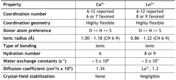
Os lantanídeos são substitutos isomórficos dos iões de cálcio, na medida em que possuem raios iónicos, preferência pelos átomos doadores e números de coordenação análogos. Devido a esta similaridade, estes elementos exibem uma acentuada atividade biológica e têm sido investigados devido às suas potenciais aplicações médicas. A afinidade dos lantanídeos pelo tecido ósseo já é reconhecida há décadas, contudo, só recentemente foi sugerido o seu poten-cial para substituir o cálcio ósseo e afetar o ciclo de remodelação óssea, através da modelação do desenvolvimento das células ósseas. O efeito dos lantanídeos em populações celulares rele-vantes para o metabolismo ósseo têm sido analisado em diversos trabalhos. Contudo, apenas um número limitado de trabalhos reportou o efeito destes elementos em células ósseas huma-nas cultivadas in vitro.
Neste contexto, analisaram-se os efeitos celulares e moleculares dos lantanídeos no desen-volvimento de células humanas precursoras de osteoblastos e osteoclastos. As culturas celulares de osteoblastos foram obtidas a partir de cabeças de fémur de pacientes (25-45 anos) subme-tidos a cirurgia ortopédica e foram caracterizadas para o conteúdo de ADN, apoptose, atividade da fosfatase alcalina (ALP), coloração histoquímica da ALP e colagénio, e coloração da F-actina. As culturas celulares de osteoclastos foram estabelecidas a partir de células percursoras isola-das de sangue periférico humano (PBMCs) e caracterizaisola-das para o conteúdo de ADN, apoptose, atividade da fosfatase ácida resistente ao tartarato (TRAP), número de células multinucleadas TRAP positivas e pela presença de células com anéis de actina e que expressam recetores de vitronectina e calcitonina. Adicionalmente, foi também analisado o envolvimento de algumas vias de sinalização associadas ao processo de osteoblastogénese e osteoclastogénese, na res-posta celular induzida pelos lantanídeos.
As culturas celulares estudadas foram cultivadas na presença de zinco e magnésio (como catiões de referência) e três lantanídeos diferentes (cério, lantânio e praseodímio) em concen-trações compreendidas entre 10-7 e 10-3 M e foram caracterizadas após 14 e 21 dias em cultura.
Os resultados obtidos indicaram que o cério (na maioria das concentrações testadas) não afetou significativamente a proliferação dos precursores osteoblásticos. Adicionalmente, foi observado que este catião, nas concentrações mais baixas testadas, induziu a diferenciação dos precursores osteoblásticos e que este efeito foi perdido nas concentrações mais altas testadas. A densidade dos precursores osteoclásticos foi regulada positivamente pelas concentrações mais baixas de lantânio, contudo, este efeito foi-se perdendo ao longo do tempo. Por outro lado, a diferenciação dos precursores osteoclásticos foi inibida pelas concentrações mais altas de lantânio em ambos os dias.
ii
O praseodímio, independentemente da concentração testada, pareceu actuar como um mo-dulador positivo da proliferação dos precursores osteoblásticos, enquanto a influência oposta foi observada relativamente à densidade dos precursores osteoclásticos. Embora este catião tenha mostrado a capacidade de regular negativamente a diferenciação tanto dos precursores osteoblásticos como dos precursores osteoclásticos, nas culturas dos precursores osteoclásticos este efeito só foi observado nas concentrações mais altas.
Todos os lantanídeos testados modularam o desenvolvimento das células osteoclásticas e osteoblásticas, embora tenham revelado diferentes perfis de modulação dos processos de os-teoblastogénese e osteoclastogénese. Adicionalmente, foi observado que o efeito promovido pelos lantanídeos depende da sua concentração e do tempo de cultura. Foi ainda observado que o mesmo lantanídeo pode afetar os dois tipos celularesde forma inversa. Em termos gerais as vias de sinalização testadas foram afetadas de forma diferente pelos lantanídeos, sugerindo que estes elementos afetam os processos de osteoblastogénese e osteoclastogénese através de diferentes mecanismos.
Em suma, este trabalho demonstrou a capacidade dos lantanídeos de modular o desenvol-vimento de células ósseas humanas e estabeleceu algumas das vias de sinalização que poderão estar envolvidas na resposta celular induzida pelos mesmos. Os resultados obtidos reiteraram a necessidade de compreender e manipular o comportamento dual destes elementos de forma a induzir o efeito pretendido. Deste modo, é necessário investigar com mais detalhe o efeito dos lantanídeos na atividade das células ósseas humanas e clarificar os seus mecanismos de ação.
iv
Abstract
Lanthanides are isomorphic replacements of calcium ions, as they possess analogous ionic radius, donor atom preferences and coordination numbers. By virtue of their resemblance to calcium, lanthanides exhibit a pronounced biological activity and have attracted intensive re-search interest for medical applications. Although the affinity of lanthanides for bone has been known for decades, their potential to exchange with calcium in bone and affect bone remod-eling cycle, by modulating osteoblast and osteoclast development, was only suggested a few years ago. Since then, several works have addressed the effect of lanthanides in cellular popu-lations with relevance to bone metabolism. Nevertheless, only a limited number of studies have reported the effect of lanthanides on human bone cells cultured in vitro. Furthermore, the signaling pathways involved on the cellular response have not yet been elucidated.
In this context, the aim of this study was to investigate the cellular and molecular effects of lanthanides on human osteoblast and osteoclast precursor cells development. Osteoblast precursor cell cultures were obtained from femur heads of patients (25-45 years old) undergoing orthopedic surgery procedures and were then studied for DNA content, apoptosis, alkaline phos-phatase (ALP) activity, histochemical staining of ALP and collagen, and F-actin staining. Oste-oclastic cell cultures were established from precursor cells isolated from human peripheral blood (PBMCs) and were characterized for DNA content, apoptosis, tartrate-resistant acid phos-phatase (TRAP) activity, number of TRAP-positive multinucleated cells, and the presence of cells displaying F-actin rings and expressing vitronectin and calcitonin receptors. Also the in-volvement of some osteoblastogenesis and osteoclastogenesis-related signaling pathways on cellular response were addressed.
Cell cultures were treated with zinc, magnesium (as reference physiological cations) and three different lanthanides (cerium, lanthanum and praseodymium) at concentrations of 10-7 M
to 10-3 M, and were characterized at days 14 and 21.
The results showed that cerium (in the majority of the concentrations tested) was not able to significantly affect the proliferation of osteoblast precursor cells. Moreover, it was observed that this cation induced the differentiation of osteoblast precursor cells at the lowest concen-trations tested, and that this effect was lost at the highest concenconcen-trations.
The density of osteoclast precursor cells was up-regulated by the lowest concentrations of lanthanum, nevertheless, this effect was also showed to be lost over time. On the other hand, the differentiation of osteoclast precursor cells was inhibited by the highest concentrations of lanthanum at both days.
Praseodymium, regardless of the concentration tested, appeared to act as a positive mod-ulator of osteoblast precursor cells proliferation, while the opposite influence was observed concerning the density of osteoclast precursor cells. Although, this cation was shown to be able to regulate negatively the differentiation of both osteoblast and osteoclast precursor cells, this effect on osteoclast cultures was only observed at the highest concentrations tested.
Hence, all lanthanides were shown to be able to affect osteoclastic and osteoblastic pre-cursor cells development although with different profiles on their osteoblastogenic and osteo-clastogenic modulation properties. Moreover, it was observed that the effect of the lanthanides is closely related to the concentration and culture time. Also, it was observed that the same lanthanide may affect osteoclastic and osteoblastic precursor cells development in an antago-nistic manner.
The signaling pathways involved in the process were overall affected differently by the different lanthanides tested, suggesting that they affect osteoblastogenesis and osteoclasto-genesis processes through different mechanisms.
In sum, the present study underlined the ability of lanthanides to modulate the develop-ment of human bone cells and outlined some of the signaling pathways involved in this cellular response. Furthermore, the results obtained reiterate the need to understand and to manipu-late the dual behavior of these elements to attain the required effect. Accordingly, further experiments are required to fully understand the effect of lanthanides on human bone cells activity and their mechanism of action.
Acknowledgments
This work was only possible thanks to the contribution of several people to which I want to express my gratitude:
To Professor João Rodrigues, my supervisor, for giving me the opportunity to undertake this work, and for his constantsupport, availability, incentive and knowledge transmitted.
To my co-supervisor, Professor Maria Helena Fernandes, who provided all the necessary conditionsfor the development of this thesis.
To Dra. Mónica Garcia for all the training, availability, support and patience.
Finally, I would like to thank my parents and sister for providing me with the conditions to take this course and for all the encouragement on the difficult days, and to João for the un-conditional love, support and patience.
Contents
Resumo ... i
Abstract... iv
Acknowledgments ... vii
List of Figures ... xii
List of Tables ... xv
Abbreviations and acronyms ...xvi
1. General Introduction ... 20 1.1 Bone Tissue ... 20 1.2 Macroscopic Morphology ... 20 1.3 Microscopic Morphology ... 21 1.4 Microscopic Organization ... 22 1.4.1 Matrix Mineralization ... 23 1.5 Bone Cells ... 25 1.5.1 Osteoblasts ... 25 1.5.2 Osteoclasts... 27
1.6 Bone Modelling and Remodelling ... 31
1.7 Bone and Calcium Homeostasis ... 32
1.7.1 Parathyroid Hormone, Calcitriol and Calcitonin ... 33
1.8 Lanthanides ... 34
1.8.1 Biochemistry of Lanthanides ... 35
1.8.2 Lanthanide Induced-Proliferation, Apoptosis and Differentiation ... 36
1.8.3 Lanthanides Distribution in the Skeleton ... 37
1.8.4 Lanthanides Interaction with Bone Matrix ... 38
x
1.9 Chapter Insights and Research Objectives ... 39
2. Materials and Methods ... 42
2.1 Materials ... 42
2.2 Methods ... 42
2.2.1 Osteoblastic Cell Cultures ... 42
2.2.2 Osteoclastic Cell Cultures... 42
2.2.3 Exposure to Lanthanides ... 43
2.2.4 Osteoblastic and Osteoclastic Cell Response to Lanthanides ... 43
3. Results ... 47
3.1 Osteoblastic Cell Response to Lanthanides ... 47
3.1.1 DNA Content ... 47
3.1.2 Apoptosis ... 48
3.1.3 ALP activity ... 50
3.1.4 Histochemical Staining of ALP and Collagen ... 51
3.1.5 F-actin Staining of Osteoblasts Precursor Cells ... 52
3.1.6 Intracellular Signalling Mechanisms ... 52
3.2 Osteoclastic Cell Response to Lanthanides ... 53
3.2.1 DNA Content ... 54
3.2.2 Apoptosis ... 55
3.2.3 TRAP activity ... 56
3.2.4 Number of TRAP-Positive Multinucleated Cells ... 57
3.2.5 PBMCs Displaying F-actin Rings and Expressing VNR and CTR ... 59
3.2.6 Intracellular Signalling Mechanisms ... 59
4. Discussion ... 63
4.1 Overview ... 63
4.2 Osteoblastic Cell Response to Lanthanides ... 64
4.3 Osteoclastic Cell Response to Lanthanides ... 68
4.4 General Discussion ... 71
5. Conclusion and Future Directions ... 74
5.1 Conclusion ... 74
5.2 Future Directions ... 75
xii
List of Figures
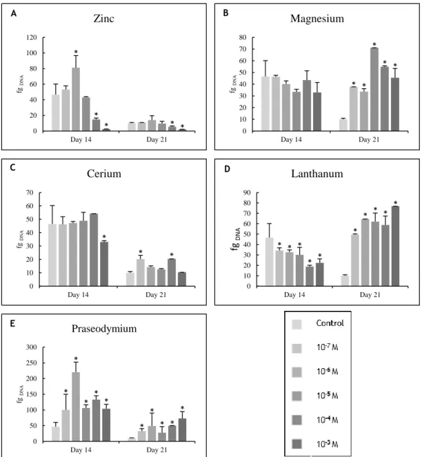
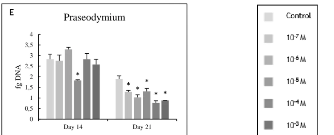
Figure 3.1 - DNA content of osteoblast precursor cells cultured in the presence of zinc,
magnesium (A and B) and lanthanides (C to E) at days 14 and 21 of culture. * Significantly different from the control. ... 48
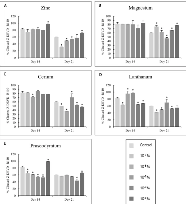
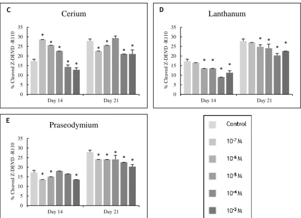
Figure 3.2 – Caspase-3 activity of osteoblast precursor cells cultured in the presence of zinc,
magnesium (A and B) and lanthanides (C to E) at days 14 and 21 of culture. * Significantly different from the control. ... 49
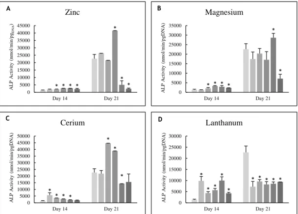
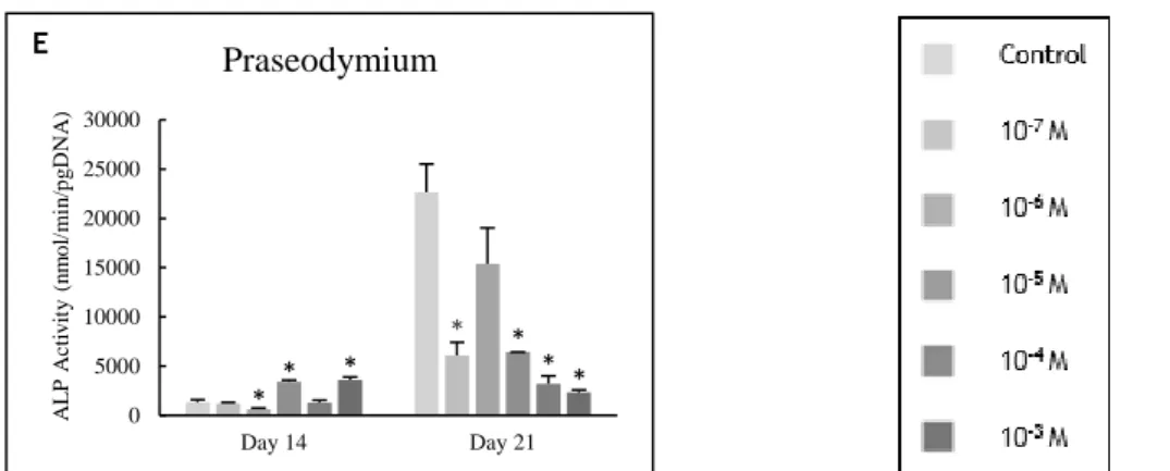
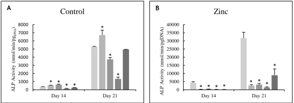
Figure 3.3 – ALP activity of osteoblast precursor cells cultured in the presence of zinc,
magnesium (A and B) and lanthanides (C to E) at days 14 and 21 of culture. * Significantly different from the control. ... 51
Figure 3.4 - Representative optical microscopy images of osteoblast precursor cell cultures at
day 14, following ALP histochemical staining: (A) control, (B) magnesium at 10-4 M and (C)
cerium at 10-6 M. Black bars represent 200 μm. ... 51
Figure 3.5 - Representative optical microscopy images of osteoblast precursor cell cultures at
day 14, following collagen histochemical staining: (A) control, (B) zinc at 10-5 M and (C) cerium
at 10-6 M. Black bars represent 200 μm. ... 51
Figure 3.6 - CLSM visualisation of osteoblast precursor cell cultures at day 21, stained blue for
F-actin: (A) control, (B) magnesium at 10-6 M and (C) praseodymium at 10-5 M. White bars
represent 70 μm. ... 52
Figure 3.7 – Osteoblast precursor cells cultured with selective inhibitors of the MEK, NFκB, PKC
and JNK signaling pathways and maintained in the absence (A) or in presence of the cations (B to F): zinc at 10-4 M, magnesium at 10-3 M, cerium, lanthanum and praseodymium at 10-5 M. Cell
responses were evaluated for ALP activity. * Significantly different from the control. ... 53
Figure 3.8 - DNA content of PBMCs cultured in the presence of zinc, magnesium (A and B) and
lanthanides (C to E) at days 14 and 21 of culture. * Significantly different from the control. 55
Figure 3.9 - Caspase-3 activity of PBMCs cultured in the presence of zinc, magnesium (A and
B) and lanthanides (C to E) at days 14 and 21 of culture. * Significantly different from the control. ... 56
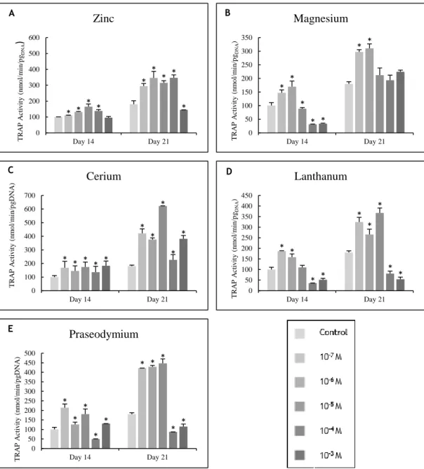
Figure 3.10 - TRAP activity of PBMCs cultured in the presence of zinc, magnesium (A and B)
and lanthanides (C to E) at days 14 and 21 of culture. * Significantly different from the control. ... 57
Figure 3.11 – Number of TRAP-positive multinucleated cells on PBMC cultures: in the presence
of zinc, magnesium (A and B) and lanthanides (C to E) at days 14 and 21 of culture. * Significantly different from the control. ... 58
Figure 3.12 - CLSM visualisation of PBMC cultures at day 21, stained blue for F-actin and green
for VNR and CTR: control (A and B), zinc at 10-6 M (C and D) and lanthanum at 10-5 M (E and F).
White bars represent 170 μm. ... 59
Figure 3.13 - PBMCs cultured with selective inhibitors of the MEK, NFκB, PKC and JNK signaling
pathways and maintained in the absence (A) or in presence of the cations (B to F): zinc at 10-4
M, mag-nesium at 10-3 M, cerium, lanthanum and praseodymium at 10-5 M. Cell responses were
List of Tables
xvi
Abbreviations and acronyms
Arp Actin-Related Proteins
Akt Protein Kinase B
ALP Alkaline Phosphatase
AP-1 Activator Protein-1
BAD Bcl-2-Associated Death Promoter
BSP Bone Sialoprotein
BMP Bone Morphogenetic Proteins
BMU Bone Multicellular Unit
BSA Bovine Serum Albumin
cAMP Cyclic Adenosine Monophosphate
CASR Calcium - Sensing Receptor
CTR Calcitonin Receptors
CLSM Confocal Laser Scanning Microscopy
Dlx5 Distal-Less Homeobox-5
DNA Deoxyribonucleic Acid
DTT Dithiothreitol
EDTA Ethylenediamine Tetraacetic Acid
ELISA Enzyme-Linked Immunosorbent Assay
ERK Extracellular Signal-Regulated Kinase
FGF Fibroblast Growth Factor
Gla Gamma-Carboxyglutamic Acid
HAp Hydroxyapatite
IGF Insulin-Like Growth Factor
IL Interleukin
IκB Inhibitory κB
JNK c-Jun N-terminal kinase
LDH Lactate Dehydrogenase
Ln Lanthanides
MAPK Mitogen-Activated Protein Kinase
M-CSF Macrophage Colony-Stimulating Factor
Mcl Myeloid Cell Leukemia
MEK Methyl Ethyl Ketone
MITF Microphthalmia-Associated Transcription Factor
MMP Matrix Metalloproteinase
MSC Mesenchymal Stem Cell
MTT 3-(4, 5- dimethylthiazol-2-yl)-2, 5-diphenyltetrazolium bromide
MV Matrix Vesicle
NFATc1 Nuclear Factor Of Activated T-Cells Cytoplasmic 1
NF-κB Nuclear Factor-Kappa β OC Osteocalcin ON Osteonectin OPG Osteoprotegerin OPN Osteopontin ON Osteonectin Osx Osterix PBS Phosphate-Buffered Saline
PBMC Peripheral Blood Mononuclear Cell
PDGF Platelet-Derived Growth Factor
PG Prostaglandin
PI3K Phosphoinositide 3-Kinase
PKC Protein Kinase C
pNPP p-nitrophenylphosphate
PTH Parathyroid Hormone
RANK Receptor Activator of Nuclear Factor Kappa-β
RANKL RANK Ligand
RGD Arginine-Glycine-Aspartate
ROS Reactive Oxygen Species
Runx2 Runt-Related Transcription Factor 2
Tcf T-cell factor
TGF Transforming Growth Factor
TNF Tumour Necrosis Factor
xviii
TRAP Tartrate-Resistant Acid Phosphatase
TNFR Tumor Necrosis Factor Receptor
VNR Vitronectin
WASp Wiskott-Aldrich syndrome protein
20
Chapter 1
General Introduction
1.1 Bone Tissue
Bone is a dynamic and specialized connective tissue, with a unique capacity to heal and remodel without leaving a scar (Salgado, Coutinho, & Reis, 2004). One of the primary functions of this tissue is to provide the mechanical integrity for both locomotion and protection of the vital organs. Metabolically, bone maintains the mineral homeostasis, regulates the acid-base balance, acts as a reservoir of growth factors and cytokines, and stores energy reserves as lipids in areas filled with yellow bone marrow. Furthermore, bone tissue encloses the red bone mar-row thus providing the environment for haematopoiesis to occur (Kini & Nandeesh, 2012).
This tissue is renewed continuously in the adult skeleton in response to a variety of stimuli by the process of bone remodelling (Datta, Ng, Walker, Tuck, & Varanasi, 2008). This process allows each bone to adapt to changes in biomechanical forces and guarantees the removal of old and microdamaged bone, replacing it with new and mechanically stronger bone which en-sures the preservation of bone strength (Clarke, 2008). Bone remodelling involves the interac-tion between different cell phenotypes and is regulated by a variety of biochemical and me-chanical factors (Hadjidakis & Androulakis, 2006). A disruption or imbalance in these processes can lead to either an increase or decrease in bone mineral density that may be detrimental to skeletal strength. Accordingly, the process of bone turnover must be carefully regulated in order to maintain bone strength (Datta et al., 2008).
1.2 Macroscopic Morphology
On a macroscopic level, bone exists in one of two forms: cortical bone, also called dense or compact bone, and trabecular bone, also called cancellous or spongy bone (Bancroft & Mikos, 2002; Sandhu, 2003). Overall the adult human skeleton is constituted of 80% cortical bone and 20% trabecular bone (Buckwalter, Glimcher, Cooper, & Recker, 1995; Hadjidakis & Androulakis, 2006). However, different types of bone and skeletal sites within the bone present different ratios of cortical to trabecular bone. For example, the ratio of cortical to trabecular bone in
the vertebra is 25 to 75 percent, in the femoral head 50 to 50 percent and 95 to 5 percent in the radial diaphysis (Clarke, 2008).
Cortical bone presents two types of surfaces. The surface on its inner side, facing the bone marrow, is known as the endosteum, and the surface facing the surrounding soft tissues, is known as the periosteum (Morgan, Barnes, & Einhorn, 2010). Cortical bone is dense and orga-nized, providing protection and mechanical support. It is only 10% porous, and therefore pro-vides space for only a low concentration of cells and blood vessels (Suárez-González & Murphy, 2008).This tissue makes up the outer tubular shell of long bones and the outer surface of small and flat bones in the skeleton (Bancroft & Mikos, 2002).
Trabecular bone is loosely organized and highly porous (50-90% porosity) and contains a functional vasculature and a bone marrow space (Suárez-González & Murphy, 2008). Due to its characteristics, trabecular bone is more elastic, has a higher turnover rate and performs a more active metabolic function than cortical bone (Downey & Siegel, 2006). Trabecular bone is found near the ends of long bones, in the interior of small bones, and between the surfaces of flat bones (Bancroft & Mikos, 2002).
1.3 Microscopic Morphology
At the microscopic level, bone is described as either woven or lamellar bone, depending on the organization of the collagen fibres (Becken, Hilger, & A.E., 2005).
Woven bone, also designated as primary bone, is characterized by a random organization of the collagen fibres. This type of bone exists mainly during embryonic and fetal development. In later years, woven bone is found at sites of fracture healing and in some metaphyseal regions of the growing skeleton since it is laid down more quickly than lamellar bone (Downey & Siegel, 2006; Kini & Nandeesh, 2012; Morgan et al., 2010). Lamellar bone, which is the major portion of both cortical and trabecular bone in adults, is more highly organized and specialized than woven bone. Collagen fibres in the lamellar bone are aligned in thin sheets, called lamellae, which are stacked in a plywood-type arrangement (Morgan et al., 2010).
Both woven and lamellar bones contain small cavities called lacunae which are connected to each other by means of tubular canals called canaliculi. The lacunae are arranged along the interfaces between the lamellae in the lamellar bone. Osteocytes, a specific type of bone cells, remain entrapped within these lacunae and receive and transmit nutrients and stimuli from and to the bone through the canaliculi (Cowin, 2007; Kamioka, Honjo, & Takano-Yamamoto, 2001). Lamellar bone is also commonly arranged in smaller cylindrical structures, called secondary osteons or Harversian systems (Gore, Unnikrishnan, Hussein, & Morgan, 2012). Osteons make up approximately two-thirds of cortical bone volume. Each osteon consists of 10 to 30 concen-tric rings of lamellae, which surround a central cavity, the Haversian canal, in which blood vessels and nerves are contained. Each osteon is in direct contact with the periosteum, the bone marrow and with other osteons through horizontally oriented canals, called Volkmann’s canals. The osteons have a typical diameter of approximately 200 µm and lengths of 1 to 3 mm. The outer border of each osteons contains a cement line that consists of a layer of mineralized matrix attaching adjacent osteons together (Gore et al., 2012; Morgan et al., 2010).
In trabecular bone, the counterpart to the osteon is the trabecular packet (Sandhu, 2003). Although, trabeculae are primarily composed of lamellar bone arranged in packets, ticker
tra-22
beculae can also contain secondary osteons (Gore et al., 2012). Trabecular osteons are semilu-nar in shape, normally approximately 35 mm thick, and composed of concentric lamellae. It is estimated that there are 14 x 106 trabecular osteons in healthy human adults, with a total
trabecular area of approximately 7 m2 (Clarke, 2008).
1.4 Microscopic Organization
Bone is a composite material containing an inorganic and an organic phase. By weight, approximately 60% of bone tissue is inorganic matter, 8 to 10% water and the remainder, is organic matter (Gong, Arnold, & Cohn, 2005).
The mineral (or inorganic) phase of bone is mainly formed by a calcium-deficient, car-bonate-containing, poorly-crystalline analogue of the naturally occurring mineral hydroxyap-atite (Ca10(PO4)6(OH)2 or HAp) (A.L. Boskey, 2005). The small plate-shaped (20-50 mm long, 15
nm wide, and 2-5 nm thick) apatite crystals contain impurities, most notably small amounts of carbonate in place of the phosphate groups. Other noticed substitutions are magnesium, potas-sium, strontium and sodium in place of the calcium ions and chloride and fluoride in place of the hydroxyl groups. These impurities reduce the crystallinity of the apatite resulting in the change of certain physical properties, such as the solubility (Zhu, Robey, & Boskey, 2010). Hence, bone mineral crystals are more soluble than geologic apatite, which facilitates bone to act as a reservoir for calcium and phosphate and other ions (Hammett-Stabler, 2004).
The organic matrix (or osteoid) is mainly composed of type I collagen that constitutes up to 85-90% of the total protein in the bone matrix. The remaining 10-15% in composed of noncolla-genous proteins, whereas the cellular content comprises less than 1% of the bone mass (Robey & Boskey, 2008). Type I collagen is the most abundant protein in vertebrates. Besides being expressed in bone, type I collagen is also expressed in most of the other connective tissues, especially in tendons, ligaments and dermis. The repetitive nature of the amino acid sequences of collagen, which consists of Gly-X-Y, where X and Y are often proline and hydroxyproline residues, allows the protein to assemble into triple helical structures. Type I collagen is se-creted as a propeptide, but the globular ends are cleaved rapidly by specific proteases, in order that shorter molecules assemble to form fibrils (Bou-Gharios & Crombrugghe, 2008). These fi-brils are gathered in bundles to form collagen fibres. Noncollagenous proteins and mineral are found within gaps, named hole zones, that exists at the ends of collagen molecules and within pores formed between the sides of parallel molecules (Gore et al., 2012; Morgan et al., 2010).
Noncollagenous proteins present in bone matrix appear to regulate the organization, turn-over, and mineralization of the bone matrix. In fact, most of these proteins appear to have more than one function (A.L. Boskey, 2005).
Osteocalcin (OC) is the major non-collagenous protein of bone, accounting for 10 to 20% of the noncollagenous protein content (van Gaalen et al., 2008). OC is exclusively synthesized by osteoblasts, odontoblasts, and hypertrophic chondrocytes and is only marginally detectable during the earlier phases of proliferation and matrix maturation (Seibel, 2001). This protein contains three vitamin-K dependent gamma-carboxyglutamic acid (Gla) residues, which are re-sponsible for the calcium-binding properties (van Leeuwen, van der Eerden, van de Peppel, Stein, & Lian, 2013). Due to its interaction with HAp it is believed that OC regulates the growth and maturation of bone crystals (Gundberg, 2003). Moreover, some investigators have suggested that OC may act as a chemoattractant for osteoblasts (Chenu et al., 1994).
Osteonectin, osteopontin and bone sialoprotein are multifunctional phosphorylated glyco-proteins with high affinity for bone mineral carrying one or more arginine-glycine-aspartate (RGD) sequences, thus capable to linking cells, mineral and matrix (A.L. Boskey, 2005). Oste-onectin (ON) is a glycoprotein secreted by the osteoblast that binds collagen, calcium and Hap (van Leeuwen et al., 2013). It is assumed that this molecule supports bone remodelling, regu-lates cell proliferation and cell-matrix interactions, and stimuregu-lates the production of matrix metalloproteinases (Sroga & Vashishth, 2012). Osteopontin (OPN) is a phosphorylated glycopro-tein that is expressed in several tissues, including bone, kidney and in the immune and vascular systems (Lund, Giachelli, & Scatena, 2009). In bone, OPN is produced by osteoblasts, osteocytes, macrophages, and osteoclasts and is found at the late stage of osteoblastic maturation, during matrix formation and mineralization (Robey, 2008; Zhu et al., 2010).OPN promotes osteoclast binding to the extracellular matrix and activates intracellular signalling pathways (McNamara, 2011; Robey, 2008). During normal bone mineralization, osteoclast‐derived OPN inhibits the formation of HAp, thus acting as an inhibitor of mineralization (Hunter, 2013). Bone sialoprotein (BSP) comprises 15% of the total noncollagenous proteins in bone (Wuttke et al., 2001). BSP expression marks a late stage of osteoblastic differentiation and an early stage of matrix min-eralization (Robey, 2008). This sialoprotein binds to collagen and promotes osteoblast attach-ment (A.L. Boskey, 2005). Additionally, it is considered that BSP also plays a role in matrix mineralizationdue to its high affinity for calcium and because of the timing of its appearance in relation to the timing of mineral emergence (Robey, 2008). Although, alkaline phosphatase (ALP) is found mainly distributed on the cell membrane and in matrix vesicles, it is also a bone matrix constituent (Bonucci, 2013). ALP is considered an important regulator of mineralization, due to its ability to enzymatically degrade pyrophosphate, a mineralization inhibitor (Gade et
al., 2011).
Proteoglycans play important roles in organizing the bone extracellular matrix, taking part in the structuring of the tissue itself as active regulators of collagen fibrillogenesis (Lamoureux, Baud'huin, Duplomb, Heymann, & Rédini, 2007). The two most abundant proteoglycans present in bone matrix are decorin, and biglycan (Zhu et al., 2010). These proteoglycans bind to type I collagen and interact with several growth factors, therefore, it is assumed that they play a part in modulating the activity of growth factors and matrix proteins (Robey, 2002). Furthermore, decorin and biglycan were both found to inhibit bone cell attachment, apparently by binding to fibronectin and inhibiting its cell-matrix binding abilities (Robey, 2002).
Growth factors and cytokines such as, transforming growth factor-β (TGF-β), insulin-like growth factor (IGF), osteoprotegerin, tumour necrosis factors (TNFs), interferon-γ and bone morphogenetic proteins (BMPs 2-10) are present in very small quantities in bone matrix. These proteins are responsible for the regulation of bone cell behaviour, namely for, cell differentia-tion, activadifferentia-tion, growth and turnover. Additionally, it is thought that these growth factors act as coupling factors linking bone resorption and formation processes (Zhu et al., 2010).
1.4.1 Matrix Mineralization
Mineralization of bone collagen fibrils occurs in an organized manner. Initially crystal ap-pear in hole zone regions within individual fibrils, separated by unmineralized regions. Gradu-ally they start to appear in an increasing number of hole zone regions. Ultimately, crystal growth, embraces the zone of the collagen fibrils between hole zones and occupies all of the available space within the fibrils (Buckwalter et al., 1995).
24
One possible mechanism of HAp crystal formation is via extracellular matrix vesicles (MVs) (New & Aikawa, 2013). MVs are small (approximately 100 nm in diameter) membrane particles, which bud off from the plasma membrane of mineralizing cells (chondrocytes, osteoblasts and odontoblasts) and are released into the pre-mineralized organic matrix (Golub, 2011; Vigorita, 2008). The signals that promote MVs release are not well understood, although concentrations of intracellular calcium and extracellular phosphate may be important (Orimo, 2010).
Mineralization occurs in two phases: an initial formation of apatite within the MV, and a subsequent propagation phase in the matrix (Golub, 2011). The first phase starts with the in-corporation of calcium into the MV by calcium-binding proteins and calcium-binding lipids pre-sent in the MV membrane. Phosphate concentration is also raised during this phase through the enzymatic activity of phosphohydrolases, such as ALP, which reside on the MV membrane (An-derson, 2003; Leonor, Azevedo, Alves, & Reis, 2005; Nair & Jagannathan, 2013). At some point, the elevation of calcium and phosphate within the protective environment of the membrane leads to the precipitation of amorphous calcium phosphate mineral. The amorphous calcium phosphate mineral is later converted into HAp via octacalcium phosphate. Subsequently, HAp crystals penetrate the MV membrane and get exposed to the extracellular fluid leading to the beginning of the second phase of mineralization process. The breakdown of MVs is assisted by the hydrolytic action of phospholipases and proteases (Anderson, 2003; Nair & Jagannathan, 2013). During the second phase the preformed apatite from the MVs migrate to collagen, be-come inserted into the aligned hole zones of the fibrils, and then undergo maturation (Golub, 2011; Leonor et al., 2005). Rates of mineralization are affected by levels of calcium and phos-phate in the extracellular fluid, pH, and by matrix components that are able to accelerate or retard mineral propagation (Vigorita, 2008).
Several families of proteins associated with the collagen matrix are involved in regulation of the mineralization process, as both inhibitors and promoters of mineralization depending on the extent of post-translational modification and/or their concentration (A.L Boskey, 2007). BSP, ON, dentin sialophosphoprotein, and ALP are nucleators or initiators of mineral crystal formation. These proteins bind to calcium and/or phosphate ions, creating a surface similar to the apatite surface that provides the start of nucleation (Clarke, 2008; Zhu et al., 2010). Pro-teins, such as aggrecan, OPN, dentin matrix protein-1, α2-HS glycoprotein, and albumin act as
inhibitors of mineral crystal formation. These proteins can chelate calcium or phosphate ions creating a protected environment around the crystal nucleus which prevents crystal growth. On the other and OC, vitronectin and matrix Gla protein bind to one or more faces of the growing crystal, blocking crystal growth in one or more directions or blocking growth beyond a specific size. Other proteins, such as decorin, thrombospondin, fibronectin, vitronectin and versican bind to the collagen backbone of the matrix and to other non-collagenous proteins, changing their conformation and their ability to affect crystal nucleation and growth (Zhu et
al., 2010). Vitamin D participates indirectly in the promotion of bone matrix mineralization.
Calcitriol, of the active form of vitamin D, maintains the appropriate serum calcium and phos-phorus concentrations required for passive mineralization, through the stimulation of intestinal absorption of these minerals (Clarke, 2008).
1.5 Bone Cells
The main cell types associated with bone homeostasis are osteocytes, osteoclasts and os-teoblasts. These cells account for approximately 90% of all cells in the adult skeleton (Morgan
et al., 2010; Sommerfeldt & Rubin, 2001). Bone cells are originated from two particular cell
lines lineages: a mesenchymal stem cell lineage and a hematopoietic stem cell lineage. The osteoblasts and osteocytes are derived from mesenchymal stem cells (MSCs), whereas preoste-oclasts and ostepreoste-oclasts are originated from hematopoietic stem cells (Buckwalter et al., 1995; Downey & Siegel, 2006).
1.5.1 Osteoblasts
Osteoblasts are the cells responsible for the synthesis and mineralization of osteoid, the protein component of bone tissue. Moreover, osteoblasts are also involved in the regulation of osteoclast differentiation and activity (Ferrari et al., 2000; Kini & Nandeesh, 2012).
As mentioned formerly, osteoblasts derive from pluripotent MSCs, which prior to osteoblast commitment can also differentiate into fibroblasts, chondrocytes, myoblasts and bone marrow stromal cells depending on the activated signalling transcription pathways (A. Yamaguchi, Komori, & Suda, 2000b).
Osteoblasts are cuboidal cells, usually found in a single layer adherent to bone surface and contain an extensive and oriented secretory organelle apparatus that includes a large Golgi complex near the nucleus, a well-developed rough endoplasmatic reticulum and a high mito-chondrial content (Kartsogiannis & Wah Nga, 2004; Mackie, 2003).
The maintenance of osteoblastic function and the ability of these cells to respond to met-abolic and mechanical stimuli is sustained by cell-matrix and cell-cell interactions that occur through a variety of transmembranous proteins, such as, integrins, conexins and cadherins and specific receptors for cytokines, hormones and growth factors. In addition, the tight junctions formed between adjacent osteoblasts have regions of the plasma membrane specialized in ve-sicular trafficking and secretion (Ferrari et al., 2000; Sommerfeldt & Rubin, 2001).
As osteoblasts differentiate from their precursors they begin to secrete bone matrix pro-teins into the region of unmineralized matrix between the cell body and the mineralized matrix (Gay, Gilman, & Sugiyama, 2000). The lifespan of human osteoblasts it is of approximately 8 weeks, and during this period osteoblasts lay down 0.5 to 1.5 μm of osteoid per day (Sommer-feldt & Rubin, 2001).
As mentioned, besides their role in osteoid synthesis, osteoblasts are also involved in oste-oid mineralization. As osteoste-oid becomes mineralized, osteoblasts follow one of three pathways: persist as active osteoblasts and disappear from the site of bone formation; become surrounded by their own calcified matrix and change their phenotype turning into osteocytes or become fairly inactive, decrease their synthetic activity and assume a flatter shape of bone lining cells (Dallas & Bonewald, 2010).
1.5.1.1 Osteoblastogenesis
Osteoblastogenesis can be divided into three stages, namely proliferation, matrix develop-ment, and maturation and mineralization (Eglence, Duivenvoorden, Ghert, & Singh, 2009). In the first stage, cells proliferate intensively and produce some extracellular matrix proteins,
26
such as fibronectin and type I collagen (Przekora & Ginalska, 2015). The second stage is char-acterized by a modification in the composition of extracellular matrix, a downregulation of proliferation and activation of genes correlated with matrix maturation and organization (Le-onor, Gomes, Bessa, Mano, & Reis, 2008). The third stage of differentiation is connected with the mineralization process. During this stage osteoblasts have moderate ALP activity, high min-eralization activity and express high levels of proteins that are correlated with mineral deposi-tion (mainly, OPN and OC) (Przekora & Ginalska, 2015).
The markers most frequently used to follow osteoblasts differentiation include type I col-lagen, ALP and OC. Overall, type I collagen and ALP are thought as early osteoblastogenesis markers, while, OC is considered an advanced marker of osteoblast differentiation due to it’s closely association with bone matrix mineralization (Eglence et al., 2009).
Two transcription factors have been demonstrated to be required for osteoblast formation and differentiation, namely, runt-related transcription factor 2 (Runx2) and Osterix (Osx).
Runx2 has been described as a master regulator of osteoblastogenesis (Baek, Choi, & Kim, 2014). Expression of Runx2 is both necessary and sufficient for MSC differentiation towards the osteoblastic lineage. This factor directly stimulates the transcription of osteoblast-related genes, such as those encoding TGF-β receptor, ALP, OC, OPN, vitamin D receptor, BSP, type I collagen and collagenase, by binding to specific enhancer regions containing the core sequence (Mackie, 2003). Hence, Runx2-deficient mice exhibit a complete lack of both intramembranous and endochondral ossification due to the absence of osteoblast differentiation (Baek et al., 2014). Runx2 can be phosphorylated and thus activated by the mitogen-activated protein kinase (MAPK) cascade via stimulation of α2β1-integrins on the osteoblast surface. Both expression and activity of Runx2 are tightly controlled by other transcription factors and by protein–DNA or protein–protein interactions. Stat1, Sox9, Sox8, Aj18, MEF, Nrf2, and YAP have been reported to repress Runx2 expression, while, Rb, TAZ, HoxA10, BAPX-1, Smad1&5, CEBP/β&δ, and Menin actively enhance the function of Runx2 (J.F.L. Chau, Leong, & Li, 2008).
The second required transcription factor for osteoblast differentiation is the zinc finger-containing transcription factor Osx (J.F.L. Chau, Leong, & Li, 2009). It has been demonstrated that Osx is necessary for bone formation and mineralization in vivo (Nakashima et al., 2002). Genetic inactivation of Osx in mice results in completely lack of mineralized bone (Baek et al., 2014).
Additional studies indicated that Osx acts downstream of Runx2 in the transcriptional cas-cade of osteoblast differentiation since Osx knock-out mice express Runx2, but Osx is not ex-pressed in Runx2 knock-out mice (Baek et al., 2014; C. Zhang et al., 2011). The mechanism underlying the regulation of Osx expression in osteoblasts is still unclear. However, it was shown that both BMP-2 and IGF-1 can induce Osx expression in undifferentiated MSCs. Furthermore, ascorbic acid and 1,25(OH)2 vitamin D3, have also been shown to up-regulate Osx expression.
On the other hand, some studies emphasized the ability of negative regulators of osteoblasto-genesis to inhibit Osx expression. For instance, TNF was demonstrated to inhibit Osx mRNA in pre-osteoblastic cells (C. Zhang 2010).
BMP, Wnt, and Notch signalling pathways play important roles in osteoblast differentiation (Lin & Hankenson, 2011).
The first indication that Wnt signalling plays a critical role in bone formation came from human genetic studies where recessive loss-of-function mutations in lipoprotein receptor-re-lated protein (LRP) 5 were linked to osteoporosis, while, dominant missense LRP5 mutations
were associated with high bone mass diseases (Lin & Hankenson, 2011; Zuo et al., 2012). Ca-nonical Wnt signalling in osteoblastogenesis has been linked to Runx2. The Runx2 gene promoter contains a Wnt-responsive T-cell factor (Tcf) regulatory element, and both β-catenin and Tcf1 are recruited to the Runx2 locus. Additionally, transient activation of Wnt/β-catenin signalling in MSCs in vitro induces expression of bone lineage genes such as Distal-less homeobox-5 (Dlx5) and Osx (Lin & Hankenson, 2011).
In vitro and in vivo studies have established that BMP-Smad signalling regulates osteoblast
differentiation from MSC, osteoprogenitor cell expansion, osteoblast bone formation activity, and its coupling to osteoclasts (J.F.L. Chau et al., 2009). BMP2 activates Smad1/5/8 signalling and regulates the transcription of osteogenic genes, including Dlx5, which is a key mediator of BMP2-induced expression of Runx2 (Jang, Kim, Lee, Son, & Koh, 2011). In vitro studies with a human marrow stroma derived cell line, demonstrated that BMP2 treatment promotes the in-crease of both Runx2 gene expression and ALP levels (Gori, Thomas, Hicok, Spelsberg, & Riggs, 1999).
MAPKs, namely, the p38, extracellular signal-regulated kinase (ERK) 1/2 and c-Jun N-ter-minal kinase (JNK) 1/2 were shown to promote the expression and activation of Runx2 (R. L. Huang, Yuan, Tu, Zou, & Li, 2014). Additionaly, the inhibition of p38 MAPK was shown to down-regulate Osx expression and reduce osteoblast differentiation (J.F.L. Chau et al., 2008).
As with BMP and Wnt signalling in osteogenesis, Runx2 function is also influenced by Notch signalling. Runx2 transcriptional activity is physically antagonized by the protein encoded by Notch target gene Hey1 (Lin & Hankenson, 2011). Transient transfection of MSCs with Notch intracellular domain, Hey decreases Runx2 transactivity. In addition, Notch intracellular do-main can interact directly with Runx2 protein and repress terminal osteoblastic differentiation
in vitro (Lin & Hankenson, 2011).
In addition, several hormones, such as, IGF-1, parathyroid hormone (PTH), and glucocorti-coids influence osteoblast differentiation. For instance, PTH has been found to regulate the expression and activity of Runx2 and to inhibit Osx expression (Barbuto & Mitchell, 2013). More-over, IGF-1 has been shown to promote osteoblast differentiation and mineralization in vitro (W. Zhang et al., 2012). Lastly, glucocorticoid have been shown to inhibit osteoblast differen-tiation through the suppression of cytokines such as interleukin (IL)-11 (Rauch et al., 2010).
1.5.2 Osteoclasts
Osteoclasts are highly specialized, multinucleated and terminally differentiated cells gen-erally regarded as the only cells in the body capable of resorbing bone (Chambers, 2000; Clarke, 2008; Itzstein, Coxon, & Rogers, 2011).
Mobilization of osteoclasts precursors involves their release from bone marrow to the blood circulation and their guidance to bone resorption sites, where these cells fuse into multinucle-ated cells to form mature osteoclasts, a process that is regulmultinucle-ated by osteoblasts and stromal cells of the bone marrow (Boyle, Simonet, & Lacey, 2003; Soysa, Alles, Aoki, & Ohya, 2012)
Osteoclasts contain large numbers of mitochondria and acidic vacuoles that carry acid phos-phatases and other lysosomal enzymes. Compared to the cytoplasmic volume of the cells, the rough endoplasmic reticulum is scarce and the Golgi apparatus consists of few flattened cister-nae around the nuclei, suggesting that mature osteoclasts do not synthesise large amounts of protein (Stenbeck, 2002).
28
Bone resorbing cells are highly migratory cells with a relatively short lifespan and consid-ered rare in bone with only two to three cell per mm (Miyazaki et al., 2012). Activated osteo-clasts are able to resorb 200000 µm3 of bone per day, an amount of bone formed by seven to
ten generations of osteoblasts (Sommerfeldt & Rubin, 2001).
1.5.2.1 Osteoclast Activity
Osteoclast activation is initiated upon cell attachment to bone matrix, an event leading to osteoclast actin cytoskeletal reorganization. Cytoskeletal organization promote osteoclast po-larization and the formation of new membrane domains, namely, the sealing zones and the ruffled membrane (Bellido, Plotkin, & Bruzzaniti, 2014).
The sealing zone appears as a ring of filamentous actin (F-actin, known as the actin ring), consisting of dynamic, densely packed small actin punctuate structures, called podosomes. These consist of a core of densely packed actin filaments and F-actin-associated proteins such as cortactin, Wiskott-Aldrich syndrome protein (WASp) and actin-related proteins (Arp) 2/3, surrounded by integrins and attachment-related proteins such as vinculin and talin (Itzstein et
al., 2011).
The attachment of osteoclast to bone matrix is controlled by the vitronectin receptor, αvβ3 receptor, which binds to RGD sequences within matrix proteins, such as vitronectin, OPN, and type I collagen (Bellido et al., 2014).
As the osteoclast prepares to resorb bone, it attaches to the bone matrix through the sealing zone and forms another specific membrane domain, the ruffled border (Väänänen, Zhao, Mulari, & Halleen, 2000). The ruffled border is a highly convoluted membrane domain formed by the fusion of targeted transport vesicles with the apical membrane (Bellido et al., 2014). This membrane, together with its underlying resorption lacuna create the bone-resorbing organelle in osteoclasts (Zhao & Väänänen, 2006).
As mentioned, the main physiological function of osteoclasts is to degrade mineralized bone matrix. This involves dissolution of crystalline HAp and proteolytic cleavage of the organic ma-trix, which is rich in collagen. Before proteolytic enzymes can reach and degrade collagenous bone matrix, tightly packed HAp crystals must be dissolved (Väänänen et al., 2000). Osteoclasts resorb bone in resorption lacunae by generating a pH gradient between the cell and bone sur-face (Hienz, Paliwal, & Ivanovski, 2014). The low pH in the resorption lacuna is achieved by the action of ATP-consuming vacuolar proton pumps both at the ruffled border membrane and in intracellular vacuoles (Sagalovsky, 2013). Protons are provided to the proton pumps by carbonic anhydrase II, which is highly expressed in osteoclast cytosol (Baron & Horne, 2005). Carbonic anhydrase II converts CO2 and H2O into H+ and HCO3- (Nakamura, 2007). The HCO3- ions are
exchanged for Cl- through an anion exchanger located in the basolateral membrane, leading to
continued availability of Cl- for acidification of the resorption lacuna (Sagalovsky, 2013). The
hydrochloric acid formed in the resorption lacuna dissolves the HAp component of bone matrix and exposes the organic matrix to the enzymatic attack of cysteine proteases and metallopro-teinases (Bellido et al., 2014; Hienz et al., 2014). The degradation of the demineralized organic component of the bone matrix is primarily due to the action of cathepsin K, a member of the papain family of cysteine proteases that is highly (although not exclusively) expressed by acti-vated osteoblasts. Cathepsin readily degrades type I collagen, with an optimal activity at ap-proximately pH 6.0 (Arnett, 2013). Thereafter, matrix metalloproteinases (MMPs), such as
gelatinase A (MMP-2), stromelysin (MMP-3), and collagenase (MMP-1), continue with the matrix degradation process (Hienz et al., 2014).
After matrix degradation, the degradation products are removed from the resorption lacuna via a transcytotic vesicle transport process oriented towards the centrally located functional secretory domain of the basolateral membrane (Mulari, Zhao, Lakkakorpi, & Väänänen, 2003).The specific enzyme tartrate-resistant acid phosphatase (TRAP) is located in cytoplasmic vesicles, which fuse to the transcytotic vesicles to destroy the endocytosed material. When the osteoclast moves away from the resorption lacuna, phagocytes clean up the debris, and osteo-blasts move in to begin bone formation (Hienz et al., 2014). Additionally, osteoclasts possess some mechanisms to guarantee the maintenance of cytoplasmic pH homeostasis, since the ex-trusion of large amounts of acid would eventually cause the rise of osteoclast intracellular pH that would lead to detrimental effects on many cellular processes. Hence, osteoclast intracel-lular alkalinisation is compensated by the action of the HCO3−/Cl− exchanger in the basolateral
membrane (Bellido et al., 2014).
1.5.2.2 Osteoclastogenesis
The first step in osteoclastogenesis is the commitment of the hematopoietic stem cell to the myeloid lineage. The transcription factor PU.1 plays a central role in this step. Its absence
in vivo results in general myeloid lineage deficiencies (Biosse-Duplan, Horne, & Baron, 2012).
Another transcription factor that is crucial to early stage of osteoclast development is microph-thalmia-associated transcription factor (MITF). MITF-deficient mice display a suitable macro-phage differentiation, but an impaired osteoclast differentiation, which might indicate that this transcription factor functions downstream of PU.1 in osteoclast differentiation, and regu-lates the balance between macrophage and osteoclast fate (Wei & Wan, 2013).
Two cytokines are essential and sufficient for osteoclastogenesis, specifically, receptor ac-tivator of nuclear factor-kappa B (NF-κB) ligand (RANKL) and macrophage colony-stimulating factor (M-CSF). M-CSF contributes to proliferation, survival, and differentiation of early osteo-clasts precursors (Ross, 2006). In turn, RANKL activates downstream signalling pathways that control osteoclast differentiation and bone resorption (Gupta et al., 2010)
M-CSF is a homodimeric glycoprotein produced by several cell types, such as granulocytes, endothelial cells, fibroblasts, osteoblasts and lymphocytes (Costa-Rodrigues, Teixeira, Sampaio, & Fernandes, 2010). Loss of function mutation in the M-CSF gene originates an osteopetrotic phenotype due to the lack of osteoclasts (Provot, Schipani, Wu, & Kronenberg, 2008). The tran-scription factor PU.1, mentioned earlier, promotes the expression of the M-CSF receptor, c-Fms, and prepares the cell to respond to M-CSF (Morgan et al., 2010).
M-CSF binding to c-Fms, a member of the receptor tyrosine kinase family, results in its dimerization and auto-phosphorylation. Phosphorylated c-Fms activates ERK via growth factor receptor bound protein 2 (Grb-2) son of sevenless (Sos) complex (Grb2/Sos) and protein kinase B (Akt) via phosphoinositide 3-kinase (PI3K) (Kikuta & Ishii, 2013). PI3K/Akt signalling pathway has been shown to be involved in osteoclasts survival and differentiation (Moon et al., 2012). Akt activity was demonstrated to be necessary and in some cases sufficient to promote cell survival. Akt target the apoptotic machinery by phosphorylating downstream molecules like Bcl-2-associated death promoter (BAD), caspase-9, glycogen-synthase kinase and forkhead fam-ily members (Glantschnig, Fisher, Wesolowski, Rodan, & Reszka, 2003). The mechanism by which Akt regulates the differentiation of osteoclasts was associated to the ability of Akt to
30
regulate nuclear factor of activated T-cells cytoplasmic 1 (NFATc1) signalling cascade, a master regulator of RANKL-induced osteoclast differentiation (Moon et al., 2012). The ERK pathway has also insightful effects on apoptosis regulation through the post-translational phosphoryla-tion of apoptotic regulatory molecules such as Bim, myeloid cell leukemia 1 (Mcl-1), and B-cell lymphoma 2 (Bcl-2) (Longo et al., 2008)
Most importantly, M-CSF induces receptor activator of NF-κB (RANK) expression in osteo‐ clast precursor cells, promoting an efficient response to the RANKL-RANK signalling pathways (Morgan et al., 2010).
RANKL is a type II homotrimeric protein of the TNF superfamily. It is expressed at highest levels in bone and bone marrow, but is also found in lymphoid tissues (Provot et al., 2008). RANKL expression is strongly stimulated by 1,25(OH)2 vitamin D3, prostaglandin (PG) E2,
inter-leukin-1 alpha (IL-1α), and TNF-α (Chambers, 2000). RANKL is a critical cytokine for the final stages of osteoblast differentiation, accordingly, the genetic deletion of RANKL leads to an osteopetrotic phenotype characterized by a complete absence of osteoclasts (Morgan et al., 2010; Weitzmann, 2013). RANKL initiates a cascade of events that leads to the activation of a nuclear genetic program orchestrated by the transcription factors NF‐kB, c‐Fos/activator pro-tein 1 (AP‐1) and NFATc1 (Indo et al., 2013).
The sole receptor for RANKL, found on osteoclast progenitors and their precursors, is RANK (Provot et al., 2008). Binding of RANKL to RANK initiates a sequence of signal transduction that lead to the differentiation of the early osteoclast precursor into a preosteoclasts. Preosteo-clasts ultimately fuse with each other into mature multinucleated bone resorbing osteoPreosteo-clasts recognized by the expression of key osteoclast markers including TRAP, calcitonin receptors, cathepsin K, pp60c-src, MMP9, and the alpha V beta 3 integrin chains (Weitzmann, 2013).
The soluble receptor, osteoprotegerin (OPG), a member of the tumor necrosis factor recep-tor (TNFR) superfamily, was shown to act as a decoy receprecep-tor, preventing association of RANKL with RANK receptor. Knock-out and transgenic overexpression studies in mice demonstrated that the deletion of the OPG gene leads to severe bone erosions. OPG–RANKL complex counter-balances the effect of the RANK–RANKL complex in order to regulate bone resorption and den-sity (Boyle et al., 2003).
RANKL activates RANK receptor through the interaction with an adaptor molecule TNF re-ceptor-associated factor (TRAF). To date, six TRAF proteins have been identified but only TRAF6 seems to have a critical role in osteoclastogenesis (Yavropoulou & Yovos, 2008). The binding of TRAF6 to RANK induces TRAF6 trimerization and leads to the activation of NF-κB and MAPKs (Takayanagi, 2008).
In human, the NF-κB transcription factor family is composed of five members, named p50, p52, RelA, c-Rel and RelB. Among NF-κB members, p50 and 52 are crucial for osteoclastogenesis, since, animals lacking both the p50 and p52 members develop severe osteopetrosis (Roodman, 1999). In resting cells, NF-κB are kept silent in the cytosol through their binding to members of the inhibitory κB (IκB) family (Remouchamps & Dejardin, 2015). NF-κB activation involves the degradation of IκB and the release of NF-kB dimers, which translocate into the nucleus and bind to specific DNA sequences triggering the transcription of specific genes (Yavropoulou & Yovos, 2008).
The transcription factor c-Fos, a component of AP-1, is essential for osteoclast differentia-tion (Matsuo & Ray, 2004). Hence, c-Fos-deficient mice exhibit severe osteopetrosis as a result of a complete block of osteoclasts differentiation (Faccio, Choi, Teitelbaum, & Takayanagi, 2011). On the other hand, the role of the Jun family proteins of AP-1 has been shown to be
redundant. Mice lacking Jun proteins are embryonically lethal, yet, although deficiencies in JunB or c-Jun lead to a significant decrease in osteoclast formation, the blockage of osteoclas-togenesis doesn’t occur, suggesting that Jun members can substitute each other (Faccio et al., 2011).
RANKL-induced activation of NF-κB and c-Fos has been shown to be essential for the induc-tion of NFATc1 (Choi et al., 2013). The role of NFATc1 in osteoclastogenesis was proposed after
in vitro observations that NFATc1-deficient embryonic stem cells fail to differentiate into
os-teoclasts, and that ectopic expression of NFATc1 in the absence of RANKL promotes bone mar-row-derived precursor cell differentiation in osteoclasts (Takayanagi, 2008).
The majority of MAPKs are activated downstream of RANK and can relay RANK stimulation to the cellular response (Wada, Nakashima, Hiroshi, & Penninger, 2006). MAPKs (particularly JNK) were shown to be involved in the activation of AP-1 components and therefore perform a role in osteoclastogenesis, however the molecular mechanisms involved are not clear to date (Faccio et al., 2011; Wada et al., 2006). Hotokezaka and his co-workers used p38-MAPKs and ERKs specific inhibitors to study the involvement of MAPKs in osteoclastogenesis. The authors suggested that osteoclastogenesis is regulated under a balance between ERK and p38 pathways and that the ERK pathway negatively regulates osteoclastogenesis while the p38 pathway does so positively (Hotokezaka et al., 2002).
1.6 Bone Modelling and Remodelling
The coordinated actions of osteoblasts, osteocytes and osteoclasts occur within two biolog-ical contexts, namely, bone modelling and remodelling (Baron & Horne, 2005).
Bone modelling is defined as either the formation of bone by osteoblasts or resorption of bone by osteoclasts on a given surface (Allen & Burr, 2014). Thus, this process is responsible for the gain in skeletal mass and the changes in skeletal size and shape taking place during the growth period (Delgado-Calle, Garmilla, & Riancho, 2012). Although the processes of formation and resorption during bone modelling are locally independent, they are not globally independ-ent since both processes occur simultaneously and must be coordinated to shape bone (Allen & Burr, 2014).
The coupling of osteoclast and osteoblast activities at one specific bone area is known as bone remodelling (Sims & Martin, 2014). Bone remodelling replaces old bone by new bone tissue, throughout life, and specifically in the adult skeleton, to maintain bone mass, repair bone microfractures, sustain mineral homeostasis, and ensure mechanical competence by modifying the microarchitecture (Delgado-Calle et al., 2012)
Bone remodelling can be classified as targeted or stochastic. Targeted remodelling is char-acterized by a local signalling event that directs osteoclasts to a specific location to begin remodelling. The most accepted signalling events are bone microdamage and osteocyte apop-tosis, which have been demonstrated to be connected (Allen & Burr, 2014). In fact, it has been proposed that osteocytes are able to sense bone deformation and to detect microdamage in old bone, and transmit signals of an unknown nature to recruit osteoclast precursors to a spe-cific bone site and trigger the remodelling process (Feng & McDonald, 2011). In contrast, sto-chastic remodelling is not site-dependent, although probably is not also a completely random process (Burr, 2002). This process occurs in response to systemic hormones, like PTH, thyroxine, growth hormone and estrogen (Eriksen, 2010). While, targeted remodelling plays an important
32
role in the maintenance of skeletal integrity, stochastic remodelling enables bone to fulfil its metabolic requirements (Fuchs, Warden, & Turner, 2009).
Remodelling takes place within temporary anatomical structures, known as bone multicel-lular units (BMUs), which consist of clusters of bone-resorbing osteoclasts and bone-forming osteoblasts (Raggatt & Partridge, 2010). The remodelling cycle encompasses four sequential phases, respectively, activation, resorption, reversal, and formation.
The activation phase is characterized by the recruitment of osteoclast precursors from blood circulation to the bone surface. This phase involves the interaction of osteoclast and osteoblast precursor cells, which leads to the differentiation, migration, and fusion of the large multinucleated osteoclasts (Kini & Nandeesh, 2012; Raisz, 1999).
Once mature osteoclasts are present, bone lining cells retract from the bone surface ex-posing the mineralized matrix to bone-resorbing cells (Bellido et al., 2014; Raisz, 1999). This process appears to be stimulated either by the osteoclasts as they approach to the surface or by the same signals that initiate the remodelling process (Bellido et al., 2014). Subsequently, osteoclasts adhere to bone and resorb this tissue by means of acidification and proteolytic digestion (Manolagas, 2000; Raisz, 1999). The regulatory mechanisms that arrest osteoclastic activity are poorly understood. It was suggested that since osteoclasts have a limited lifespan, they eventually undergo apoptosis after an extensive resorptive activity. Additionally, it was also considered that TGF-β or related peptides released from the matrix during the resorption phase could inactivate osteoclasts (Hill, 1998).
When resorption has been completed, the reversal phase begins. In this phase, mononuclear cells of an undetermined lineage remove the undigested demineralized collagen matrix from the resorption lacunae and prepare bone surface for subsequent osteoblast-mediated bone mation (Raggatt & Partridge, 2010). The reversal phase couples bone resorption to bone for-mation (Delaisse, 2014). Bone matrix-derived factors released during bone resorption, such as, TGF-β, IGF-1, IGF-2, BMPs, platelet-derived growth factor (PDGF), and fibroblast growth factors (FGF) were suggested to link these two phases, namely by recruiting osteoblasts into the reab-sorbed area (Kini & Nandeesh, 2012; Raggatt & Partridge, 2010).
During formation phase of the remodelling cycle, the cavity created by resorption can be completely filled in by successive layers of osteoblasts, which differentiate from their mesen-chymal precursors and deposit a mineralizable matrix (Raisz, 1999).
Imbalances in bone remodeling can result in severe perturbations in skeletal structure and function, leading to conditions such as osteoporosis, osteosclerosis, and osteopetrosis (Boyle et
al., 2003).
1.7 Bone and Calcium Homeostasis
The total amount of calcium in the human body ranges from 1000 to 1200 g (Blaine, Chonchol, & Levi, 2014). Calcium accretion begins throughout the third trimester of fetal life and increases during childhood, adolescence, and adulthood before peaking in early adulthood and declining thereafter (about 1 to 2% per year) (Unnanuntana, Gladnick, McArtur, McCarthy, & Lane, 2010).
Over 99% of total body calcium is found as HAp crystals in bone. Bone calcium contributes to the mechanical weight-bearing properties of the skeleton and, provides a dynamic store to maintain the intra- and extracellular calcium pools (Peacock, 2010; Review, 2011). Non-bone
calcium, which accounts for less than 1% of total body calcium is found in the intracellular and extracellular fluid compartments, including blood (Favus, Bushinsky, & Lemann, 2006).
Approximately 0.9% of total body calcium content is intracellular (Shahay, 2013). Intracel-lular calcium is a crucial regulator of numerous celIntracel-lular events, including muscle contraction, hormone secretion, glycogen metabolism, and cell division (E.M. Brown, 1991). The intracellu-lar concentration of calcium is approximately 0.15 µM (Hruska, Levi, & Slatopolsky, 2007). At the intracellular level, calcium homeostasis is maintained through the influx of extracellular calcium or from release of intracellular calcium stores (Comission, 2003).
Extracellular calcium accounts for roughly 0.1% of the total body calcium content (Calvi & Bushinsky, 2008; Shahay, 2013). Extracellular calcium not only provides a steady supply of cal-cium for intracellular use but also plays an important role in clotting and membrane integrity (E.M. Brown, 1991). Serum calcium concentration is held in a very narrow range, usually from 2.2 to 2.6 mM (or 8.8 to 10.4 mg/dl) (Peacock, 2010). Extracellular calcium exists in three forms, protein-bound, complexed, and ionized. Approximately 40% of serum calcium is bound to proteins, namely albumin. Another 10% is complexed with various chelators, such as citrate, phosphorus and sulfate. The remaining 50% of serum is found in the ionized form (Calvi & Bushinsky, 2008; Favus et al., 2006).
The biological activity of calcium is attributed to the ionized fraction, which is readily ex-changeable with pools of calcium in bone, blood, and intracellular sites (Favus et al., 2006). To avoid calcium toxicity, the concentration of serum ionized calcium is tightly maintained within a physiologic range of 1.10 to 1.35 mM (4.4 to 5.4 mg/dl) (Peacock, 2010).
The extracellular ionized calcium concentration is tightly controlled by the concerted ac-tion of calcium absorpac-tion in the intestine, reabsorpac-tion in the kidney, and exchange from bone, which are all under the control of the calciotropic hormones, such as PTH, 1,25-dihydroxyvita-min D (1,25(OH)2D; calcitriol) and calcitonin (Blaine et al., 2014; Flynn, 2003; Mundy & Guise,
1999).
During bone remodeling, osteoclasts and osteoblasts function in local environments charac-terized by dramatic fluctuations in extracellular calcium concentrations (Silver, Munflls, & Etherington, 1988). Elevation of extracellular calcium has been shown to inhibit osteoclastic bone resorption and to stimulate proliferation and chemotaxis of osteoblasts. Therefore, cal-cium released by bone resorption may perform an important role in the coupling of bone re-sorption and bone formation (Sugimoto et al., 1993).
1.7.1 Parathyroid Hormone, Calcitriol and Calcitonin
Parathyroid hormone (PTH) is secreted by the parathyroid glands as a polypeptide (Juppner, Gadella, Brown, Kronenberg, & Potts, 2010). Secretion of PTH is highly dependent on the ion-ized calcium concentration and represents a simple negative feedback loop. Calcium level is sensed by the calcium-sensing receptor (CASR) in the parathyroid chief cells. Hence, low cal-cium levels stimulate PTH secretion, whereas, high calcal-cium levels supress, although not entirely, PTH secretion (V. David & Quarles, 2011; Mundy & Guise, 1999).
Elevated levels of PTH target the PTH receptor on kidney to increase renal reabsorption of calcium and to promote the conversion of 25(OH)D into the active state of 1,25(OH)2D, by
increasing the activity of the renal 1-α-hydroxylase (Blaine et al., 2014; V. David & Quarles, 2011; Moe, 2008). An elevated level of 1,25(OH)2D increases intestinal absorption of calcium