American Journal of Immunology 4 (1): 8-13, 2008 ISSN 1553-619X
© 2008 Science Publications
Corresponding Author: Kazem Ahmadi, Department of Immunology, Research Center of Molecular Biology, Baqiyatallah University of Medical Sciences, Tehran, Islamic Republic of Iran
T-2 Toxin Regulated
Ganoderma lucidum
Induced Cytokine Release
Kazem Ahmadi and Majid Riazipour
Department of Immunology, Research Centre of Molecular Biology,
Baqiyatallah University of Medical Sciences. Tehran, Islamic Republic of Iran
Abstract: The water-soluble extract of Ganoderma lucidum (Reishi) has been used as immunomodulator to stimulate spleen cells proliferation and cytokine expression. It has also been shown that at some level of exposure, T-2 toxin typically act as immunosuppressive agent and can increase disease susceptibility. The aim of this study was to investigate the effect of T-2 toxin on cytokine production by Ganoderma lucidum (G. lucidum) treated-cells. Mice peritoneal macrophages and lymphoid T cells were prepared by usual manner and plated out at 1×10⁶ or 1×10⁴ cell/well respectively in RPMI 1640 supplemented with 10% FCS, 50 µg streptomycin and 50U penicillin. Cells were incubated with different concentrations of G. lucidum in the presence or absence of 1 ng mL−1 T-2 toxin at 37°C and 5% CO2 for 48 h. Cell free medium was removed and used for cytokine assay by
ELISA method. The results showed that T-2 toxin in the absence of G.lucidum enhanced IL-2, IFN-γ
release compared with control group, but it reduced the production of other cytokines. G. lucidum
enhanced the production of IL-1β, TNF-α, IL-12, IL-2 and IFN-γ compared with control group, but reduced IL-4 and IL-10 release. T-2 toxin, up regulated the enhancement effect of G. lucidum on
IFN-γ, IL-2 and TNF- α, but it down regulated its effect on the production of other cytokines. In conclution our results indicate that T-2 toxin at 1 ng mL−1 may augment the immunomodulating effects of
G. lucidum on cytokine release.
Key words: Ganoderma lucidum, T-2 toxin, IFN-γ, IL-2, IL-4, IL-10, IL-1 , IL-12,
TNF-INTRODUCTION
T-2 toxin is one of the mycotoxins of type a trichothecenes produced by several fungal genera including Fusareium species[1]. This toxin has been shown to cause a variety of toxic effect in both animal and human experimental. These include inhibition of DNA, RNA and protein synthesis in several cellular systems[2], membrane and lipid peroxidation[3], hamatotoxicity[4] and immunotoxicity[5]. It has also been shown that mycotoxins suppressed phogocytic activity and increased susceptibility to bacterial infections in animal models[6,7].
On the other hand, Trichothecenes are extremely toxic to leukocytes and other rapidly dividing cells. Since these mycotoxins can be acquired via food or air, they have the potential to cause human and animal immune dysfunction and disease[8]. Leukopenia, granulopenia, exhaustion of bone marrow, progressive lymphocytosis and thymic apoptosis are the most important pathological symptoms[9]. T-2 toxin has also been found to produce significant immunosuppression and suggested that effect of T-2 toxin might be related
to apoptosis of immune cells[9]. On the other hand, depending on the dose, timing and route of exposure, mycotoxins can also have stimulatory effects on immune cells. It can affect both cell mediated and humoral immune compartments[10,11]. Specific effects ascribed to the trichothecenes include suppressed mitogenic response in human T and B lymphocytes[12]. Furthermore, mycotoxin has also been shown to super induce IL-2, IL-4 and IL-5 cytokine mRNA expression and production[13]. Mycotoxin modulated kinetic of IL-2, IL-4, IL-6 production[14] and also suppressed reovirus-induced INF- elevation in bronchial alveolar lavage fluid, but enhanced production of IL-6[15,16].
9 believe that Ganoderma lucidum acts as adjuvant so that it has also been used as a natural adjuvant for immunotherapy[23]. The purpose of this study was to elucidate the effect of T-2 toxin on cytokine production by G. lucidum treated-cells.
MATERIALS AND METHODS
Cell preparation: Eight weeks old BALB\c male mice were maintained in a dust free bedding cages in the animal unit of Baqiyatallah University of Medical Sciences. The animals were anesthetized and subjected to 5 mL intra-peritoneal ice cold PBS. Then, the peritoneal cells were extracted immediately and kept at 2-8°C ice cold to avoid non-specific attachments before being washed at 4°C for three times (1500g for five minutes) with cold RPMI 1640 (Tissue culture medium with L-Glutamin, without phenol Red, Gibco Co.). Lymph node were also removed and lymphoid cells were extracted immediately[24]. Cells were kept at 2-8°C ice cold before being washed at 4°C for three times (1500 g for 5 min) with cold RPMI 1640. After that, cells were passed through a siring containing special sponge to omit non T cells. The cells were re-suspended in RPMI 1640 supplemented with 10% FCS (Fatal Cow Serum- Gibco Co.), 50µg mL−1 streptomycin and 50 U mL−1 penicillin (Gibco Co.). Next, the cells were counted and finally, cell viability was checked by trypan blue (Sigma Co.) exclusion test. Sample of cell suspension was mixed with an equal volume of 0.4% (W/V) Trypan blue in PBS and incubated for 10 min. The cells failing to exclude the dye were counted and expressed as a percentage of the total cells present.
[24] (number of viable cells)
Cell viability = 100
(sum of viable and dead cells)×
Cell culture: Peritoneal macrophages were plated out at 1×10⁶cells/well in 24 well plates (Nunclon ®DELTA- Denmark) and allowed to adhere for 2 h (n = 6). Non-adherent cells were then removed by washing with warm PBS. Next, the adherent cells were cultured at 37°C at 5% CO2 in air for 48h in 1 mL
RPMI 1640, containing 10% FCS, 50 µg streptomycin and 50 U penicillin and different concentrations of
G. lucidum (JHS natural products- Reishi extract with 12% polysaccharide and 6% triterpenes- USA)ranging from 5 µg to 160 µg mL−1 in the presence or absence of 1ng mL−1 T-2 toxin (Sigma Aldrich).
Lymphoid T cells were also plated out at 1×10⁴ cells/well (96 well plate-Nunclon ®DELTA- Denmark) in 0.1 mL RPMI 1640, supplemented with 10% FCS, 50 µg streptomycin and 50 U penicillin, 2.5 µg mL−1
concanavalin-A and different concentrations of
G. lucidum ranging from 5 µg-160µg mL−1 in the presence or absence of 1 ng mL−1 T-2 toxin (Sigma Aldrich). All plates (24 wells and 96 wells plates) were incubated at 5% CO2, 37°C for 48 h. The selection of
the concentration of T-2 toxin for the evaluation of the effect of this toxin on the cytokine production was based on cytotoxicity assays, whereby at concentrations below 10 ng mL−1 was associated to noncytotoxic effect, 100 ng mL−1 to partial cytotoxic effect and at concentrations over 100 ng mL−1 to full cytotoxic effects.
Culture medium of both above mentioned tissue culture plates was subsequently removed from each well, placed into 1.5 mL micro centrifuge-tubes (Citolab, BRAND) and centrifuged at 13000 g for 10 min at room temperature. Finally, the supernatants from the centrifuged tubes were transferred to clean tubes and were stored at –70°C until they were analysed for cytokine assay using the ELISA kit (Bender Med System Company, USA).
Statistical analysis: Data expressed as the mean±S.E.M. An analysis of variance (ANOVA) was used to determinate differences between the control and test wells. When statistically significant differences (p<0.05) were found between the groups, unpaired t-test were used to determine the level of significant difference between the two groups.
RESULTS AND DISCUSSTION
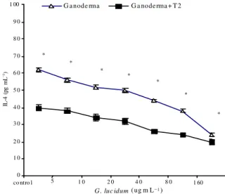
The results showed, when T-2 toxin was added to cells without any G. lucidum, it significantly enhanced IFN- and IL-2 release (Fig. 1 and 2, Control wells, p<0.025), but reduced the production of remaining cytokines in the same conditions (Fig. 3-7). The results also show that G. lucidum enhanced IFN- , IL-2 release (Fig. 1-2), but reduced IL-4, IL-10 production (Fig. 3-4) by T cells in a concentration dependent manner. When, 1ng mL T-2 toxin was added to any concentration of G. lucidum, it augmented both stimulatory and inhibitory effects of G. lucidum on cytokine release by T cells (Fig. 1-4).
The results also showed, that G. lucidum enhanced IL-1 , TNF- release in a concentration dependent manner (Fig. 5-7), but in case of IL-12, the highest enhancement was induced in response to 5 µg mL
G. lucidum (Fig. 6). In contrast to the effect of T-2 toxin on cytokine release by Th1 cells, here, it reduced
IL-1 , IL-12 and TNF- release by peritoneal macrophages comparing with control groups (Fig. 5-7). T-2 toxin augmented the stimulatory effect of
10
* * *
* *
* * * ** * **
0 1 0 2 0 3 0 4 0 5 0 6 0 7 0 8 0 9 0 1 00
5 1 0 2 0 4 0 8 0 1 60
G. luc idum (ug m L−1)
IN
F
-?
(
pg
m
L
−1)
G anoderma Ga noderma + T2
Con trol
Fig. 1: Effect of different concentrations of G. lucidum
on IFN- production by T cells in the presence or absence of 1 ng mL−1 T-2 toxin
* * *
*** *** **
*
0 10 20 30 40 50 60 70 80 90 100
control 5 10 20 40 80 160
G. lucidum (ug mL−1)
IL
-2
(
pg
m
L
−1)
Ganoderma Ganoderma+T2
Fig. 2: Effect of different concentrations of G. lucidum
on IL-2 production by T cells in the presence or absence of 1 ng mL−1 T-2 toxin
effect on IL-1 , IL-12 (Fig. 5 and 6) release by peritoneal macrophages. We have previously demonstrated the effect of T-2 toxin and G. lucidum on nitric oxide and cytokine release by peritoneal mΦ of mice[22,25]. In this study, the results showed that T-2 toxin without any G. lucidum supplementation significantly enhanced IFN- and IL-2 release (Fig. 1 and 2, Control wells, p<0.025), but reduced the production of remaining cytokines in the same conditions (Fig. 3-7), demonstrating both enhancement and inhibitory effects of T-2 toxin on cytokine production depending on cell sources.
Our data presented here in, is supported by a study on human lymphocyte where 1 and 5 ng mL−1 of T-2 toxin induced 50% inhibition of PHA-stimulated proliferation, but had no statistically significant effect on the viability of unstimulated lymphocytes[26].
* * * * * * *
0 1 0 2 0 3 0 4 0 5 0 6 0 7 0 8 0 9 0 1 00
co ntro l 5 1 0 2 0 4 0 8 0 1 60
G . luc idum ( ug m L−1)
IL
-4
(
pg
m
L
−1)
G a node rma G a node rma + T 2
Fig. 3: Effect of different concentrations of G. lucidum
on Il-4 production by T cells in the presence or absence of 1 ng mL−1 T-2 toxin
* *
*
**
**
**
*
0 10 20 30 40 50 60 70 80 90 100
control 5 10 20 40 80 160
G. lucidum (µg mL−1
)
Ganoderma Ganoderma+T2
IL
-1
0
(p
g
m
L
−1
)
Fig. 4: Effect of different concentrations of G. lucidum
on Il-10 production by T cells in the presence or absence of 1 ng mL−1 T-2 toxin
11
*
*
* * * * *
0 5 10 15 20 25 30 35 40 45 50
Control 5 10 20 40 80 160
G. lucidum (ug m L−1) Ganoderma Ganoderma+T2
IL
-I
β
(
p
g
m
L
−1)
Fig. 5: Effect of different concentrations of G. lucidum
on IL-1 production by peritoneal macrophages in the presence or absence of 1 ng mL−1 T-2 toxin
* * **
* *
** * *
*
0 50 1 00 1 50 2 00 2 50 3 00
Con trol 5 1 0 20 4 0 80 160
G. lucidum (ug m L−1)
IL
-1
2
(p
g
m
L
−
1)
Ganoderma Ganoderma+T2
Fig. 6: Effect of different concentrations of G. lucidum
on IL-12 production by peritoneal macrophages in the presence or absence of 1 ng mL−1 T-2 toxin
acute exposure of animals or human to T-2 toxin resulted in severe damage to actively dividing cells in tissues such as bone marrow, lymph nodes, spleen, thymus and intestinal mucosa. This is also in agreement to other studies that reported the exposure to T-2 toxin either enhanced or suppressed B and T lymphocyte mitogen proliferation in a dose dependent manner[29,30].
*
*
*
*
**
**
**
0 20 40 60 80 100 120 140 160 180 200
Control 5 10 20 40 80 160
G. lucidum (ug m L−1)
T
N
F
-a
(
p
g
m
L
−
1)
Ganoderma
Ganoderma+ T-2
Fig. 7: Effect of different concentrations of G. lucidum
on TNF- production by peritoneal macrophages in the presence or absence of 1 ng mL−1 T-2 toxin
Therefore, it is not surprising if T-2 toxin, depending on dose, time of administration and cell origin can induce different effects on immune cell function[31-34]. In contrast to our results in which T-2 toxin reduced IL-1 production by peritoneal m , a significant increase of IL-I and IL-6 in supernatants of chondrocytes cultivated for 24 h with T-2 toxin at 8 ng mL−1 after PMA stimulation is reported previously[31]. Present results also are in favour of Hymery et al.[35] who demonstrated that trichothecenes have adverse effects on dendritic cells maturation process. Therefore, Effect of T-2 toxin on peritoneal macrophages and lymphoid T cells of mice observed in this study in some parts is different that those described in the literature[36,37], which could be contributed to the dose and condition used in this study. Thus, it seems that the effects of T-2 toxin and G. lucidum on cell function are different in single usage or using them in combination form. On the other hand, this effect of G. lucidum might be related to its adjuvant property, as Chan et al.[23] reported
12 CONCLUSION
Based on data presented here in and the finding of others, it is possible to suggest that T-2 toxin may modulate the effect of G. lucidum on immune function.
ACKNOWLEDGMENTS
Thanks are due to the Research Centre of Molecular Biology for financial assistance. This project was approved by the office of Vice Chancellor for Research of the Baqiyatallah University of Medical Sciences (grant number 756 dated May 14th 2006).
REFERENCES
1. Chen, J., Y. Chu, J. Cao, Z. Yang, X. Guo and Z. Wang, 2005. T-2 toxin induces apoptosis and selenium partly blocks, T-2 toxin induced apoptosis in chondrocytes through modulation of the Bax/Bcl-2 ratio. Food Chem. Toxol., 44: 567-573.
2. Ji, X., Y. Ke, T. Ning, Y. Liang, D. Wang and G. Shi, 1994. Effects of sterigmatocystin and T-2 toxin on the inducion of unscheduled DNA synthesis in primary cultures of human gastric epithelial cells. Nat. Toxins., 2 (3): 115-119. 3. Myriam, L., S. Armando, R. Felipe and G. Mejia,
1999. Effect of lycopene on lipid peroxidation and glutathione- dependent enzymes induced by T-2 toxin in vivo. Toxicol. Lett., 109: 1-10.
4. Tiefenbach, B., G. Hennighausen and J. Merkord, 1995. Trichothecenes T-2 toxin (T-2) induces toxic effects on the developing immune system in rats. Toxicol. Lett., 78: 78-79.
5. Tiefenbach, D., 2004. Haematotoxicity of trichothecenes. Toxicol. Lett., 53: 75-81.
6. Thuvander, A., C. Wikman and I. Gadhasson, 1999. In vitro exposure of human lymphocytes to trichothecenes individual variation in sensitivity and effects of combined exposure of lymphocyte function. Food Chem. Toxicol., 37: 639-648. 7. Sugita-Konishi, Y., Y. Kara-Kudo, F. Kasuga,
M. Yamamoto and S. Kumagai, 1997. Effect of deoxynivalenol on humoral and cellular immune-responses to salmunella infection. Mycotoxins, 44: 37-40.
8. Yang, G.H., B.B. Jarvis, Y.J. Chung and J.J. Pestka, 2000. Apoptosis induction by the satratoxins and and other trichothecence mycotoxins: Relationship to ERK, P38 MAPK and SAPK/JNK activation. Toxicol. Applied Pharmacol., 164: 149-160.
9. Islam, Z., M. Nagase, T. Yoshizawa, K.E. Yamauchi and N. Sakato, 1998. T-2 toxin induces thymic apoptosis in vivo in mice. Toxicol. Applied Pharmacol., 148: 205-214.
10. Pestka, J.J. and G.S. Bondy, 1990. Alteration of immune function following dietary mycotoxin exposure. Can. J. Physiol. Pharmacol., 68: 1009-1016.
11. Pestka, J.J. and G.S. Bondy, 1994. Mycotoxin-Induced Immune Modulation. In: Immunotoxicology and Immunopharmacology, Dean, J.H, M.I. Luster, A.E. Munson and I. Kimber (Eds.). 2nd Edn. Raven Press, New York. pp: 163-182.
12. Mekhancha-Dahel, C., C. Lafarge-Frayssinet and C. Frayssinet, 2001. Immunosuppressive effects of four trichothecene mycotoxins. Food Additives Contaminants, 7: S94-S96.
13. Ouyang, Y.L., J.I. Azcona-Olivera and J.J. Pestka, 1995. Effects of trichothecenec structure on cytokine secretion and gene expression in murine CD4+ T cells. Toxicology, 104: 187-202.
14. Meky, F.A., L.J. Hardie, S.W. Evans and C.P. Wild, 2001. Deoxynivalenol-induced immunomodulation of human lymphocyte proliferation and cytokine production. Food Chem. Toxicol., 39: 827-836.
15. Li, M., C.F. Cuff and J. Pestka, 2006. T-2 toxin impaiment of enteric reovirus clearance in the mouse associated with suppressed immunoglobulin and IFN- responses. Toxicol. Applied Pharmacol., 214: 318-325.
16. Li, M., J.R. Harkema, Z. Islam, C.F. Cuff and J.J. Pestka, 2006. T-2 toxin impairs murin immune response to respiratory reovirus and exacerbates viral bronchiolitis. Toxicol. Applied Pharmacol., 217: 76-85.
17. Chen, H.S., Y.F. Tsai, C.C. Lin, K.H. Khoo, C.H, Lin and C.H. Wong, 2004. Studies on the immuno-modulating and anti-tumor activities of Ganoderma lucidum (Reishi) polysaccharides. Bioorganic Med. Chem., 12: 5595-5601.
18. Chien, C.M., J.L. Cheng, W.T. Chang, M.H. Tien, C.M. Tsao and Y.H. Chung, 2004. Polysaccharides of Ganoderma lucidum alter cell immune phenotypic expression and enhance CD56+NK-cell cytotoxicity in cord blood. Bioorganic Med. Chem., 12: 5603-5609.
13 20. Wang, S.Y., M.L. Hsu, H.C. Hsu, C.H. Tzeng and
S.S. Lee, 1997. The antitumor effect of Ganoderma Lucidum is mediated by cytokines released from activated macrophages and T-lymphocyes. Int. J. Cancer., 70: 699-705.
21. Zhou, S.F. and Y.H. Gao, 2002. The immunomodulating effects of Ganoderma Lucidum. Int. J. Med. Mushroom, 4: 1-11.
22. Ahmadi, K. and M. Riazipour, 2007. Effect of Ganoderma lucidum on cytokine release by peritoneal macrophages. Iran J. Immunol., 4 (4): 220-226.
23. Chan, W.K., D.T. Lan, H.K. Law, W.T. Wong, M.W. Koo and As. Lau, 2005. G. lucidum
mycelium and spore extracts as natural adjuvant for immunotherapy. J. Altern Complement Med., 11: 1047-1057.
24. John Stone, A. and R. Thorp, 1996. Immunochemistry in Practice. 3rd Edn. Black Well Science.
25. Ahmadi, K. and Riazipour, M., 2007. Effect of Mycotoxin T-2 on Cell death and nitric oxide release by mice peritoneal macrophages. J. Military Med., 9 (1): 1-6.
26. Islam, Z., M. Nagase, T. Yoshizawa, K.E. Yamauchi and N. Sakato, 1998. T-2 toxin induces thymic apoptosis in vivo in mice. Toxicol. Applied Pharmacol., 48: 205-214.
27. Jaradat, Z.W., B. Vila and R.R. Marquardt, 2006. Adverse effect of T-2 toxin on chicken lymphocytes blastogenesis and its protection with Vitamin E. Toxicology, 225: 90-96.
28. Jaradat, Z.W., 2005. T-2 Mycotoxin in the Diet and its Effects on Tissues. In: Reviews in Food and Nutrition Toxicology, Preedy, V.R. and R.R. Watson (Eds.). CRC Press, Boca Raton, pp: 174-214.
29. Berek, L., I.B. Petri, A. Mesterhazy, J. Teren and J. Molner, 2001. Effects of mycotoxin on human immune functions in vitro. Toxicol. Vitr., 15: 25-30.
30. Muller, G., P. Kielstein, H. Rosner, A. Berndt, M. Heller and H. Kohler, 1999. Studies on the influence of combined administration of ochratoxin A, Fumonisin B1, deoxynivalenol and T-2 toxin on immune and defense reaction in weaner pigs. Mycoses, 42: 485-493.
31. Fu, Y.T., W.G. Lin, Z. BaoCheng and G. Quan, 2001. The effect of T-2 toxin on IL-1beta and IL-6 secretion in human fetal chondrocytes. Int. Orthop., 25 (3): 199-201.
32. Rocha, O. and F.M. Ansari-Doohan, 2005. Effects of trichothecene mycotoxins on eukaryotic cells: Areview. Food Addit. Contam., 22 (4): 369-378. 33. Chung, Y.J., H.R. Zhou and J.J. Pestka, 2003.
Transcriptional and posttranscriptional roles for p38 mitogen-activated protein kinase in upregulation of TNF-a expression by deoxynivalenol (Vomitoxin). Toxicol. Applied Pharmacol., 193: 188-201.
34. Albarenque, S.M., K. Suzuki, H. Nakayama and K. Doi, 2001. Kinetics of cytokines mRNAas expression in the dorsal skin of WBN/ILA-HT rats following topical application of T-2 toxin. Exp. Toxicol. Pathol., 53 (4): 271-274.
35. Hymery, N., Y. Sibiril and D. Parent-Massin, 2006.
In vitro effects of trichothecenes on human dendritic cells. Toxicol. In vitro, 20: 899-909. 36. Tammer, B., I. Lehmann, K. Nieber and
R. Altenburger, 2007. Combined effects of mycotoxin mixtures on human T cell function. Toxicol. Lett., 170: 124-133.