Ecotoxicological Effects of Gold Nanoparticles and
Microplastics in Daphnia magna
Alexandre Pacheco Lopes
Dissertação realizada no âmbito do Mestrado Integrado em Bioengenharia
Ramo de Biotecnologia Molecular
Orientador: Professora Doutora Lúcia Maria das Candeias Guilhermino
i
Ecotoxicological Effects of Gold Nanoparticles and
Microplastics in Daphnia magna
Dissertação de Candidatura ao Grau de Mestre em Bioengenharia submetida à Faculdade de Engenharia da Universidade do Porto.
Orientador – Professora Doutora Lúcia Maria das Candeias Guilhermino Categoria – Professora Catedrática Afiliação – Instituto de Ciências Biomédicas de Abel Salazar da Universidade do Porto, Departamento de Estudos de Populações, Laboratório de Ecotoxicologia.
iii
Resumo
Os nanomateriais (NMs) têm recebido uma crescente atenção tanto em contextos académicos como industriais. As suas propriedades particulares, consequentes do seu tamanho diminuido, apresentam um enorme potencial para aplicações num largo leque de áreas tais como a eletrónica, biomedicina, produtos de higiene pessoal, entre outras. Isto tem motivado um aumento da produção de NMs, tanto para investigação académica como para aplicações industriais e comerciais. À medida que a sua produção aumenta, aumenta também a probabilidade de exposição a estes materiais. Em simultâneo, a consciencialização em relação à presença de microplásticos (MP) no ambiente tem também aumentado. Os MP resultam da degradação de objetos de plástico de maiores dimensões, após uma exposição prolongada a, por exemplo, radiação ultra-violeta e abrasão física. Enquanto que já existe um conhecimento extensivo sobre o potencial tóxico de plásticos à escala macroscópica, os MP só recentemente têm ganho a atenção das entidades reguladoras. Tanto os NMs como os MP apresentam um potencial tóxico considerável que permanece maioritariamente por explorar. O tamanho nanométrico dos NMs poderá conceder-lhes a abilidade de ultrapassar várias barreiras biológicas, infiltrar e danificar elementos sub-celulares, interagir com vários processos celulares ou causar outros tipos de efeitos detrimentais ainda desconhecidos. Alguns destes fenómenos já foram reportados, principalmente em modelos in vitro ou modelos in vivo com mamíferos. Também já foi mostrado que os MP podem interferir com organismos vivos. A acumulação de MP ingeridos por vários organismos marinhos já foi reportada, embora os efeitos dessa acumulação permaneçam desconhecidos.
Durante o presente trabalho, o potencial tóxico de nanopartículas de ouro (AuNP) e MP de polietileno foi avaliado no cladócero de água doce Daphnia magna. Este organismo foi escolhido porque é um dos organismos modelo mais usados na investigação em ecotoxicologia, uma vez que é considerado representativo do zooplâncton e dos consumidores primários em água doce. Com este fim, um ensaio ecotoxicológico crónico foi conduzido com AuNP, MP e com misturas de ambos os tipos de partículas. Numa primeira fase, as metodologias associadas à manutenção de culturas de D. magna foram aprendidas e treinadas. Isto incluiu não só o manuseamento e manutenção dos animais, mas também a manutenção de culturas de Chlorella
vulgaris, uma vez que estas microalgas foram usadas para alimentar as dáfnias durante o trabalho.
A segunda fase consistiu na caracterização das AuNP e MP. Foram determinadas curvas de calibração e foram conduzidos ensaios de degradação das partículas em água ultra-pura e no meio de cultura de D. magna, usando espectrofotometria ou espectrofluorometria. Finalmente, o ensaio para avaliar a toxicidade crónica em D. magna foi conduzido, usando a reprodução como critério de toxicidade. O ensaio teve uma duração de 21 dias, durante os quais a mortalidade dos animais
iv
e vários parâmetros reprodutivos foram monitorizados diariamente. As hipóteses nulas testadas foram: i) concentrações na ordem das ppm de AuNP não são tóxicas para D. magna; ii) concentrações na ordem das ppb não são tóxicas para D. magna e iii) a precença de MP na água não influencia a toxicidade das AuNP em D. magna. Os resultados obtidos sugerem que tanto as AuNP como os MP individualmente podem induzir toxicidade em D. magna, causando mortalidade e reduzindo as capacidades reprodutivas dos indivíduos expostos. Também se observou que as AuNP e MP, quando presentes em misturas, parecem interagir entre elas, intensificando o potencial tóxico observado em D. magna. Consequentemente, as três hipóteses nulas testadas foram rejeitadas.
Palavras-chave: Daphnia magna,, nanopartículas de ouro, nanotecnologia, microplásticos, toxicidade crónica, toxicidade de misturas,
vii
Abstract
Nanomaterials (NMs) have been receiving increased attention in both academic and industrial contexts. Their particular properties, which are a consequence of their small size, present a huge potential for applications in a wide range of areas such as electronics, biomedicine, personal care products, among many others. This has been driving an increase in NM manufacture, either for academic research or for industrial and commercial applications. As their manufacture increases, so does the probability of exposure to these materials. Simultaneously, the awareness about the presence of microplastics (MP) in the environment is raising as well. MP result from the degradation of larger plastic items after prolonged exposure to, for example, ultra-violet radiation and physical abrasion. While an extensive knowledge exists about the toxic potential of macro-sized plastics, MP are only recently beginning to gain attention from regulatory entities. Both NMs and MP present a considerable toxic potential that is still largely unexplored. The nano-size of NMs could confer them the ability to bypass many biological barriers, infiltrate and disrupt sub-cellular elements, interact with several cellular processes or cause other types of detrimental effects still unknown. Some of these phenomena have already been reported, mainly in in vitro models or in vivo mammalian models. MP have also been shown to interfere with living organisms. The accumulation of ingested MP in the digestive tract of several marine organisms has been reported, even though the effects of such accumulation remain largely unknown.
During the present work, the toxic potential of gold nanoparticles (AuNP) and polyethylene MP was evaluated in the freshwater cladoceran Daphnia magna. This organism was chosen because it is one of the most widely used model organisms in ecotoxicology research, as it is considered representative of freshwater zooplankton and primary freshwater consumers. To achieve this, a chronic ecotoxicological bioassay was conducted with AuNP, MP and mixtures of both types of particles. In a first phase, the methodologies associated with D. magna culture maintenance were learned and trained. This included not only the handling and maintenance of the animals, but the maintenance of Chlorella vulgaris cultures as well, as these microalgae were used to feed the daphnids throughout the duration of the work. The second phase consisted on the characterization of the AuNP and MP. Calibration curves were determined and degradation assays were conducted in ultra-pure water and D. magna culture medium, using spectrophotometry or spectrofluorimetry. Finally, the bioassay to access chronic toxicity in D.
magna was conducted, using reproduction as the effect criterion. The bioassay had a duration of
21 days, during which animal mortality and reproduction parameters were monitored daily. The null hypotheses tested were: i) AuNP at concentrations in the low ppm are not toxic to D. magna;
viii
ii) MP at concentrations in the low ppb are not toxic to D. magna and iii) the presence of MP in the water does not influence the toxicity of AuNP to D. magna. The obtained results suggest that both AuNP and MP alone can induce toxicity in D. magna, causing mortality and reducing reproductive fitness of the exposed individuals. It was also observed that AuNP and MP, when in mixture, appear to interact with each other, intensifying the toxic potential observed in D.
magna. Therefore, all three null hypotheses tested were rejected.
Keywords: Daphnia magna, chronic toxicity, gold nanoparticles, microplastics, mixture toxicity, nanotechnology.
xi
Agradecimentos
Em primeiro lugar, deixo um agradecimento à orientadora científica deste trabalho, Professora Doutora Lúcia Guilhermino, pela disponibilidade, orientação e apoio prestado ao longo do trabalho.
Não posso também deixar de dar um agradecimento enorme à Mestre Alexandra Martins, por toda a ajuda, amizade e transmissão de conhecimentos e experiência que foram essenciais durante o desenvolvimento do trabalho.
Agradeço às colegas do laboratório de Ecotoxicologia, nomeadamente à Doutora Beatriz Lavorante e à Joana Prata, pelo apoio e por todas as pequenas conversas e momentos de descontração que me ajudaram a ir sobrevivendo aos momentos mais alucinantes do trabalho.
Deixo também um agradecimento enorme à minha mãe, por sempre me ter apoiado em todas as minhas escolhas e pelo apoio constante.
O trabalho de investigação conducente à presente Tese foi desenvolvido no âmbito do projeto EPHEMARE – Ecotoxicological effects of microplastics in marine ecosystems. JPI Oceans – Microplastics., financiado pela Fundação para a Ciência e a Tecnologia (JPIOCEANS/0004/2015) e por verbas do Instituto de Ciências Biomédicas Abel Salazar da Universidade do Porto, atribuídas ao Departamento de Estudos de Populações e ao Laboratório de Ecotoxicologia.
xiii
Table of Contents
Resumo... iii
Abstract ... vii
Agradecimentos ... xi
Table of Contents ... xiii
Figure Index ... xv
Table Index ... xvii
Abbreviations List ... xix
1. Introduction ... 1
1.1 Nanotechnology ... 1
1.1.1. Materials’ Properties at the Nano Scale ... 2
1.1.2. NMs' Applications ... 3
1.2. Gold Nanoparticles... 5
1.3. Microplastics ... 6
1.4. Nanotoxicology ... 7
1.5. Toxicity Assessment ... 8
1.6. Daphnia magna as Model Organism ... 11
1.7. Toxicological Potential of AuNP ... 12
1.8. Toxicological Potential of MP ... 14
1.9. Aims of the Study, Hypotheses to be Tested and Structure of the Thesis ... 17
2. Methodologies and Materials ... 19
2.1. Chlorella vulgaris Cultures ... 19
2.2. Daphnia magna Cultures ... 21
2.3. AuNP Description ... 22
2.3.1. AuNP Calibration Curve ... 24
2.4. MP Description ... 25
2.4.1. MP Calibration Curve ... 25
2.5. Bioassays ... 26
2.5.1. Test Validation With a Reference Substance ... 26
xiv
2.6. Statistical Analysis ... 28
3. Results and Discussion ... 30
3.1. Daphnia magna Cultures ... 31
3.1.1 Number of Juveniles per Parental Individual ... 31
3.1.2. Acute Assay With a Reference Substance ... 31
3.2. Characterization of AuNP in Ultra-Pure Water and ASTM ... 32
3.2.1. AuNP Calibration Curve ... 34
3.2.2 Behaviour of 5 nm AuNP Along 48 h ... 38
3.3 MP Calibration Curves ... 41
3.3.1 Behaviour of 5 µm MP Along 48 h... 43
3.4. Chronic Assay with AuNP and MP ... 45
3.4.1. General Conditions, and Actual Concentrations and Decay of AuNP and MP in Test Media ... 45
3.6.2 Mortality ... 49
3.4.2. Effects on Reproduction ... 50
3.4.3. Effects on Somatic Growth ... 56
3.4.4. AuNP and MP Interactions and General Discussion ... 57
4. Conclusions and Future Prospects ... 59
xv
Figure Index
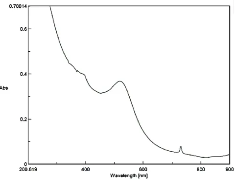
Figure 1 – Absorption spectrum of a 20 mg/L AuNP solution prepared in ultra-pure water. Abs = absorbance in optical density (OD) units. The image was obtained through the Spectra
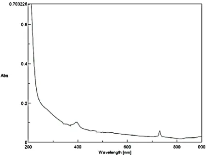
Manager software. ... 35 Figure 2 - Absorption spectrum of a 20 mg/L AuNP solution prepared in ASTM. The image was obtained through the Spectra Manager software. Abs = absorbance in optical density (OD) units. ... 35 Figure 3 - Absorption spectrum of a 0.078 mg/L AuNP solution prepared in ultra-pure water. The image was obtained through the Spectra Manager software. Abs = absorbance in optical density (OD) units. ... 36 Figure 4 - Absorption spectrum of a 0.078 mg/L AuNP solution prepared in ASTM. The image was obtained through the Spectra Manager software. Abs = absorbance in optical density (OD) units. ... 36 Figure 5 - AuNP calibration curves constructed by linear regression analysis of AuNP
concentration vs absorbance at 520 nm. The solid line and the triangle markers refer to the model fitted to the solutions prepared in ultra-pure water. The dashed line and square markers refer to the model fitted to the solutions prepared in ASTM. ... 37 Figure 6 - MP calibration curves constructed by linear regression analysis of MP concentration vs fluorescence. The solid line and the triangle markers refer to the model fitted to the solutions prepared in ultra-pure water. The dashed line and square markers refer to the model fitted to the solutions prepared in ASTM. ... 42 Figure 7 – Total percentage of mortality recorded in parental animals over the 21-day exposure period per treatment. 0.2AuNP+0.02MP = mixture of 0.2 mg/L AuNP + 0.02 mg/L MP. 0.2AuNP+0.2MP = mixture of 0.2 mg/L AuNP + 0.2 mg/L MP. 2AuNP+0.02MP = mixture of 2 mg/L AuNP + 0.02 mg/L MP. 2AuNP+0.2MP = mixture of 2 mg/L AuNP + 0.2 mg/L MP. 49 Figure 8 – Average and standard deviation of the total number of viable juveniles, immobile juveniles and aborted eggs produced per parental animal, per treatment, at the end of the assay. The lowercase letters above the error bars indicate treatments whose effects were not
statistically different among themselves. Mix1 = mixture of 0.2 mg/L AuNP + 0.02 mg/L MP. Mix2 = mixture of 0.2 mg/L AuNP + 0.2 mg/L MP. Mix3 = mixture of 2 mg/L AuNP + 0.02 mg/L MP. Mix4 = mixture of 2 mg/L AuNP + 0.2 mg/L MP. ... 51 Figure 9 - Average and standard deviation of the age (in days) of the release of the first brood and total number of broods produced per parental animal, per treatment, at the end of the assay. The lowercase letters above the error bars indicate treatments whose effects not statistically different among themselves. Mix1 = mixture of 0.2 mg/L AuNP + 0.02 mg/L MP. Mix2 = mixture of 0.2 mg/L AuNP + 0.2 mg/L MP. Mix3 = mixture of 2 mg/L AuNP + 0.02 mg/L MP. Mix4 = mixture of 2 mg/L AuNP + 0.2 mg/L ... 52 Figure 10 - Average and standard deviation of the total somatic growth per parental animal, per treatment, at the end of the assay. The lowercase letters above the error bars indicate treatments whose effects not statistically different among themselves. Mix1 = mixture of 0.2 mg/L AuNP
xvi
+ 0.02 mg/L MP. Mix2 = mixture of 0.2 mg/L AuNP + 0.2 mg/L MP. Mix3 = mixture of 2 mg/L AuNP + 0.02 mg/L MP. Mix4 = mixture of 2 mg/L AuNP + 0.2 mg/L. ... 56
xvii
Table Index
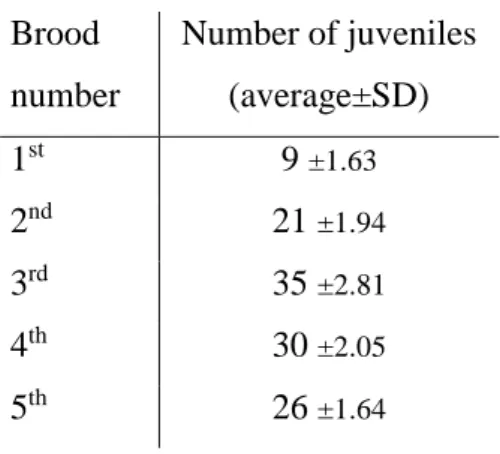
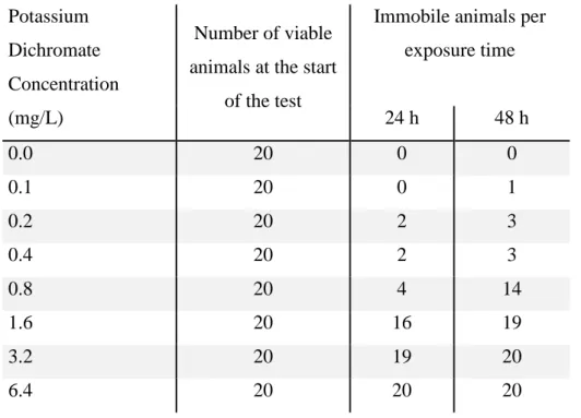
Table 1 - Qualitative and quantitative chemical composition for MBL culture medium. ... 19 Table 2 - Qualitative and quantitative chemical composition for ASTM culture medium. ... 21 Table 3 - Average number of juveniles per animal per brood. SD = Standard Deviation ... 31 Table 4 - Immobile animals per group after 24 h and 48 h of exposure to different
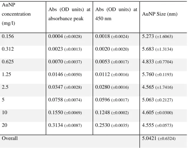
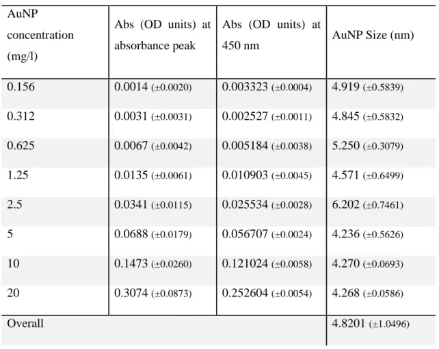
concentrations of potassium dichromate. ... 32 Table 5 - estimated diameter of 5 nm AuNP in solutions prepared in ultra-pure water. The values presented are the average of 3 solutions prepared independently, with the corresponding standard deviation (within brackets). Absorbance peak = absorbance at 520 nm. Overall = overall average and standard deviation. OD = Optical Density. ... 33 Table 6 - estimated diameter of 5 nm AuNP in solutions prepared in ASTM. The values
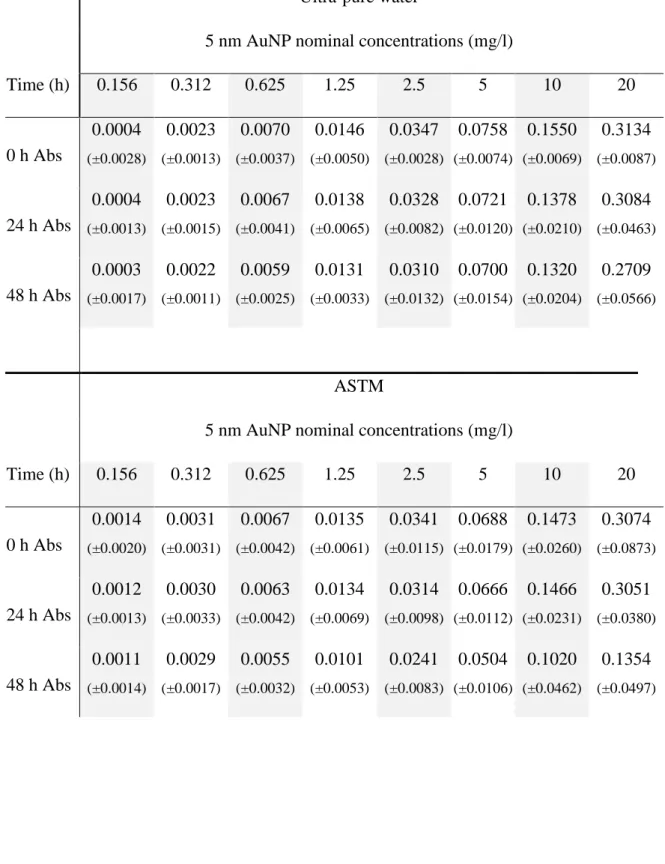
presented are the average of 3 solutions prepared independently, with the corresponding standard deviation (within brackets). Absorbance peak = absorbance at 520 nm. Overall = overall average and standard deviation. OD = Optical Density. ... 34 Table 7 - Absorbance values at 520 nm for the average of the three replicates prepared in ultra-pure water or ASTM, measured 24 h and 48 h after the preparation of the solutions. Values are the average of absorbance at 520 nm (3 replicate solutions) with the corresponding standard deviation within brackets. Abs = absorbance at 520 nm. ... 40 Table 8 – Decay of 5 nm AuNP (%) in solutions prepared in ultra-pure water or ASTM, at 24 h and 48 h. The decay was calculated as: Decay (%) = 100 - (abs2 x 100/abs1). Abs1 =
absorbance at 520 nm at 0h. Abs2 = absorbance at 520 nm at 24 or 48h. ... 41 Table 9 - Fluorescence values at 575 nm for the excitation wavelength and 607 nm for the emission wavelength for the average of the three replicates prepared in ultra-pure water or ASTM, measured 24 and 48h after the preparation of the solutions. Values are the average of fluorescence (3 replicate solutions) with the corresponding standard deviation within brackets. Fl = fluorescence at 575 nm for the excitation wavelength and 607 nm for the emission
wavelength. ... 44 Table 10 – Decay of 5 µm MP (%) in solutions prepared in ultra-pure water or ASTM, at 24h and 48h. The decay was calculated as: Decay (%) = 100 - (Fl2 x 100/Fl1). Fl1 = fluorescence (575 nm for the excitation wavelength and 607 nm for the emission wavelength) at 0h. Fl2 = fluorescence (575 nm for the excitation wavelength and 607 nm for the emission wavelength) at 24 h or 48h. ... 45 Table 11 - Absorbance at 520 nm of the different test solutions from the chronic assay. The AuNP actual concentrations were estimated using the linear regression model fitted to ultra-pure water presented in section 3.2.1 Values are presented as the average of 2 to 10 replicates with corresponding standard deviation. Only the values referring to the treatments containing AuNP are presented. Abs = absorbance value at 520 nm in optical density (OD) units. Old media = media prepared in the previous day. Fresh media = freshly prepared media. The decay was calculated as: Decay (%) = 100 - (absOld x 100/absFresh). AbsFresh = absorbance at 520 nm of
the fresh media. AbsOld = absorbance at 520 nm of the old media. The deviation from the actual
xviii
100/NominalConc). ActualConc = concentration estimated using the linear regression model.
NominalConc = nominal concentration of AuNP. Conc. = concentration. ... 47
Table 12 – Fluorescence intensity (FI) values at 575 nm for the excitation wavelength and 607 nm for the emission wavelength, of the different test solutions from the chronic assay. The MP actual concentrations were estimated using the linear regression model fitted to ultra-pure water presented in section 3.3.1 Values are presented as the average of 2 to 10 replicates with
corresponding standard deviation. Only the values referring to the treatments containing MP are presented. Fluorescence = fluorescence at 575 nm for the excitation wavelength and 607 for the emission wavelength in FI units. Old media = media prepared in the previous day. Fresh media = freshly prepared media. The decay was calculated as: Decay (%) = 100 - (FlOld x 100/FlFresh).
FlFresh = fluorescence (at 575 nm for the excitation wavelength and 607 for the emission
wavelength) of the fresh media. FlOld =fluorescence (at 575 nm for the excitation wavelength
and 607 for the emission wavelength) of the old media. The deviation from the actual concentration to the nominal ones was calculated as: Deviation (%) = 100 - (ActualConc x
100/NominalConc). ActualConc = concentration estimated using the linear regression model.
NominalConc = nominal concentration of MP. Conc. = concentration. NA = Not applicable ... 48
Table 13 – Summary of the statistical parameters obtained in the two-way analysis of variance (2-ANOVA). Only the biological parameters that didn’t show differences between the control and citrate treatment after the one-way ANOVA (1-ANOVA) analysis are presented. df = degrees of freedom. AuNP = Gold nanoparticles concentration. MP = Microplastics
xix
Abbreviations List
ANCOVA - Analysis of Co-Variance 1-ANOVA - One-Way Analysis of Variance 2-ANOVA - Two-Way Analysis of Variance ANOVA - Analysis of Variance
ASTM - American Society for Testing and Materials
AuNP - Gold Nanoparticle
C. Vulgaris - Chlorella vulgaris
CI - Confidence Interval
CTAB - Cetyltrimethylammonium Bromide
D. magna - Daphnia magna
EC - Effective Concentration FI - Fluorescence Intensity
LC - Lethal Concentration
LOEC - Lowest Observed Effect Concentration EPA - Environmental Protection Agency MBL - Marine Biological Culture
MP - Microplastic
MPA - Mercaptopropionic Acid
NM - Nanomaterial
NOEC - No Observed Effect Concentration
NP - Nanoparticle
OD - Optical Density
OECD - Organisation for Economic Co-operation and Development PAH - Polyallylamine Hydrochloride
PMMA - Poly(methylmethacrylate) ROS - Reactive Oxygen Species
UV - Utraviolet
1
1. Introduction
1.1 Nanotechnology
Nanotechnology is a multidisciplinary science that studies the properties, manufacture, applications and interactions of systems and materials at the nano (<10-9 m) scale. According recent definitions, a nanomaterial (NM) is a structure that has at least one dimension between 1 and 100 nm (Nel et al., 2006). Due to their small size, NMs exhibit interesting properties that are not found in their corresponding bulk counterparts (Nel et al., 2006) that could be applied to create new products or to enhance and update existing products in numerous industrial, academic and personal contexts. Even though the concept of “nanotechnology” is a recent one, there are some known uses of NMs throughout the human history. One of the most famous examples of NM usage in ancient times, albeit probably an unintentional one, is the Lycurgus Cup, a roman chalice with gold and silver nanoparticles (NPs) dispersed throughout the glass. The cup appears to be red when lit from behind or green when lit from the front as a result of the NPs’ different absorption and reflection properties (Freestone et al., 2007). However, it was only in 1857 that Michael Faraday described for the first time how the nano size of gold particles in colloidal gold suspensions influences their behavior and macroscopic properties (Faraday, 1857). NMs can have natural or anthropogenic origins (Dhawan and Sharma, 2010). Some examples of natural sources of NMs include volcano fumes, forest fires, dust storms and viruses (Navarro et al., 2008; Doak et al., 2009). Anthropogenic NMs can be of primary origin or secondary origin. Primary origin NMs are those that are designed and manufactured to be used in academic or industrial context, while secondary origin NMs are accidentally generated by human activity, mainly air pollution and waste degradation (Doak et al., 2009). Current technology allows for the creation and manipulation of structures at the molecular and atomic scale (Singh et al., 2011). In recent years, the volume of anthropogenic NMs has been increasing (Navarro et al., 2008), both in quantity as well as in diversity. It is estimated that hundreds of different NMs have already been developed (Doak et al., 2009) in a wide selection of sizes, shapes, compositions and surface modifications. The nanotechnology industry is expanding every year, with new NMs being developed at a fast rate (Doak et al., 2009). This increase in NMs manufacture is motivated by the enormous potential for applications, which
2
brings not only academic interest but also investments in the field of nanotechnology.
1.1.1. Materials’ Properties at the Nano Scale
It is thought that at the nano scale, quantic interactions prevail over the conventional chemical interactions (The Royal Academy of Engineering, 2004; Handy
et al., 2008) conferring the NMs their particular optical, magnetic and electric properties.
Their small size results in an increase of the surface area to volume ratio, meaning that for the same volume of material, NMs offer a higher surface area compared to bulk materials (Nel et al., 2006). This increases the amount of atoms and molecules readily available to interact with the particles' surroundings and makes NMs highly reactive (Nel
et al., 2006). It is also possible that, because of this, they adsorb and concentrate high
quantities of surrounding substances, modifying their bioavailability (Handy et al., 2008; Navarro et al., 2008). Their small size means that NMs might be able to interact with biological systems in unpredictable ways (Doak et al., 2009). There might exist several physical and chemical factors that, while not relevant to conventional toxicology studies, could be crucial to address in nanotoxicology studies. Therefore, it’s imperative to adapt current methodologies and experimental designs in order to account for their unique properties (Doak et al., 2009).
One step that must be present in every nanotoxicology study is particle characterization (Burleson et al., 2004). This can be achieved through different methods and techniques. It is often advisable to use multiple methods of characterization, as each method provides different and complementary types of information, which can be useful to later interpret the obtained results (Cho et al., 2013). Particle characterization is also important to allow the comparison between different studies and to facilitate the consolidation of knowledge as new studies are published. The most frequently used methods for characterization of NPs include ultraviolet–visible spectroscopy (UV-Vis) to determine NP concentration and size (Haiss et al., 2007), dynamic light scattering to determine the particles' zeta potential, surface charge and hydrodynamic diameter (Cho
et al., 2013), transmission electron microscopy to evaluate the particles' shape (Wang,
2000) and X-ray photoelectron spectroscopy to determine the particles' surface composition (Zhang et al., 2004).
3
Some types of NPs are prone to form aggregates in aqueous solutions (Handy et
al., 2008), so another important aspect to consider is their aggregation state and size
distribution (Navarro et al., 2008). Zeta potential measures the electrostatic potential of a particle at the double layer relative to the bulk fluid away from the particle and is an indicator of the stability of a NP solution (Patil et al., 2007). As the absolute value of the zeta potential approaches zero, the stability of the solution decreases and the tendency to form aggregates increases (The Royal Academy of Engineering, 2004; Navarro et al., 2008). The most common method to control NP aggregation is to use ultrasounds to disperse the particles (Bihari et al., 2008). It has been shown that the tendency of NPs to form aggregates varies with the pH and ionic force of a given solvent (Navarro et al., 2008) so, alternatively, changing the solvent might also work, but this may also alter other properties of the NP solution. A factor that can significantly alter NPs properties and behavior is their external coat (Christian et al., 2008). This can modulate the particles' surface charge, total size (as opposed to the core size), reactivity, stability (Navarro et al., 2008) and determines the nature of the interface between the NPs and their surrounding environment (Christian et al., 2008). Some NPs are relatively easy to coat and can be modified in order to give them new properties and functionalities (Ghosh et al., 2008; Zhou et al., 2010). These properties give NMs a wide range of potential applications, as well as new challenges in understanding their mechanisms of action, toxicity and interactivity with other materials, biological systems and ecosystems.
1.1.2. NMs' Applications
The nanotechnology industry is expected to continue growing and expanding its range of applications in several areas. The properties of NMs open new opportunities to design completely new products, processes and techniques or to significantly improve upon those that already exist. Some areas that could benefit even more from nanotechnology include:
Medicine: NMs can be used as both diagnostic and treatment options in a variety of conditions and procedures. For example, iron oxide NPs are already being used as contrast agents for magnetic resonance imaging (Doak et al., 2009), allowing for a better contrast with lower concentrations of the contrast agent. Silver NPS are known to have antibiotic activity and could be applied in catheters to lower
4
the risk of infections (Samuel and Guggenbichler, 2004); silver NPs are already being used in wound dressings and surgical instruments with the same purpose (The Royal Academy of Engineering, 2004; Chen and Schluesener, 2008). NPs are also being studied as targeted drug delivery agents (Oberdörster et al., 2005), NP arrays could find use in new detection and diagnostic tools (Pattani et al., 2008; Pek et al., 2008) and nanoporous scaffolds show promising results in bone tissue regeneration (Pek et al., 2008).
Environmental remediation: some NPs are capable of absorbing large quantities of inorganic contaminants, making them useful for both in situ and ex situ soil and water remediation (Zhang, 2003). Organic pollutants can also be addressed using highly reactive NPs that are capable of degrading them, mainly by promoting photooxidation (Tratnyek and Johnson, 2006). For example, gold NPs (AuNP) can be used to selectively remove mercury from waters containing multiple pollutants (Ojea-Jiménez et al., 2012), and iron oxide NPs are being studied to be used for soil remediation of arsenic (Shipley et al., 2011). Polymeric NPs can be used to enhance the in situ biodegradation rate of organic pollutants such as phenanthrene and other polycyclic aromatic hydrocarbons (Tungittiplakorn et al., 2005).
Electronics: the first piece of nanoelectronic technology was a field effect transistor measuring less than 100nm, designed and manufactured in 2000 (Gargini, 2004). Nowadays, several common devices such as computer processors or memory chips include nanoelectronic technology. More recently, quantum dots are being studied as light emitting diodes (LED), presenting advantages over organic LEDs such as their thermic stability and pure emission color (Molaei et al., 2012). The electronic and optical properties of carbon nanotubes are being studied for applications in photovoltaic cells (Wang et al., 2015).
Personal care products: NPs are already used in cosmetics and personal care products. For example, titanium (TiO2) and zinc (ZnO) NPs are used in some sunscreens due to their high ultraviolet (UV) absorption and low reactivity with the human skin (Nel et al., 2006). Silver NPs are being incorporated in underarm deodorants due to their antibacterial properties (Raj et al., 2012), and
5
nanocapsules - NPs containing an active ingredient on their interior - are being used to overcome some problems in cosmetic dermatology, such as the incompatibility of different ingredients used in creams and gels (Morganti, 2010) This wide range of application fields is driving an increasing interest and investments on nanotechnology (Nel et al., 2006). However, knowledge about their toxicological potential lags behind their development and application, and the consequences of acute or chronic exposure of humans, wildlife and ecosystems to NMs are still largely unknown, despite the increasing number of studies that have been made in the last years focused in a relatively reduced amount of substances in a limited variety of species and biological systems.
1.2. Gold Nanoparticles
AuNP, also known as colloidal gold, are one of the most studied NMs (Eustis and El-Sayed, 2006; Khlebtsov and Dykman, 2011) mainly due to their presumed biocompatibility and unique optical and electronic properties (Eustis and El-Sayed, 2006). In recent years, AuNP have been receiving increasing attention, with the number of publications relating to these particles rising exponentially every year (Eustis and El-Sayed, 2006). These particles are included in the “List of Representative Manufactured Nanomaterials” published by the OECD in 2010, which includes manufactured NMs that are already on the markets or are predicted to be commercialized soon (OECD, 2010). AuNP already find applications in several areas of physics and chemistry (Daniel, 2004), but recently there has been an increasing interest in exploring their potential biomedical applications (Dykman and Khlebtsov, 2012). AuNP can be modified with several surface ligands (Sperling et al., 2008), allowing them to be functionalized with biologically active molecules such as antibodies, providing them with precisely tuned targeting (Dreaden et
al., 2012). This makes AuNP very interesting from a therapeutic point of view, as they
could become a “magic bullet” type of drug delivery agent, targeting exclusively the cells or tissues of interest (Pissuwan et al., 2011; Kumar et al., 2013). Furthermore, their small size could allow for the delivery of molecules in biological sites that remain inaccessible to current drug delivery agents (Jong et al., 2008). Biodistribution studies with mice have shown that NPs pass through several biological barriers and tend to accumulate on the
6
liver and spleen, but smaller particles can be found in other places such as the heart, testis, lungs and even in the brain, passing through the blood-brain barrier (Jong et al., 2008; Sonavane et al., 2008, Lasagna-Reeves et al., 2010). It is also known that AuNP are capable of converting light into heat - a phenomenon called “photothermal effect” (Huang and El-Sayed, 2011). In order to explore this property, some studies have been emerging about the usage of AuNP as agents for targeted thermal ablation of cancer cells (O'Neal
et al., 2004, Letfullin et al., 2011). AuNP are also being studied as miniaturized biological
sensors, being capable of detecting oligonucleotides, proteins and other biomolecules (Saha et al., 2012). This can be used to create new diagnostic tools. Their photophysical properties can also be exploited to enhance bioimaging techniques (Dreaden et al., 2012). Furthermore, AuNP have a low environmental background, making them easy to track within a living organism (Skjolding et al., 2014) and are resistant to dissolution in typical environmental and biological conditions (Bozich et al., 2014), which facilitates their study in biological systems.
1.3. Microplastics
Ever since they began being mass produced during the 1940s (Cole et al., 2011) plastics production has been growing at a fast pace every year (Fossi and Depledge, 2014). Due to the wide diversity of plastic polymers, they have been introduced in nearly all contexts, from household items, construction materials, several industries and even medical devices (Andrady and Neal, 2009). Reports show that 311 million tonnes were manufactured worldwide in 2014, accounting for 4-6% of all oil production (Plastics Europe, 2015). Polypropylene, polyethylene, polyvinyl chloride and polyurethane were the plastic types with greater demand in Europe in 2014, accounting for 66.3% of the total demand (Plastics Europe, 2015). Plastic items are considered to be safe for human health as they are largely biologically inert and non-toxic (Lithner, 2011). However, the presence of additives, catalyst remnants or polymerization solvents that remain in the plastic items after their manufacture can become problematic in certain biological contexts. One of the major reasons that make plastics such a widely used material is not only their versatility but also their resistance to degradation (Fossi and Depledge, 2014). However, their resistance to degradation is also the main reason why plastics pollution is becoming an increasing problem as they are able to persist in the environment during
7
thousands of years (Barnes et al., 2009). Their persistence in the environment causes larger plastic items to slowly degrade into micro sized particles (Barnes et al., 2009) commonly referred to as “microplastics” (MP). This degradation can be the result of prolonged exposure to UV radiation, heat, oxidation, ionic radiation as well as physical abrasion (Lithner 2011; Besseling et al., 2014).
1.4. Nanotoxicology
Nanotoxicology presents a new challenge as the study of NMs has some particularities that must be addressed in order to accurately study their toxic potential. Rather than their mass, is thought that particle size and surface area are the main factors that modulate the NMs' toxic potential (Handy et al., 2008). This creates doubts when trying to define the best metric to assess what constitutes a "dose" of NMs. This means that nanotoxicology might not be as straightforward as classical toxicology, as the dose-response dynamics are probably more complex to define and understand (Elsaesser and Howard, 2012).
NMs not only show properties considerably different from their bulk counterparts, but their fate and toxicity in organisms and ecosystems can also change drastically among NMs with the same composition but with different sizes, coatings and surface charges (Elsaesser and Howard, 2012). Because of this, NM interactions with living organisms are very difficult to predict or to extrapolate from other nanotoxicity studies, even when the particle composition and size are similar (Ginzburg and Balijepalli, 2007; Handy et
al., 2008; Elsaesser and Howard, 2012). In addition, a considerable lack of adequate and
standardized methodologies for NM toxicity testing exists (Dhawan and Sharma, 2010, Khlebtsov and Dykman, 2011). Considering the wide use that some NMs already have and the expected increase of NMs use, including new substances, in the next future, both technology for toxicity testing and knowledge on the toxic effects of NMs are urgently needed to improve the bases for human and environmental risk assessments. Unlike most other materials, due to their small size, NMs might be able to penetrate biological barriers and easily gain access to the intracellular environment (Lynch et al., 2007). It has already been shown that NPs are capable of crossing the bilipidic cellular membrane (Verma and Stellacci, 2010; Yacobi et al., 2010) either by simple diffusion of the particles through the biological barrier or by active transportation through endocytosis (Nabiev et al., 2007;
8
Sengstock et al., 2011; Pietroiusti et al., 2013). It is also possible that NP entrance follows the disruption of cellular membranes. Cellular barriers are crucial in order to maintain homeostasis and intracellular compartmentalization of the different organelles. NPs might cause direct physical damage to the membranes or cause lipid oxidation (Kruis et
al., 1998, Handy et al., 2008), possibly leading to severe impairment of cellular function
or even death. After being internalized by a living organism, NPs can induce a variety of chemical or physical responses. It is thought that reactive oxygen species (ROS) production (Xia et al., 2006; Auffan et al., 2008; Gou et al., 2010), release of toxic ions (Kittler et al., 2010), lipid peroxidation (Kamat et al., 2010), protein folding modifications (Xia et al., 2008), genotoxicity (Gou et al., 2010) and disruption of biological membranes (Handy et al., 2008; Gou et al., 2010) are among the effects that NPs can induce once inside a cell. Potential subcellular targets of NP toxicity include the mitochondria, lysosomes, cellular membranes, nucleus (Williams et al., 2009; Sengstock
et al., 2011), golgi apparatus or other vesicles (Foley et al., 2002; Sengstock et al., 2011;
Elsaesser and Howard, 2012).
However, NPs rarely exist in the environment as naked (without any coat) particles. Most NPs are either manufactured with a known coating or immediately adsorb other particles from their surrounding environment (Lynch et al., 2007). In biological context, this means that several biological molecules can adsorb to the NPs, forming what is known as a corona - a layer of proteins, lipids or other biological molecules adsorbed strongly or weakly to a particle. It is that external corona that interacts with the NPs’ surrounding environment, effectively “camouflaging” them in biological context (Lynch
et al., 2007), further granting them access to subcellular locations otherwise inaccessible.
This phenomenon can also cause protein unfolding or fibrillation at the NP surface (Xia
et al., 2008), with consequent loss or modification of protein activity. The presence of
coats or a corona can modify the particles' toxic potential (Lundqvist et al., 2008). Surface charge, catalytic properties and aggregation potential are among the properties that can vary widely with different coatings, core composition notwithstanding (Christian et al., 2008, Navarro et al., 2008).
1.5. Toxicity Assessment
9
exposure to NMs, and the necessity to assess their toxic potential becomes evident. Toxicity and ecotoxicity studies are crucial in order to understand how different substances (either naturally occurring or man-made) interact with living organisms. The information generated during these studies can then be used by governmental institutions to regulate manufacture, distribution and disposal conditions of chemical substances (Zbinden and Flury-Roversi, 1981). Generally, toxicity testing is designed around the principle that, all other factors being equal, the response of living organisms to a given substance is dependent on the dose or the concentration to which the organisms are exposed (Klaassen, 2013). From this principle, a dose-response (or concentration-response) relationship can be obtained by plotting the values of the concentration of the studied substance against the measured effect (the response being studied). To aid the organization and consolidation of knowledge generated through different laboratories, as well as to increase the replicability of the results, a number of organizations developed standardized test methods. The Organisation for Economic Co-operation and Development (OECD), the American Society for Testing and Materials (ASTM) and the United States' Environmental Protection Agency (EPA) are some entities that have published several documents describing standardized methodologies for toxicological testing of chemical substances.
During acute toxicity testing with aquatic organisms, the response under study often is the survival of the individuals exposed to the toxic agent during a short period of their life cycle through the water (Klaassen, 2013). From the toxicity curve obtained (probit transformed percentage of mortality versus log of the chemical concentration), the median lethal concentration (LC50) can be estimated (ASTM, 1980). This is the
concentration of the chemical agent estimated to induce 50% of mortality on the population tested in the specific conditions used (Klaassen, 2013). If an effect criteria other than mortality is used, the ecotoxicological parameter to be estimated is the median effective concentration (EC50). From the toxicity curve other LC and EC concentrations
can be estimated, such as LC10 or EC10 (ASTM, 1980). Ecotoxicity chronic toxicity
testing aims at assessing the effects induced by relatively low concentrations of a chemical substance over a longer period of time than in acute toxicity testing. In general, this period should cover a considerable part of the life cycle or at least particularly sensitive stages (ASTM, 1980). It is often useful to determine the NOEC (No Observed Effect Concentration - the highest concentration for which there is no statistical
10
significant differences relatively to the control group) or the LOEC (Lowest Observed Effect Concentration - the lowest concentration for which there are statistical significant differences relatively to the control group) for a given substance under specific testing conditions. The most common effect criteria are somatic growth and reproduction reduction, and/or decrease of the population growth rate (ASTM, 1980). However, other effect criteria may be used, such as morphology, behavior, enzymatic activities and other sub-individual alterations, and/or other relevant endpoints.
Throughout the duration of both acute and chronic bioassays it is possible that the toxic agent being studied changes some of its properties over time and it’s important to monitor those changes and how they can affect the obtained results (Meent 2007). Degradation, volatilization, sedimentation and adsorption of the toxic agent to the test beacker are some examples of phenomena that can happen over time and that should be taken in consideration during the bioassays (Meent, 2007; la Farré et al., 2008). When using standard toxicity testing guidelines (e.g. OECD guidelines for chemical toxicity testing), in general the concentration of the tested substance should be maintained constant and its degradation should not exceeded a certain value (e.g. 80%) (OECD, 2012). A strategy that can be used in order to minimize this problem is to design the assay as a semi-static renewal test or to use a flow-through method of medium renewal (OECD, 2004). During semi-static renewal tests, the testing medium is renewed periodically, either by moving the test organisms to a new test-chamber with freshly prepared medium of by removing the medium in the test-chamber and replacing it with freshly prepared medium (Meent, 2007). The periodicity of the medium renewal should be determined in accordance with the known degradation rate of the chemical being tested. If this information is not known, degradation assays should be conducted prior to the toxicity tests. The flow-through method is ideal in order to assure that the concentration of the tested chemical remains constant over time but requires a large volume of test solution and generates more toxic residuals (Meent, 2007). With this method, the test chamber is continuously provided with new test medium, keeping the test concentration stable throughout the duration of the assay.
11
1.6. Daphnia magna as Model Organism
A significant part of toxicology research relies on the study of model organisms and how potentially toxic substances interact with them (Nikinmaa, 2014). In Ecotoxicology, often a good model organism to use in laboratory bioassays must fulfil some criteria such as having a reasonable size, be relatively easy to maintain in laboratory conditions and have a short generation time (Nikinmaa, 2014). It is also of crucial importance to have in-depth knowledge about their anatomy, physiology, life-cycle and ecology. Only then it becomes possible to understand how any findings done of a particular model organism can be generalised and what broader conclusions can be derived from those findings (Nikinmaa, 2014).
Daphnids, commonly known as "water fleas", are a genus of fresh-water crustaceans that have been used in biology research for a long time, dating back to 1933 (Martínez-Jerónimo, 1994). They are small filter-feeders that usually live in lakes and ponds and have a cyclical parthenogenetic reproduction cycle (Lampert, 2006). Daphnia
magna is one of the most readily available species for ecotoxicology research (Mark and
Solbé, 1998) and a large volume of publications relating to this species exist (Martínez-Jerónimo et al., 1994) because it was one of the first organisms used in aquatic toxicity testing and is one of the most used species. D. magna exhibit several characteristics that fulfil the aforementioned criteria for good model organisms in ecotoxicology research (Pérez and Beiras, 2010). For example, they are small in size, reaching 5 mm maximum (Ebert, 2005), can be cultivated in simple media and can easily be kept individually in small flasks or in group cultures with a high number of individuals in larger recipients (Martínez-Jerónimo et al., 1994; Lampert, 2006). Their life cycle is relatively short, and they reproduce by cyclic parthenogesis (Shaw et al., 2008). Neonates are born as juveniles, which are identical to an adult daphnid, but smaller in size (Ebert, 2005). The adult phase begins when the first eggs are laid into the brood pouch, marking the start of their reproductive cycle (Kee and Ebert, 1996). Under favourable conditions they reproduce by parthenogenesis, originating a population of almost genetically identic females over several generations (Decaestecker et al., 2009), usually known as clones. Under adverse conditions, females produce two haploid resting eggs instead of a large quantity of parthenogenetic diploid eggs. These resting eggs are encapsulated by a melanised protective structure called the ephippium (Ebert, 2005) and must be fertilized
12
by a male. These eggs are extremely resistant to adverse conditions and can remain viable up to 150 years (Decaestecker et al., 2009). D. magna is known to be highly sensitive to slight variations of their environmental water - such as the introduction of a test substance (Mark and Solbé, 1998) - and to substandard food items (Martínez-Jerónimo et al., 1994). Their diet consists mainly of planktonic algae, but other particles such as bacteria can also be caught by their filtering apparatus (Ebert, 2005). D. magna in general plays an important role in the freshwater ecosystems where it occurs, as primary consumer. It preys over the phytoplankton, preventing algal blooms and contributing to water clearance, and is an important prey for higher level predators (Lampert, 2006). D. magna is widely spread across different geographical areas worldwide and it’s considered representative of primary freshwater consumers and zooplankton (Lampert, 2006).
1.7. Toxicological Potential of AuNP
The knowledge about the potential toxicity of AuNP in D. magna is still extremely limited. Wray et al. studied the uptake and depuration rates of AuNP with different sizes (6nm, 20nm and 30nm) and concluded that the main factors contributing to the total body burden of NPs in exposed daphnids were the core size and surface charge (Wray and Klaine, 2015). Skjolding et al. arrived to the same conclusion while studying 10nm and 30nm AuNP with two different coatings (mercaptoundecanoic acid and citrate) but noted that size and surface charge seemed to be inconsequential for the depuration of NPs from the organism (Skjolding et al., 2014). This seems to be related to other studies that compared AuNP with different coatings and surface charges. Bozich et al found that positively charged AuNP (cetyltrimethylammonium bromide (CTAB) and polyallylamine hydrochloride (PAH) coatings) were significantly more acutely toxic to
D. magna than negatively charged (citrate and mercaptopropionic acid (MPA) coatings)
AuNP. Chronic toxicity also varied with the different coatings, but didn't seem to be related to the particles surface charge. The authors noted that negatively charged AuNP were less stable and would form aggregates easier, which might be related to their acute toxicity (Bozich et al., 2014). Dominguez et al. used AuNP with the same coats (MPA, Citrate, PAH and CTAB) and sizes as the aforementioned study during 24 h acute toxicity essays with adult D. magna and observed that positively charged coats induced elevated levels of ROS in the gastrointestinal tract and caused an overexpression of 4 genes
13
associated with oxidative stress compared with negatively coated AuNP and with the control group (Dominguez et al., 2015). Other group reported that gene expression analysis suggested that PAH-coated AuNP could induce toxicity through damage in the cytoskeleton during 21-day chronic exposure times (Qiu et al., 2015).
It is possible that AuNP are ingested and accumulate in D. magna's guts. Lee at al. observed that 21 nm citrate stabilized AuNP seemed to decrease their concentration in the testing media in the presence of D. magna juveniles after 12 h of exposure (Lee and Ranville, 2012), which suggests that the AuNP were internalized by the living organisms. Once ingested, AuNP were found by numerous studies to accumulate in the daphnids gut (García-Cambero et al., 2013; Gilroy et al., 2014; Khan et al., 2014; Skjolding et al., 2014). D. magna is only capable of filtering particles in the size range of 0.4-40 µm (Baun
et al., 2008) and smaller particles such as NPs should not be ingested by the daphnids.
However, not only are NPs internalized, but smaller particles could have higher uptake rates than larger particles, as was observed in a study by Wray et al. where 6 nm particles had higher uptake and depuration rates that 20 nm and 30 nm AuNP (Wray and Klaine, 2015). There isn't yet a clear explanation for these observations, but a hypothesis is that these NPs could be internalized by two pathways: passive diffusion of smaller particles through biological membranes or active ingestion of aggregates by filtration. AuNP aggregation seems to be a common occurrence and can happen very soon (<1 h) after the addition of the particles to the testing media (García-Cambero et al., 2013; Gilroy et al., 2014; Skjolding et al., 2014; Lee et al., 2015). After being internalized, AuNP seem to induce toxic effects on the exposed organisms. Naked AuNP with a diameter of 21 nm exhibited a EC50 of 70 mg/L after 48 h of exposure with D. magna juveniles while ionic gold (in the form of aurochloric acid) had, for the same exposure time, as EC50 of 2 mg/L (Li et al., 2010). As for chronic exposure, after 8 days of exposure to naked 21 nm AuNP at 10 mg/L concentration no effects were found on D. magna birth rates or embryo development (Li et al., 2010). However, the exposed individuals molted more often and had a shorter life span comparing to the control group. These results show that AuNP are capable of being internalized by D .magna and can cause acute and chronic toxicity in these organisms.
14
1.8. Toxicological Potential of MP
It’s been shown that plastics have been accumulating on aquatic environments such as oceans and rivers over the years (Barnes et al., 2009; Wright et al, 2013b) as a result of the increase in their usage and disposal. Although there is an abundance of studies and reports about the impact of macro-sized plastic items in the environment, micro-sized plastics are only recently gaining the attention of academic and regulatory entities (Barnes et al., 2009). Because of their small size and consequent ability to evade sewer filtration facilities (Cauwenberghe et al., 2013), it's assumed that MP exist in aquatic environments in significant amounts and that their presence can induce toxicity in several aquatic organisms (Galgani et al., 2014). Even though most research is focused on the MP presence and effects in marine environments, an increasing number of publications have been emerging about their interactions with freshwater ecosystems. In recent years, the presence of MP in freshwater environments has become evident. Imhof
et al. reported that low density MP (mainly polystyrene and polyethylene) were found in
the beach sediments of Lake Garda, one of the main tourist spots in Italy (Imhof et al., 2013). In the same country, other authors found significant amounts of micro-sized polyethylene and polypropylene fragments in ten different samples of sediments from the lagoon of Venice (Vianello et al., 2013). MP were also found in St. Lawrence River (Canada) (Castañeda et al., 2014) and in an urban river in Chicago (United States of America) (McCormick, 2014). Eriksen et al. studied the presence of MP along the Laurentian Great Lakes (United States of America) and detected a sharp increase in MP abundance on the samples collected in a location downstream from two major cities (Eriksen et al., 2013), showing a clear correlation between human population density and MP abundance in the environment. This was also shown during a study conducted by Free et al., where MP density decreased with the distance from the most populated areas along Lake Hovsgol (Mongolia) (Free et al., 2014). The main sources of MP are thought to be the degradation of larger plastic items (Besseling et al., 2014) that reach the environment through incorrect and uncontrolled disposal of plastic consumer items (Hopewell et al., 2009) or industrial processing plants that remain unregulated due to a regulatory gaps regarding MP handling and disposal (Lechner and Ramler, 2015).
Plastic microbeads have been used in feeding research with filter feeders such as sponges or some copepods such as Eurytemora affinis (Powell and Berry, 1990). More
15
recently, it has been found that MP are capable not only of being ingested, but also accumulate rapidly on the digestive track of freshwater filter feeders such as the blue mussel (Mytilus edulis) (Browne et al., 2008). This raises concerns about the potential toxic effects that these particles might induce after ingestion by freshwater organisms. A particular concern related to MP ingestion is not about the particles per se but about their ability to adsorb and concentrate other particles and molecules in their surrounding environment. It has already been shown that MP concentrate several waterborne pollutants like metals or persistent organic pollutants such as dichlorodiphenyltrichloroethane, polycyclic aromatic hydrocarbons or polychlorinated biphenyls (Frias et al., 2010). The presence of additives is yet another concern related to MP ingestion, as they can be leached out of the particles and cause several negative effects (Talsness et al., 2009; Andrady, 2011). This could make the MP a vector for all sorts of contaminants, increasing their bioavailability by concentrating them and, consequently, exacerbating their toxic potential. These concerns are intensified when considering the potential accumulation and amplification of MP ingestion throughout the food web (GEF, 2012).
Regarding D. magna, MP could cause internal abrasion and blockades throughout the daphnids’ gut or could interfere directly with the filtering apparatus (Wright et al, 2013b). Some MP are in the same size range as microscopic algae, which could cause them to be confounded as prey and to be actively ingested by the daphnids, causing a false sensation of fullness and reducing their effective feeding rates (Gophen and Geller, 1984). Rosenkranz et al. compared the uptake rates of differently sized polystyrene particles (20 nm and 1000 nm) by D. magna and reported that particle uptake and depuration was higher in mass for larger sized particles. However, when comparing the total number of particles and total surface area, the uptake of smaller 20 nm particles was significantly higher (Rosenkranz et al., 2009). Booth et al. showed that both poly(methylmethacrylate) (PMMA) and poly(methylmethacrylate-co-stearylmethacrylate) (PMMA–PSMA) MP with sizes ranging from 86 to 125 nm are ingested by D. magna after 24 h of exposure. However, only PMMA-PSMA MP induced mortality after 24h (EC50 = 1550 mg/L) and 48h (EC50 = 879 mg/L) of exposure (Booth
et al., 2015). Nasser et al. reported much lower values of EC50 for 80 nm polystyrene
16
36.3 mg/L for a 24 h exposure, and amino acid coated plastic MP had an EC50 value of 25.8 mg/L for the same exposure time. These authors also showed that pre-conditioning of the MP with proteins excreted by D. magna juveniles created a proteic corona that coated the MP and affected their uptake and depuration rates (Nasser and Lynch, 2015). In another study, Besseling et al reported that 70 nm polystyrene particles didn't affect D.
magna mortality rates in concentrations up to 150 mg/L, but affected reproduction.
Broods produced by exposed D. magna had a decreased number of neonates that were smaller in size and had higher rates of incidence of malformations. These effects were exacerbated when the MP were left for 5 days in D. magna culture medium with algae that were subsequently fed to the daphnids. The authors suggested that the MP could adsorb to the algae during those 5 days, which then served as a vehicle for MP delivery and increased the total number of MP ingested (Besseling et al., 2014). This could be a significant factor that is currently being overlooked, as in real-life scenarios MP are sure to interact with a variety of biological molecules that could affect their behavior and toxic potential.
The presence of anthropogenic MP in lakes and rivers is increasingly becoming clear. There isn't yet clear evidence of MP induced toxicity in D. magna, and there is still a gap in the knowledge about chronic exposure times to these particles. Furthermore, the fact that MP exist in the environment among many other chemicals and pollutants cannot be ignored as it can play an important role in defining the real toxic potential of MP.
17
1.9. Aims of the Study, Hypotheses to be Tested and Structure of the
Thesis
The main goal of the present study was to assess the chronic toxicity of AuNP and MP alone and in mixture, to the freshwater cladoceran D. magna, using somatic growth and reproduction as effect criteria. The following null hypotheses were tested:
H01: AuNP (4-7 nm diameter) at concentrations in the low ppm range are not toxic
to D. magna.
H02: MP (1-5 µm diameter) at concentrations in the low ppb range are not toxic
to D. magna.
H03: The presence of MP in the water does not influence the toxicity of AuNP to
D. magna
This thesis is structured in 4 chapters. In the first chapter, corresponding to the introduction, a short review of NMs, MP and their toxicological potential in D. magna is given and the hypothesis to test are proposed. In the second chapter, the materials and methodologies used during the execution of the bioassays are described. In the third chapter, the results are presented and discussed. In the fourth chapter, the hypothesis proposed are rejected or confirmed and a general overview of the work done and future prospects are presented.
19
2. Methodologies and Materials
2.1. Chlorella vulgaris Cultures
Cultures of the microalga Chlorella vulgaris were maintained as a food source for
D. magna. Cultures were maintained in Marine Biological Culture (MBL) medium (Stein et al., 1973) as described in Table 1 supplemented with vitamins B12, H and B1, for a
maximum period of 30 days.
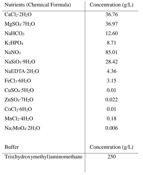
Table 1 - Qualitative and quantitative chemical composition for the nutrient stock solutions used for MBL culture medium.
Nutrients (Chemical Formula) Concentration (g/L)
CaCl2∙2H2O 36.76 MgSO4∙7H2O 36.97 NaHCO3 12.60 K2HPO4 8.71 NaNO3 85.01 NaSiO3∙9H2O 28.42 NaEDTA∙2H2O 4.36 FeCl3∙6H2O 3.15 CuSO4∙5H2O 0.01 ZnSO4∙7H2O 0.022 CoCl2∙6H2O 0.01 MnCl2∙4H2O 0.18 Na2MoO4∙2H2O 0.006 Buffer Concentration (g/L) Tris(hydroxymethyl)aminomethane 250
All chemicals used to prepare the culture media were of analytical grade and were purchased from Sigma-Aldrich (USA) or Merck (Germany). The vitamin stock solutions
20
were prepared in ultrapure water (MILLI-Q, Merck Millipore, Germany) and vacuum filtered with a 0.2 µm filter (Millex-GS, Merck Millipore, Germany). The prepared solutions were kept at -20ºC and thawed as needed. The nutrient stock solutions were prepared in ultra-pure water and stored at 4ºC. Culture medium was prepared by first adding the appropriate amount of each nutrient stock solution to deionized water. After the addition of the nutrients, the medium was sterilized in the autoclave (Uniclave 88, AJC, Portugal) for 60 minutes at 120ºC. The medium was left to cool off for 24 h, after which the vitamin solutions were added.
The cultures were maintained in aerated 5 L glass erlenmeyer flasks with 4 L of sterile MBL medium. The flasks were kept at 20ºC with 24h photoperiod with continuous air supply. Culture medium was partially renewed three times per week by removing 2 L of the culture and adding 2 L of fresh medium. The flasks were monitored daily to detect any signs of bacterial contamination.
D. magna food preparation was made three times per week, at the same time as
media renewal. A volume of 2 L of C. vulgaris culture was collected and centrifuged at 3500 rpm for 7 minutes (Heraeus Megafuge 16, ThermoFisher Scientific, USA). The resulting supernatant was discarded and the pellet was ressuspended in ASTM (refer to section 2.2 for a detailed description of the ASTM medium). This algae suspension was used to feed the daphnids and was stored in a refrigerator at 4ºC for a maximum period of 3 days.
To calculate the algae density in the food preparation, a sample from the algae suspension was diluted with a factor of 1:10 (v/v) and the optical density (OD) was measured at 440 nm (V-630 Spectrophotometer, Jasco, USA). According to the obtained OD, the volume corresponding to 3.00*105 cells per 100 mL was calculated using the relationship presented in Equation 1:
21
Where:
Algae concentration = concentration of the algae at cells per mL Abs = absorbance value of the food preparation solution at 440 nm Df = dilution factor
2.2. Daphnia magna Cultures
D. magna (clone A) cultures have been maintained in the Laboratory of
Ecotoxicology of ICBAS and CIIMAR for more than 15 years. The parental cultures that were used to provide the individuals used during the assays were maintained in American Society for Testing and Materials hard water (hereafter indicated as “ASTM”) (ASTM, 2010) as described in Table 2, enriched with Ascophyllum nodosum extract (Baird et al., 1989) and vitamins B1, B12 and H (Bradley et al., 1993).
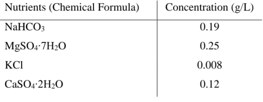
Table 2 - Qualitative and quantitative chemical composition for the nutrient stock solutions used for ASTM culture medium.
Nutrients (Chemical Formula) Concentration (g/L)
NaHCO3 0.19
MgSO4∙7H2O 0.25
KCl 0.008
CaSO4∙2H2O 0.12
All chemicals used to prepare the culture media were of analytical grade were purchased from Sigma-Aldrich (USA) or Merck (Germany). Nutrient stock solutions were prepared in ultra-pure water and stored at 4ºC for all nutrients except for CaSO4∙2H2O, which had to be freshly prepared at the time of medium preparation.
Culture medium was prepared by adding the appropriate amount of each nutrient and vitamin solutions and to deionized water.
D. magna cultures were maintained in parthenogenetic reproduction under