Defining novel stages in
medullary thymic epithelial cell
differentiation: Implications for
tolerance induction
Maria Camila Trindade Ribeiro
Mestrado em Bioquímica
Faculdade de Ciências e Instituto de Ciências Biomédicas Abel Salazar da Universidade do Porto
2018
Orientador
Doutor Nuno Miguel de Oliveira Lages Alves Investigador Principal
Instituto de Investigação e Inovação em Saúde (i3S); Instituto de Biologia Molecular e Celular (IBMC)
Coorientador
Doutor Pedro Miguel do Mar Ferreirinha Investigador Pós-Doc
Instituto de Investigação e Inovação em Saúde (i3S); Instituto de Biologia Molecular e Celular (IBMC)
Coorientadora
Doutora Maria Salomé Custódio Gomes Professora Associada
Instituto de Ciências Biomédicas Abel Salazar da Universidade do Porto (ICBAS); Instituto de Investigação e Inovação em Saúde (i3S); Instituto de Biologia
Molecular e Celular (IBMC)
Todas as correções determinadas pelo júri, e só essas, foram efetuadas. O Presidente do Júri,
Acknowledgements
I would like to take advantage of this space to thank all of those who, in various ways, have contributed to the accomplishment of the work here presented.
First of all, I would like to express my sincere gratitude to my supervisor Nuno Alves, for all the opportunities and entrusted responsibilities, and for the continuous guidance and motivation. I must also thank my cosupervisor Pedro Ferreirinha for his immense support, knowledge and patience. I would also like to recognize the important role of everyone at the Thymus Development and Function lab, for their crucial support and advice in all science-related (and unscience-related) matters, and for always providing a welcoming and light-hearted environment for my workdays. For these reasons, I thank Pedro, Laura, Leonor, Rute, Chiara, Ruben and Gema.
I must also acknowledge the key contributions from the i3S/IBMC scientific platforms: Catarina Meireles and Emília Cardoso from Translational Cytometry, and Sofia Lamas from the Animal Facility.
I also wish to thank everyone from Room 22, for always listening to my rants and for the countless pep talks. Most importantly, I am grateful to my mother, Martinha, and my aunt Helena, for always being my source of strength and inspiration.
This study was supported by the European Research Council (ERC) under the European Union’s Horizon 2020 research and innovation programme (grant agreement No 637843 - TEC_Pro) - starting grant attributed to NLA, and by NORTE-01-0145-FEDER-000012 - Structured program on bioengineered therapies for infectious diseases and tissue regeneration, supported by Norte Portugal Regional Operational Programme (NORTE 2020), under the PORTUGAL 2020 Partnership Agreement, through the European Regional Development Fund (FEDER).
Abstract
A competent immune system requires a functionally diverse repertoire of T cells that is able to respond to a variety of foreign antigens while being tolerant to self-constituents. These principles are in part imposed in the thymus through the action of highly specialized subtypes of thymic epithelial cells (TECs). In particular, medullary TECs (mTECs) have a critical role in tolerance induction, due to their unique ability to present tissue-restricted antigens (TRAs). The establishment of the medullary epithelial compartment is fostered by thymocyte-derived signals, through the engagement of signalling via the tumour necrosis factor receptor superfamily (TNFRSF) members RANK, CD40 and LTbR, expressed on mTECs and their precursors. The described mTEC developmental program includes immature CD80lowMHCIIlow precursors (mTEClow) that give rise to mature CD80high MHCIIhigh cells (mTEChigh), including cells that express Aire, an important regulator of TRA expression. Additionally, terminally differentiated CD80lowMHCIIlow post-Aire cells also reside within the mTEClow. Further heterogeneity can also be defined by the existence of cells expressing Cld3/4, SSEA and podoplanin, which were shown to define mTEC-committed progenitors, or cells expressing CCL21, a key inducer of thymocyte migration into the medulla. As such, the mTEC niche encompasses highly diverse subsets, whose lineage and functional relationships remain difficult to characterize, mainly due to a lack of suitable markers to define existent subsets and further dissect new developmental stages.
In this thesis, we incorporate the study of CD24 and SCA1 expression in the standard flow cytometry analysis of TECs. With this strategy, we reveal three distinct subpopulations of mTECs that emerge from the perinatal period into early adulthood: mTECI (CD24+SCA1-), mTECII (CD24+SCA1+) and mTECIII (CD24-SCA1-). We find that mTECI include a mixture of both mTEClow and mTEChigh, while the mTECII and mTECIII subsets are enriched for mTEClow and mTEChigh, respectively. The production of CCL21 is specifically detected in a fraction of mTECII, while Aire expression is largely confined to the mTECIII subset. Using an in vitro lineage-tracing system, we show that mTECI and mTECII of mTEClow type have the potential to generate all subsets, including Aire+ mTEChigh. Lastly, we show that while TNFRSF-induced mTEC maturation does not directly induce mTECI-III, signals provided by thymocytes at early stages of their development are sufficient to establish the mTECI-III differentiation profile. Collectively, these results reveal a novel dimension of mTEC diversity defined by the differential expression of CD24 and SCA1. These newly-defined subpopulations provide a platform to further characterize their specific intrathymic functions. Ultimately, these findings extend our comprehension of the mTEC developmental program and introduce new markers that will allow a better insight of the mechanisms underlying mTEC development and tolerance induction.
Resumo
O sistema imunitário necessita de um vasto repertório de células T capazes de responder a inúmeros antigénios e que sejam simultaneamente tolerantes aos antigénios do próprio organismo. Estes princípios são impostos no timo pela ação das células epiteliais tímicas (TECs). Em particular, as TECs residentes na medula (mTECs) tem um papel crítico para o estabelecimento do estado de tolerância, devido à sua capacidade única de expressar antigénios restritos de tecidos (TRAs). O desenvolvimento das mTECs é dependente de sinais provenientes dos timócitos, que ativam os membros da superfamília de recetores de fator de necrose tumoral (TNFRSF) RANK, CD40 e LTbR, expressos pelas mTECs e as suas células progenitoras. O programa de desenvolvimento das mTECs atualmente descrito inclui células imaturas CD80lowMHCIIlow (mTEClow), precursoras das células maturas CD80highMHCIIhigh (mTEChigh), que por sua vez incluem células que expressam Aire, um importante regulador da expressão de TRAs. Por último, mTECs num estado pós-Aire são CD80lowMHCIIlow e estão também inseridas na população mTEClow. É ainda possível reconhecer uma maior heterogeneidade de mTECs através dos marcadores Cld3/4, SSEA e podoplanina, que identificam células progenitoras específicas da linhagem mTEC, ou CCL21, uma citocina que induz migração de timócitos para a medula. Deste modo, as mTECs possuem uma elevada diversidade, sendo que a relação entre as suas subpopulações e as respetivas funções permanecem pouco caracterizadas devido à escassez de marcadores para definir subpopulações existentes e elucidar novas fases de diferenciação.
No presente trabalho, foram introduzidos os marcadores CD24 e SCA1 na análise clássica de TECs por citometria de fluxo. Deste modo, mostramos que as mTECs se subdividem em três populações no período embrionário e pós-natal: mTECI (CD24+SCA1 -), mTECII (CD24+SCA1+) e mTECIII (CD24-SCA1-). Revelamos que as mTECI incluem mTEClow e mTEChigh. Enquanto as mTECII e mTECIII abrangem maioritariamente mTEClow e mTEChigh, respetivamente. As mTECII possuem células produtoras de CCL21, enquanto as mTECIII incluem a maioria das células Aire+. A análise in vitro da descendência das subpopulações mTECI ou mTECII caracterizadas como mTEClow revelou que ambas são capazes de gerar as outras subpopulações, incluindo células mTEChigh Aire+. Por fim, apesar da maturação induzida in vitro através dos recetores TNFRSF não ter gerado diretamente as populações mTECI-III, os sinais provenientes de timócitos em fase inicial de desenvolvimento foram suficientes para induzir o perfil de diferenciação mTECI-III. Em conclusão, estes resultados revelam uma nova diversidade de mTECs definida pelo padrão de expressão de CD24 e SCA1. As novas populações descritas neste trabalho constituem uma importante ferramenta para a continuação da caracterização das suas funções no
timo. Por último, estas observações permitem expandir o atual modelo de diferenciação das mTECs e fornecem novos marcadores para o estudo dos mecanismos que controlam o seu desenvolvimento e a indução de tolerância.
Key words
Thymus medulla, Thymic epithelial cells, TEC progenitors, TEC differentiation, T cell development, Tolerance induction
Table of Contents
Acknowledgements ... V Abstract ... VII Resumo ... VIII Key words ... IX List of Figures ... XIII List of Abbreviations ... XIV
Introduction ... 1
An outline of the immune system ...3
The thymus - Structural and functional elements ...4
Thymus organogenesis ...5
TEC progenitors ...6
Phenotypic markers of TECs ...8
T cell development and its interdependence on thymic stroma ...8
Early stages of differentiation ...8
Positive selection ...10
Negative selection and regulatory T cell development ...11
Thymic involution ...13
Cellular and molecular aspects of mTEC development ...14
mTEC diversity ...14
Thymic crosstalk ...15
Aims ...18
Materials and Methods ... 19
Mice ...21
Isolation and flow cytometry analysis of TECs ...21
Fetal thymus organ culture (FTOC) ...22
Reaggregate thymus organ culture (RTOC) ...22
Results ... 23
mTEC and mTEC subsets are respectively enriched for mTEC and mTEC ... 26
Lineage relationship between mTECI, mTECII and mTECIII ... 29
Differentiation of mTECI-III is uncoupled from in vitro TNFRSF-induced maturation and independent of post-b-selected thymocytes ... 32
The frequency of mTECI-III is regulated by Aire-dependent differentiation ... 34
CCL21-expressing cells are confined to the mTECII subset ... 35
Discussion and Final Remarks ... 37
References ... 45
List of Figures
Introduction
Figure 1 - Organization and cellular composition of the thymus………4 Figure 2 - TEC progenitors and their contribution to the development and maintenance of
cTECs and mTECs………...7
Figure 3 - mTEC differentiation stages possess cells with varying levels of MHC class II
and CD80 expression and include subpopulations with other specific molecular markers………....14
Figure 4 - RANK, CD40 and LTβR signalling lead to NF-κB activation pathways………....16
Results
Figure 5 - Differential expression CD24 and SCA1 defines novel subsets of mTECs……25
Figure 6 - Correlation between the mTECI-III subsets and mTEClow and mTEChigh…………27
Figure 7 - Aire expression is largely confined to the mTECIII subset………...28
Figure 8 - mTECICD80low give rise to the remaining mTEC subsets………...30
Figure 9 - In vitro lineage potential of mTECI-III subsets……….31
Figure 10 - Establishment of mTECI-III is uncoupled from in vitro TNFRSF-induced
maturation and independent of crosstalk with post-β-selected thymocytes………...33
Figure 11 – Aire deficiency affects mTECI-III differentiation……...………...35
Figure 12 - CCL21 production is mapped to the mTECII………..……….36
Supplemental Information
Supplemental Figure 1 - Flow cytometry gating strategy for analysis of TECs………….59 Supplemental Figure 2 - Proliferating mTECs in the postnatal thymus are enriched for
List of Abbreviations
Aire - Autoimmune regulator APC - Antigen-presenting cell BCR - B cell receptor
BM - Bone marrow
CCL - Chemokine (C-C motif) ligand CCR - C-C Chemokine receptor CD - Cluster of differentiation cDC - Conventional dendritic cell
CD40L - Cluster of differentiation 40 ligand
Cld - Claudin
CMJ - Cortico-medullary junction cTEC - Cortical thymic epithelial cell CXCL - Chemokine (C-X-C motif) ligand CXCR - C-X-C Chemokine receptor DC - Dendritic cell dGuo - 2-deoxyguanosine DLL4 - Delta-like 4 DN - Double negative DP - Double positive
DTR - Diphtheria toxin receptor E - Embryonic day
EpCAM - Epithelial cell adhesion molecule
ETP - Early thymic progenitor Fezf2 - FEZ family zing finger 2 FGF - Fibroblast growth factor FoxP3 - Forkhead box P3 FoxN1 - Forkhead box N1
FTOC - Fetal thymus organ culture Gcm2 - Glial cells missing homolog 2 GFP - Green fluorescent protein HSC - Hematopoietic stem cell
ICAM - Intercellular adhesion molecule
IFN - Interferon
IGF - Insulin-like growth factor IκB - Inhibitor of kappa B IKK - IκB kinase
IL - Interleukin
ILC - Innate lymphoid cell
iNKT - Invariant natural killer T cell Ivl - Involucrin
K - Cytokeratin KI - Knockin
KLF2 - Kruppel-like factor 2 KO - Knockout
LTi - Lymphoid tissue inducer LTβR - Lymphotoxin beta receptor MHC - Major histocompatibility complex mTEC - Medullary thymic epithelial cell mTEP - Medullary thymic epithelial progenitor
MTS - Mouse thymic stroma NC - Neural crest
NF-κB - Nuclear factor kappa B NIK - NF-κB-inducing kinase NK - Natural killer
OPG - Osteoprotegerin
pGE - Promiscuous gene expression P - Postnatal day
PAMP - Pathogen-associated molecular pattern
pDC - Plasmacytoid dendritic cell PDPN - Podoplanin
Plet-1 - Placenta-expressed transcript-1 PRR - Pattern recognition receptor PSGL-1 - P-selectin glycoprotein ligand 1 RA - Retinoic acid
RAG - Recombination-activating gene
RANK - Receptor activator of nuclear factor kappa B
RANKL - Receptor activator of nuclear factor kappa B ligand RelA - V-Rel reticuloendotheliosis viral oncogene homolog A RelB - V-Rel reticuloendotheliosis viral oncogene homolog B RFP - Red fluorescent protein
RTE - Recent thymic emigrant
RTOC - Reaggregate thymus organ culture SCA1 - Stem cell antigen-1
S1P - Sphingosine-1-phosphate
S1P1 - Sphingosine-1-phosphate receptor 1 SM - Semimature
SCF - Stem cell factor SP - Single positive
SSEA - Stage-specific embryonic antigen A TCR - T cell receptor
TEC - Thymic epithelial cell
TEP - Thymic epithelial progenitor
TNFRSF - Tumour necrosis factor receptor superfamily TRA - Tissue-restricted antigen
TRAF - Tumour necrosis factor receptor-associated factor Treg - Regulatory T cell
TSSP - Thymus-specific serine protease UEA - Ulex europaeus agglutinin
VCAM - Vascular cell adhesion molecule WT - Wild Type
An outline of the immune system
A competent immune system must confer an individual the ability to survive under the threat of constant and ever-evolving pathogens, including viral, bacterial, fungal and protozoa, as well as dysfunctional host cells. To do so, it must act rapidly and efficiently to recognize threats and activate different effector functions while still maintaining the flexibility to adapt to the changes in the internal and external environment. Importantly, recognition must include discrimination between “self” and “non-self” in order to maintain an immunological self-tolerant state. All the while, it must be efficient in its use of genes and cells, and not impose an excessive energetic burden to the host. Such requirements are met through features of both innate and adaptive immunity.
Innate immune defences constitute the first line of response to pathogens and are primarily mediated by germline-encoded receptors, termed pattern recognition receptors (PRRs), that have evolved to recognize highly conserved pathogen-associated molecular patterns (PAMPs). PRR engagement will rapidly induce a cell response characterized by the expression of different genes involved in cell activation, inflammation and production of microbicidal species [1].
In addition to the innate immune system, jawed vertebrates, beginning with cartilaginous fish, have developed an adaptive immune system mediated primarily by lymphocytes [2]. The lymphocyte receptor repertoire is highly diverse, being able to recognize antigens of any potential pathogen or toxin. This capability is the virtue of somatic recombination through the random rearrangement of variable (V), diversity (D) and joining (J) gene segments, which upon assembling produce antigen receptors expressed by T (for thymus-derived) and B (for bursa de Fabricius-derived) lymphocytes [3]. The random nature of this recombination process results in a vast clonally diverse repertoire of T cell receptors (TCRs) and B cell receptors (BCRs). However, this process inevitably generates receptor specificities against antigens of the host. Elimination or regulation of self-reactive lymphocytes and maintenance of immunological self-tolerance is safeguarded by central and peripheral tolerance mechanisms [4, 5].
Upon maturation in the bone marrow (BM) and thymus, T and B cells, respectively, migrate to secondary lymphoid tissues, which specialize in spatially organizing interactions between immune cells, such as antigen-presenting cells (APCs) and lymphocytes. With subsequent triggering by antigen recognition, these cells may undergo clonal expansion and differentiation into effector T lymphocytes or antibody-producing plasma cells or otherwise become memory cells that can act in case of reexposure to their specific antigens [3].
The thymus - Structural and functional elements
This thesis is centred on the thymus, a bilobed organ located in the central compartment of the thoracic cavity above the heart. It is the primary lymphoid site for the generation and selection of T cells bearing a TCR repertoire capable of responding to countless foreign antigens while being tolerant against self-antigens. The ability to accommodate such an essential and complex process is attributed to the unique thymic microenvironments, specialized in providing the necessary signals and cell-cell interactions to support thymocyte migration, survival, commitment, proliferation, and selection.
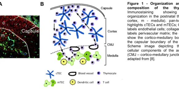
The thymus is an encapsulated and lobulated organ. The capsule consists of mesenchymal cells and connective tissue and penetrates into the thymus to form trabeculae. Below this layer is found the sub-capsular epithelium which overlies the outer cortex [6]. A peripheral cortex and central medulla are formed by three-dimensional networks of epithelial cells surrounded by developing T cells, or thymocytes, which make up for over 95% of the cellularity of the postnatal thymus (Figure 1A) [7].
The thymic stroma is mainly comprised of thymic epithelial cells (TECs), divided into two phenotypically and functionally distinct subtypes named according to their spatial location: cortical thymic epithelial cells (cTECs) and medullary thymic epithelial cells (mTECs). Other stromal elements include endothelial cells, mesenchymal cells, and BM derived cells, such as dendritic cells (DCs) , macrophages, and B cells (Figure 1B) [6]. The principles underlying TEC development are the study subject of this thesis and will be reviewed in the following chapters.
Figure 1 - Organization and cellular
composition of the thymus. (A)
Immunostaining showing cellular organization in the postnatal thymus. (c – cortex, m - medulla). pan-keratin (red) highlights cTECs and mTECs; CD31 (blue) labels endothelial cells; collagen IV (green) labels perivascular matrix; the dotted lines show the cortico-medullary boundary and the capsular boundary of the cortex. (B) Scheme image depicting the general cellular components of the adult thymus (CMJ – cortico-medullary junction). Images adapted from [8].
In the cortex, cTECs have an important role during the early stages of T cell development by promoting lineage commitment and expansion of early T cell progenitors and by providing the environment required for positive selection of thymocytes bearing
major histocompatibility complex (MHC)-restricted TCRs. In the medulla, mTECs and DCs mediate negative selection of autoreactive T cells and the generation of regulatory T cells [9]. The function of cTECs and mTECs in thymopoiesis will be described in more detail later on.
Ultimately, TECs are of vital importance in the establishment and maintenance of a functional T cell repertoire and population. This is evidenced by a clear link between TEC dysfunction and the emergence of multiple disorders, ranging from immunodeficiency to autoimmunity.
Thymus organogenesis
The establishment of the fully organized and competent thymic microenvironments is the result of a complex and tightly orchestrated process of thymus development. Thymus organogenesis occurs in the pharyngeal region of the embryo, initiating between embryonic day (E) 9 and 10. It is generated from the endoderm of the third pharyngeal pouch, in a shared primordium with the parathyroid gland [10]. Expression of the transcription factors Forkhead box N1 (Foxn1) and Glial cells missing homolog 2 (Gcm2) delineates the two non-overlapping organ-specific domains, orchestrating the differentiation of the thymus and parathyroid, respectively [11]. At E12.5, the two discrete organs begin to resolve and migrate to their final anatomical locations: the parathyroid locates adjacent to the thyroid gland, and the thymus migrates further into the chest cavity, where the two lobes meet above the heart [8]. Interestingly, an ectopic cervical thymus can also be identified in both mice and humans. The ectopic thymus emerges later, at E15.5, as a small patch of Foxn1-expressing cells [12] and possesses the same general organization as the thoracic thymus, as well as the ability to support T cell differentiation [13].
Foxn1 is the earliest thymus-specific marker and is an indispensable regulator of thymus organogenesis [14]. Foxn1-/- mice display the classical nude phenotype of congenital athymia and hairlessness. In nude mice, formation of the primordial organ is arrested between E11.5 and E12.0 and the primordium is not colonized by lymphocyte precursors, causing a severe primary T cell immunodeficiency [15]. Several other transcription factors and signalling pathways have been implicated in early thymus development [8, 15]. Still, thymic fate specification results from a complex molecular network that remains, for the most part, unknown.
The fetal thymus is seeded by hematopoietic stem cell (HSC)-derived lymphocyte precursors prior to vascularization, between E11 and E12. Lymphocytes provide key initial instructive signals for TEC development and proliferation [16]. Furthermore, the third pharyngeal pouch cells are surrounded by neural crest (NC)-derived mesenchymal cells
that will later form the capsule [17]. Proliferation and differentiation of the thymic epithelium is also highly dependent on signals supplied by the surrounding NC mesenchymal cells, such as fibroblast growth factor (FGF)-7 and -10 and insulin-like growth factor (IGF)-1 and -2 [18, 19]. In fact, NC cells provide several essential signals for thymus organogenesis. They are important regulators of the patterning of the third-pouch endoderm into the two organ-specific domains and subsequent migration and positioning of the thymus [20, 21]. Mesenchymal cells have also been shown to be the major source of retinoic acid (RA), an important regulator of TEC homeostasis, both in the embryonic and adult thymus [22]. Lastly, NC-derived cells stabilize blood-vessel structures by differentiating into perivascular cells [17].
In conclusion, thymic organogenesis is a dynamic process that involves cells from all three embryonic germ layers: endoderm-derived epithelium, ectoderm-derived NC cells and mesoderm-derived HSCs. Each undergoing tightly co-dependent and synchronised developmental programs.
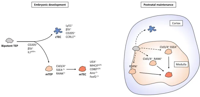
TEC progenitors
During thymus organogenesis, the first immature TECs appear at E12.5 and develop from thymic epithelial progenitors (TEPs), which have the ability to differentiate into both cortical and medullary lineages. Bipotent TEPs are essential building blocks for thymus development and maintenance and have been detected in both the embryonic and postnatal thymus [23, 24]. The identification of TEPs is an area under intense investigation, however, a full consensus has not yet been reached. Some studies have suggested Placenta-expressed transcript-1 (Plet-1) as a TEP marker, which can be recognized by the monoclonal antibodies MTS20 and MTS24 [25]. Plet1+ TECs can be detected at E12 and progressively become rarer during thymus development. These cells were shown to possess the capacity to generate functional cortical and medullary thymic microenvironments [26, 27]. In contrast, a following report argued that both MTS24+ and MTS24– cells have bipotent progenitor ability, challenging the view of Plet-1 as a TEP-specific marker [28]. Hence, the defining phenotype of bipotent TEPs remains unclear, as well as their location, abundance, and physiological contribution to the maintenance of the thymic compartments throughout life.
Follow-up reports showed that embryonic TEPs expressing several cTEC-associated markers, including β5t, CD205 and IL7, are able to generate both cTECs and mTECs [29-31]. Such findings argue against a model of TEC development in which bipotent TEPs synchronously diverge into cTEC and mTEC progenitors. Instead, they have led to the delineation of a serial progression model of TEC development, in which progenitors
pass through a transitional phase where they express cTEC traits prior to commitment to the cTEC or mTEC lineage [32]. Yet, the molecular principles controlling this cell-fate decision are still to be discovered (Figure 2).
Other studies have focused on the identification of mTEC lineage-committed progenitors (mTEPs) [33]. These cells emerge as early as E13 and can be defined by the expression of tight junction components claudin-3 and claudin-4 (Cld3/4) [34]. In addition, stage-specific embryonic antigen A (SSEA) is expressed in a small fraction of total Cld3/4+ cells and marks a more defined mTEP population whose self-renewing and progenitor potential, although decreased, can still be detected in adulthood [35]. Moreover, receptor activator of NF-κB (RANK)+ unipotent mTEC progenitors arise from the Cld3/4+SSEA -fraction between E14 and E15 [36]. Lastly, a -fraction of adult mTECs can also descend from a lineage-committed podoplanin (PDPN)+ progenitor located at the cortico-medullary junction (CMJ) [37]. It remains however unclear whether these mTEP subsets are distinct stages of the same linear differentiation or if they represent divergent mTEC lineages (Figure 2).
Figure 2 - TEC progenitors and their contribution to the development and maintenance of cTECs and mTECs. Bipotent
TEPs express cTEC traits prior to commitment to the cTEC or mTEC lineage and have been suggested to have a reduced contribution to the maintenance of the postnatal thymus. Cld3/4+SSEA+ and Cld3/4+RANK+ cells constitute two
mTEC-restricted progenitors that rise in the embryonic thymus, while PDPN+ mTEPs can also contribute to the maintenance of the
mTEC compartment in the adult thymus. The lineage relationship between the represented mTEPs is not yet defined.
The cellular and molecular mechanisms controlling the maintenance and regeneration of the adult mTEC compartment are still under investigation [38]. Possibly, throughout life, the medullary epithelium could be maintained solely by embryonic derived-mTEPs, eventually leading to an exhaustion of this limited cell pool. Alternatively, new supply of mTEPs from bipotent progenitors could still occur in the adult thymus. In this
mTEP Bipotent TEP CD205+ β5t+ IL7YFP+ PDPN+ Medulla Cortex
Embryonic development Postnatal maintenance
Cld3/4+SSEA+ mTEC cTEC Ly51+ β5t+ CD205+ CCRL1hi UEA+ MHCIIlo/hi CD80lo/hi Aire-/+ Fezf2-/+ Cld3/4+ SSEA-/+ RANK/+ Cld3/4+RANK+
regard, bipotent β5t TEPs can generate both Cld3/4 SSEA1 and PDPN mTEPs, however, this contribution to the mTEP pool fades during postnatal life, suggesting that the maintenance of the adult medullary epithelium is likely assured by mTEPs rather than bipotent progenitors [39, 40] (Figure 2).
Phenotypic markers of TECs
The development of novel antibodies and reporter mice has significantly aided the ongoing efforts to unveil all the developmental and functional complexity of TECs. Some markers are shared between cTECs and mTECs, for example, both are defined by the expression of epithelial cell adhesion molecule (EpCAM)/CD326 and MHCII, within the nonhematopoietic (CD45−) fraction of the thymus [41]. Several phenotypic traits are routinely used to discriminate cTECs and mTECs and to identify subpopulations within each. Across studies, there is some variability in the method employed to discriminate TEC subsets, which is also dependent on the analytical tools used, such as flow cytometry and immunohistochemical analyses. cTECs are commonly defined by the expression of cytokeratin (K) 8 and 18, Ly51 (CD249), CD205 and ER-TR4, as well as more recently identified functional molecules, such as CCRL1, β5t, DLL4 and high levels of IL-7 [42]. On the other hand, mTECs are distinguished by the expression of K5 and 14, MTS10, ER-TR5 and the binding to lectin Ulex europaeus agglutinin (UEA) 1. Further discrimination of mTEC heterogeneity can be achieved based on the combined levels of expression of MHCII, CD40, CD80, Aire and CCL21 [32, 41]. This thesis will further explore the diversity within the mTEC compartment, as such, a more in-depth discussion of the described mTEC markers and subpopulations will be provided in the chapter “mTEC diversity”.
T cell development and its interdependence on thymic stroma
Early stages of differentiation
All blood cell types, including T cells, originate from HSCs. These cells are characterized by continuous progenitor ability and self-renewal, and are phenotypically negative for all lineage markers [43]. HSCs undergo a series of intermediate stages where they undergo progressive lineage restriction, and ultimately differentiate into cells of the lymphoid and myeloid lineage. Lymphoid cells include T, B, natural killer (NK) and innate lymphoid cells (ILCs), while the myeloid lineage gives rise to erythrocytes, megakaryocytes, granulocytes and macrophages [44]. Plasmacytoid DCs (pDCs) and conventional DCs (cDCs) differentiate from lymphoid- and myeloid-committed progenitors, respectively [44].
During fetal development, hematopoiesis occurs primarily in the liver, whereas in postnatal life, it resides within the bone marrow [43]. In order for HSCs to differentiate into competent self-tolerant T cells, they require the specialized thymic microenvironments. Therefore, these cells must egress from the adult bone marrow, traffic through the blood, and enter the thymus. Thymus homing of bone marrow-derived progenitors is guided by the expression of the chemokine receptors CCR7, CCR9 and CXCR4, while the corresponding chemokine ligands CCL19/CCL21, CCL25 and CXCL12, are provided by multiple stromal cell types, including mTECs, cTECs and non-TEC stroma [45, 46]. Other molecules involved in this trafficking are P-selectin glycoprotein ligand 1 (PSGL-1), α4β1 integrin, and αLβ2 integrin, present on bone marrow-derived progenitors, while the respective receptors, P-selectin, VCAM-1 and ICAM-1, are expressed on the endothelial cells of the thymus vasculature [47, 48]. Early migration to the unvascularized fetal thymus also relies on chemotaxis mediated by the three chemokine receptors CCR7, CCR9 and CXCR4 [45].
Upon entry through the blood vessels localized around the cortico-medullary junction [49], T cell precursors are commonly termed early thymic progenitors (ETP). ETPs lack the expression of CD4 and CD8, hence being referred to as double negative (DN) thymocytes. Within the DN stage, the developmental program can be subdivided into 4 stages, characterized by the coordinated expression of cell surface proteins CD44 and CD25: DN1 (CD44+CD25-), DN2 (CD44+CD25+), DN3 (CD44-CD25+) and DN4 (CD44 -CD25-). DN1 cells are uncommitted, as they retain the potential to differentiate into T cells but also DCs, NKs, and possess some vestigial B cell and myeloid lineage potential [50, 51]. Engagement of the Notch signalling pathway is crucial at this stage to inhibit multiple cell fate potentials and gradually induce the genetic program for T cell fate determination. cTECs provide the essential and non-redundant ligand for Notch signalling, DLL4 [52]. The DN2 stage is characterized by a migratory movement to the subcapsular cortex. [49]. In this region, cTECs further support T lineage specification, differentiation and expansion by providing the necessary growth factors and cytokines, such as stem cell factor (SCF or kit ligand) and IL-7 [53, 54]. During this stage, alongside active proliferation, cells undergo recombination-activating gene (RAG) 1 and RAG2-mediated V(D)J rearrangements of the
Tcrb, Tcrg and Tcrd loci, which are required for the assembly of the TCR [52]. Transition
into the DN3 stage marks the complete and irreversible commitment to the T cell lineage [50]. Although during the DN3 stage, thymocytes can produce TCRγ and TCRδ chains and differentiate along the γδ T cell lineage, most cells undergo αβ TCR development pathways [53]. A successful Tcrb rearrangement will give rise to a functional TCRβ chain, which associates with an invariant TCR α-chain and CD3 signalling molecules to form the pre-TCR complex [55]. The autonomous pre-pre-TCR signalling, in non-redundant cooperation with Notch signalling, mediates a key developmental checkpoint, the β-selection event, which
rescues DN3 cells from apoptosis, promotes proliferation and ceases the recombination of the Tcrb locus [56]. Following β-selection, cells downregulate CD25, becoming DN4 or pre-double-positive cells, while initiating Tcra gene rearrangements. DN cells differentiate into CD4+CD8+ double positive (DP) thymocytes, which are the first cells to express a functional αβ TCR [57].
Positive selection
In the cortex, cTECs are self-antigen-presenting cells responsible for inducing positive selection. This process decides the fate of thymocytes (survival/differentiation versus apoptosis) according to the specificity and binding strength of the αβ TCR relatively to the self-peptide-MHC complex presented by cTECs. DP thymocytes carrying TCRs that interact with intermediate avidity are induced to survive and allowed to further differentiate [58]. Most commonly, cells express TCRs unable to interact with self-MHC molecules and undergo death by neglect [59]. On the other hand, high affinity interactions with the self-peptide-MHC complexes can lead to negative selection carried out by cTECs [59]. Ultimately, this checkpoint ensures a repertoire of cells carrying appropriate self-MHC restricted TCRs.
During positive selection, thymocytes pass through a brief intermediate CD4+CD8low stage before differentiating into either CD4+CD8- or CD4-CD8+ single positive (SP) thymocytes [57]. The divergence into the CD4 helper or CD8 cytotoxic lineage depends on whether the TCR is restricted to MHC class II or MHC class I, respectively [60]. The production of the positively selecting peptides presented on the MHC molecules is also a determinant of positive selection, and is highly dependent on a set of proteolytic enzymes that are specifically expressed in cTECs. For instance, cTEC-restricted cathepsin L and thymus-specific serine protease (TSSP) mediate the production of peptides that are loaded onto MHC class II molecules in late endosomes and are crucial for CD4+ T cell selection [61, 62]. Macroautophagy, a bulk protein degradation process, is also a source of unique peptides, important for the generation of certain MHC class II-restricted specificities [63]. On the other hand, MHC class I molecules present cytoplasm-derived peptides, which are produced by proteolytic complexes termed proteasomes. Proteasomes are formed by a set of three b catalytic subunits. While the b1, b2 and b5 subunits generate the constitutively expressed proteasome, interferon (IFN)-g-stimulated cells and antigen presenting cells (such as dendritic cells and mTECs) express the b1i, b2i and b5i subunits, which form the immunoproteasome [54]. In contrast, cTECs exclusively express the proteasomal subunit
b5t, which is incorporated with b1i and b2i to form the thymoproteasome, resulting in the ability to process a unique repertoire of peptides important for CD8+ T cell selection [64].
In conclusion, cTECs play a critical role in multiple stages of T cell development, including homing of T cell progenitors, lineage commitment and T cell expansion and development. Furthermore, they possess exclusive proteolytic machinery that allows them to present a unique self-peptide array for positive selection of αβ T cells.
Negative selection and regulatory T cell development
The random generation of the αβ TCR repertoire can produce receptors capable of recognizing self-antigens. The generation and escape of these potentially destructive autoreactive T cells is prevented through specialized functions of the medullary compartment. Positively selected thymocytes upregulate CCR7 on the cell surface, while CCR7 ligands CCL19 and CCL21 are expressed by mTECs. Consequently, CCR7-mediated chemotaxis attracts thymocytes to the medulla, where they will undergo further selection and development [65]. Once in the medulla, SP thymocytes interact with antigen-presenting cells, including mTECs, DCs and thymic B cells, that are able to screen them for self-reactive specificities [59, 66]. Cells bearing TCRs that bind to self-antigen-MHC complexes above a certain threshold are deleted through negative selection. Alternatively, CD4+ SP cells bearing high binding capacity to self-antigens can be redirected to the FoxP3+ regulatory T cell (Treg) lineage [67]. Tregs are essential for immune homeostasis and prevention of spontaneous autoimmunity [68]. Thus, clonal deletion of self-reactive T cells and Treg development are two complementary modes to impose self-tolerance.
Pivotal to both these processes, is the unique ability of mTECs to ectopically express and present tissue-restricted antigens (TRAs), a phenomenon termed promiscuous gene expression (pGE) [69]. TRA loci, whose expression is otherwise limited to specific tissues, become accessible in mTECs to allow their expression and subsequent exposure to developing T cells.Remarkably, mature mTECs express approximately 85% of the coding genome, in contrast to other tissues that typically express 60-65% [70]. The ectopic transcription of TRAs in mTECs is controlled in part by the Autoimmune regulator (Aire) protein [71], together with the more recently described FEZ family zinc finger 2 (Fezf2) [72]. Aire regulates the expression of nearly 4,000 genes by recruiting multi-protein complexes to induce transcription [73]. Still, a significant fraction of TRA transcripts is expressed in mTECs independently of Aire, which implicates the contribution of other factors to pGE [70, 74]. Interestingly, single-cell RNA sequencing revealed that, at a given time point, each TRA is expressed by 1-3% of mTECs, thus each individual mTEC expresses only a fraction of
the TRA repertoire and they ultimately add up at the population level to present a complete and stable TRA set [75]. This is thought to be a mechanism to avoid undesired physiological stress within the cell and also to increase the density of each TRA presented on the cell surface [76]. Although pGE reflects a stochastic mosaic pattern of TRA expression, it is subject to regulation. TRAs expressed at a single-cell level appear to cluster into co-expression groups, and a model has been suggested in which single mTECs can sequentially shift through distinct co-expression groups throughout their lifespan [77]. Central tolerance is highly dictated by the pool of TRAs presented in the medulla at a given time. Several mutations of the Aire gene have been shown to compromise TRA expression and negative selection of autoreactive T cells and are linked to the clinical manifestation of severe autoimmune symptoms [74, 78].
Tolerance induction is achieved through the collaboration of mTECs and DCs [79]. Thymic DCs localize predominantly in the medulla and are subdivided into three major subsets: two subtypes of cDCs, either of intrathymic or extrathymic origin, and pDCs [80]. Intrathymically-derived (or resident) cDCs are able to cross-present TRAs originally expressed by mTECs and play non-redundant roles in shaping the T cell repertoire through clonal deletion and Treg induction [81, 82]. Extrathymically-derived (or migratory) cDCs and pDCs broaden the range of self-antigens displayed in the thymus due to their ability to present peripherally-acquired self-antigens not covered by mTECs, and that otherwise would not be presented to developing thymocytes [83, 84].
During their residency in the medulla, thymocytes go through the selection process while also undergoing their final maturation steps. These last stages of thymocyte development before thymus exit are characterized by several cell surface protein changes. Initially, the recent positively selected or semimature (SM) SP thymocytes are phenotypically characterized as CD69+CD24+CD62L-Qa2- [85] and are susceptible to negative selection [86]. As they upregulate MHC class I and downregulate CD69, they become competent to proliferate in response to TCR stimulation and express molecules associated with egress [85]. The transcription factor Kruppel-like factor 2 (KLF2) promotes CD62L and sphingosine-1-phosphate receptor 1 (S1P1) expression [87]. As they mature, SP thymocytes become CD69-CD24-CD62L+Qa2+ and thus acquire the functional competences to enter the peripheral T cell pool. S1P1 regulates the exit from the thymus into the circulation, where the chemotactic gradient of the corresponding S1P ligand is higher [88]. Alternatively, in the fetal thymus, thymocytes egress via a S1P-independent mechanism. Two known contributors to fetal thymus emigration are CCR7 and CXCR4, and the corresponding ligands CCL19 and CXCL12 [89, 90].
In sum, the thymic medulla is a key site in αβ T cell development, in which mTECs and DCs act in concert to provide different layers of tolerance induction, including deletion
of autoreactive T cell clones and Treg development. All the while, it provides the specific microenvironment to nurture the final maturation of the functionally competent and self-tolerant naive T cells.
Thymic involution
At the time of birth, mice possess a virtually empty peripheral T cell pool [91]. During early postnatal life, the thymus increases in size and operates at an extremely high rate of thymopoiesis, critical to establish a functional and diverse T cell compartment. Following this highly productive period, at around 6 weeks in mice and 1 year in humans, thymic size and output begin to reduce [92, 93]. This process, termed thymic involution, is evolutionarily conserved among all thymus-bearing species and is characterized by severe architectural alterations, with loss of true thymic tissue, and an increase in fat, connective tissue, and perivascular space [92]. As a result, fewer T cells are produced and exported as aging progresses. Consequently, the peripheral naive T cell compartment has a reduced frequency of recent thymic emigrants (RTEs), and a concomitant increase occurs in the memory T cell pool [94].
Thymic involution is a contributing factor to the age-related decline in immune system competence, which is associated with several detrimental effects, such as reduced effectiveness and response to vaccination, increased infection susceptibility, autoimmunity, and increased incidence of cancer [95]. Naturally, identifying the genetic or physiological programs and the cell types responsible for involution is of great interest. Several studies have indicated the ageing thymic stroma as the primary driving force of thymic atrophy, rather than changes in the T cell progenitors supplying the thymus [96]. With age, intrinsic and extrinsic factors contribute to the downregulation of several genes that are key in TEC function and homeostasis. Particularly, it has been shown that the downregulation of Foxn1, a key regulator of TEC development, is associated with the onset of thymic involution [97]. In fact, whereas attenuation of Foxn1 in the postnatal thymus leads to premature involution, up-regulation of Foxn1 in the fully involuted thymus improves architecture, gene expression and thymopoietic ability of TECs, which in turn regenerates thymic activity [98, 99].
Cellular and molecular aspects of mTEC development
mTEC diversity
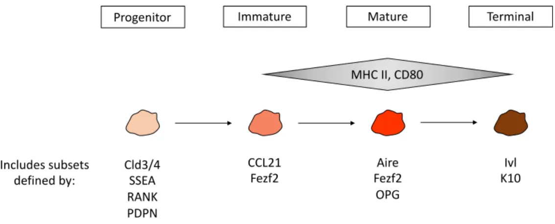
The studies described earlier highlight the role of mTECs in safeguarding self-tolerance. As such, substantial efforts have been directed into the identification and characterization of mTEC diversity. A significant degree of phenotypic and functional heterogeneity has been described, and currently, different mTEC subpopulations are routinely identified by the expression of several molecular markers. The first level of diversity can be defined according to the expression of cell surface MHCII and CD80 molecules, which subdivides mTECs into MHClowCD80low (mTEClow) and MHChighCD80high (mTEChigh) [100]. The mTEClow subset includes cells at distinct developmental stages. They were first shown to contain cells with the ability to give rise to mature mTEChigh, however, it was later revealed that a fraction of mTEClow expresses markers associated with terminal differentiation such as involucrin (Ivl) and keratin 10 (K10) and constitute a population of previously Aire-expressing cells, also termed post-Aire cells [101, 102]. Additionally, a fraction of mTEClow was shown to be responsible for the production of the chemokine CCL21, essential in the recruitment of thymocytes from the cortex to the medulla [103]. Thus, mTEClow are a heterogenous population, containing immature precursors of mTEChigh, but also terminally differentiated mTECs and a CCL21-producing subset with a specific role in T cell development (Figure 3).
Figure 3 - mTEC differentiation stages possess cells with varying levels of MHC class II and CD80 expression and include subpopulations with other specific molecular markers. Several markers are associated with mTEC precursors
(as discussed in “TEC progenitors”), including Cld3/4, SSEA, RANK and PDPN. mTECs characterized as MHCIIlowCD80low
(mTEClow) include precursors of mature MHCIIhighCD80high (mTEChigh), CCL21-producing cells, cells expressing Fezf2, and
also terminally differentiated post-Aire cells expressing Ivl and K10. The mTEChigh subset is also heterogeneous, with
subpopulations defined by expression of Aire, Fezf2 and OPG.
CCL21
Fezf2 Fezf2Aire OPG Cld3/4 SSEA RANK PDPN Ivl K10 Progenitor Immature Mature Terminal
MHC II, CD80
Includes subsets defined by:
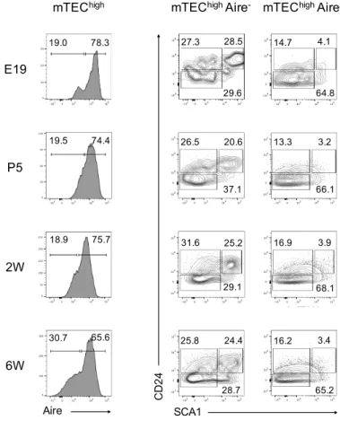
Regarding the mTEChigh subset, these cells are characterized by a high turnover rate (2 to 3 weeks), while mTEClow include only a small fraction of cycling cells [104]. At around E16, a fraction of mTEChigh begins expressing Aire, a crucial regulator of TRA gene expression for self-tolerance induction in thymocytes [100, 104]. Within Aire+ mTEChigh, further subdivision is possible based on the differential expression of osteoprotegerin (OPG), a regulator of mTEC cellularity and proliferation [105]. mTEChigh are also characterized by expression of the transcription factor Fezf2, which directly regulates TRA expression in an Aire-independent manner. Although at a lower frequency, Fezf2 is also detected in mTEClow [72] (Figure 3).
Due to the rapid turnover of Aire+ mTECs, it has been speculated that Aire expression might result in increased apoptosis [100]. This mechanism would allow the mTEC-derived TRAs to be passed onto DCs and improve the efficiency of cross-presentation. This notion was supported by the finding that in the absence of Aire, mTEChigh increase in proportion [100]. Additionally, it has been argued that Aire might not impact mTEC lifespan, but instead be required for the completion of the mTEC differentiation program and final transition into the mTEClow post-Aire stage [106]. Thus, two different models are currently contemplated for the role of Aire in mTEC maturation. On one hand, Aire expression could interrupt mTEC maturation and only in its absence the cells would reach terminal differentiation. Alternatively, Aire could be a promoter of mTEC maturation, and thus, Aire deficiency would inhibit cells from completing their differentiation program [107].
Thymic crosstalk
During early thymic ontogenesis, the cortical and medullary areas are not yet organized in the typical three-dimensional arrangement observed in the adult thymus. Growth and organization of both compartments begins with colonization of the thymus by hematopoietic progenitors and is induced by instructive signals from the developing thymocytes. As such, a symbiotic relationship exists between thymocytes and TECs. These thymocyte-TEC interactions, commonly termed “thymic crosstalk”, are important to regulate TEC development, maintenance and function [16]. In the cortex, for instance, thymocyte-derived signals control the expression of Notch ligand DLL4 and IL-7 by cTECs, which are key factors during early T cell development [108, 109].
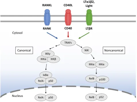
Crosstalk between thymocytes and mTECs is far more characterized and is an essential requirement for the development and maintenance of the thymic medulla. Thymocytes express ligands for several members of the tumour necrosis factor receptor
superfamily (TNFRSF), including receptor activator of NF-κB (RANK), CD40 and lymphotoxin β receptor (LTβR). These receptors are expressed in mTECs (and their precursors) and their engagement induces activation of the canonical and noncanonical nuclear factor kappa B (NF-κB) signalling pathways, which activate the transcriptional programs required for mTEC lineage specification and differentiation (Figure 4) [110]. As such, interference with elements of the NF-κB signal transduction pathways compromises mTEC development and induces autoimmunity [111]. For example, deficiency in NF-κB inducing kinase (NIK) or tumour necrosis factor receptor-associated factor 6 (TRAF6) results in a disorganized thymic medulla and absence of mature mTECs [112, 113]. Moreover, disruption at a more downstream point in the pathway further aggravates the phenotype, with deficiency in the RelB (V-Rel reticuloendotheliosis viral oncogene homolog B) subunit of the NF-κB complex resulting in a highly hypomorphic medulla (Figure 4) [114].
Figure 4 - RANK, CD40 and LTβR signalling lead to NF-κB activation pathways. Interaction of TNFRSF members with
their respective ligands induces activation of NF-κB pathways. TNF receptor-associated factor (TRAF) family proteins bind to the cytoplasmic domains of TNF receptors and activate a downstream kinase cascade, which includes the NF-κB-inducing kinase (NIK) and IκB kinase (IKK) complexes. These kinases trigger the degradation of inhibitor of κb (IκB) proteins or the processing of p100, which sequester NF-κB in the cytosol, thereby leading to NF-κB nuclear translocation and transcriptional activation. In the canonical pathway, the NF-κB complex consists predominantly of RelA and p50, while the NF-κB subunits in the noncanonical pathway are typically RelB and p52 [115].
The TNFRSF ligands are differentially expressed by hemopoietic cells and their cellular source varies between developmental periods. In the embryonic thymus, prior to αβ T cell positive selection, innate CD4+3− lymphoid tissue inducer (LTi) cells induce the development of the first Aire+ mTECs through the expression of RANK ligand (RANKL) [116]. Invariant Vγ5+ thymocytes accumulate in the embryonic medulla and are also able to trigger mTEC maturation through RANKL expression [117]. These findings support an initial role for innate LTi cells and γδ T cell progenitors in inducing Aire+ mTECs to ensure
TRAFs IκBα RelA p50 RelB IKKα IKKβ NIK IKKα IKKα p100 RelA p50 RelB p52 Noncanonical Canonical Cytosol Nucleus RANK CD40 LTβR RANKL CD40L LTα1β2, Light IKKγ
tolerance induction in the emergent αβ T cells. In adult mice, medulla formation is mainly maintained by positively selected CD4+ and CD8+ thymocytes and iNKT cells through RANKL/RANK signals [118, 119]. Furthermore, intrathymic CD40L is provided by CD4+ SP T cells and CD40L-mediated antigen-specific interactions with self-reactive CD4+ thymocytes are important to control mature mTEC cellularity [120].
While in adult RANKL-deficient thymus, Aire+ mTECs are present at a drastically reduced frequency, CD40 deficiency results in a more modest reduction. Still, mice with combined deficiency in RANKL and CD40 show a more profound reduction in Aire+ mTECs compared to mice lacking only RANK-mediated signalling [121]. Thus, CD40-mediated signalling appears to partially compensate for the absence of RANKL. Concluding, whereas RANK alone can induce Aire+ mTEChigh during embryogenesis, in adult mice, the establishment of the medullary microenvironment results from the cooperation between CD40 and RANK signals.
LTβR signalling is required for the organization of the medulla and export of mature T cells from the thymus [122]. Although it is not required for a normal Aire+ mTEC frequency [123], it has been shown to control functional properties in mTEClow. Particularly, it regulates the expression of medullary chemokines CCL19 and CCL21 by mTEClow, which are required for thymocyte migration to the medulla and consequent negative selection [124]. Additionally, it is important for the expression of Aire-independent TRAs in mTEClow [125]. LTβR was also shown to be important in later mTEC development, particularly in regulating the transition into the terminally differentiated post-Aire stage [102].
It is possible that TNFRSF-mediated signalling may act stepwise during mTEC development. Embryonic Cld3/4+SSEA1+ mTEC progenitors are LTβR+RANK- and were shown to emerge in the absence of RelB. On the other hand, production of temporally downstream Cld3/4+SSEA-RANK+ mTEC progenitors is RelB-dependent, implicating the noncanonical NF-κB pathway [36]. Coincidently, LTβR signalling can elicit RANK expression and condition mTECs to receive the early RANK signal for differentiation [126]. Still, more insight is required to assume a direct lineage relationship between these two subsets. Lastly, an initial RANK signalling is in turn necessary to upregulate CD40 expression on developing mTECs [127].
Aims
mTECs represent a dynamic niche with precursor-product relationships between subsets with distinct functional abilities, regulated by several cooperative but non-redundant thymocyte-derived signals. Valuable past studies have established the foundations of a mTEC developmental program that is believed to be far more intricate. In this thesis, we aimed to further dissect mTEC diversity and provide new insights into the lineage and functional relationships between their subpopulations.
Taking advantage of multicolour flow cytometry analysis, we began by investigating the expression pattern of CD24 and stem cell antigen-1 (SCA1) in murine mTECs, in conjunction with well-established mTEC markers such as MHCII, CD80 and Aire, throughout thymic ontogeny. We next sought to investigate the lineage potential and molecular requirements of the novel subpopulations defined by the expression of CD24 and SCA1 using thymic organotypic cultures. Furthermore, we studied the requirement for Aire expression in the emergence of the mTEC subpopulations by analysing Aire-/- and Aire reporter mouse models. Lastly, using a CCL21 reporter mouse, we mapped CCL21 expression in the newly-defined mTEC diversity.
Characterizing the cellular and molecular determinants of mTEC development, as well as their functional contributions within this compartment, can ultimately elucidate and highlight the key steps involved in thymic medulla development and tolerance induction.
Mice
C57BL/6 and BALB/c mice were kept and bred at the animal facility of Instituto de Investigação e Inovação em Saúde (i3S) under specific pathogen-free conditions. For fetal studies, the day of vaginal plug detection was designated as embryonic day (E) 0.5. ActinRFP mice were purchased from The Jackson Laboratory and bred at the animal facility of i3S under specific pathogen-free conditions. Aire-/- and Aire/DTR KI [128] mouse models were analysed in collaboration with Mitsuru Matsumoto’s laboratory and CCL21 reporter mice [129] were analysed in collaboration with Graham Anderson’s laboratory. All studies were conducted in accordance with institutional guidelines.
Isolation and flow cytometry analysis of TECs
Thymi were dissected at the indicated time points and digested for 30 minutes at 37°C in trypsin (Sigma). A syringe was used to mechanically disrupt the thymic fragments every 5 minutes. Single-cell suspensions were enriched through the depletion of thymocytes using a MACS-based CD45+ cell depletion kit (Miltenyi Biotec) according to the manufacturer’s instructions. Cell concentration was determined in a haemocytometer. Cell surface staining was carried out for 30 minutes at 4°C with the following antibodies: PerCP-Cy5-conjugated anti-CD45.2 (clone 104), PE-conjugated anti-Ly51 (clone 6C3), APC-eFlour 780-conjugated anti-I-A/I-E (clone M5/114-15-2), Alexa eFluor 647-conjugated anti-EpCAM (clone G8.8), PE-conjugated anti-CD80 (clone 16-10A1) (all from eBioscience); BV421-conjugated anti-EpCAM (clone G8.8), BV650-conjugated anti-CD80 (clone 16-10A1), BV510-conjugated CD24 (clone M1/69), BV786-conjugated anti-SCA1 (clone D7), APC-Fire-conjugated anti-CD24 (clone M1/69), Alexa 488-conjugated anti-SCA1 (clone D7) (all from Biolegend). Biotinylated UEA (vectorshield) was revealed with BV711-conjugated (Biolegend) or PE-Cy7-conjugated streptavidin (eBioscience). For intracellular staining, and upon cell surface labelling, cells were washed, fixed and permeabilized using the Foxp3 / Transcription Factor Staining Buffer Set (eBioscience) according to the manufacturer’s instructions. Intracellular staining was then carried out with eFlour 660-conjugated anti-AIRE and FITC-conjugated anti-Ki67 (both from eBioscience) for 30 minutes at 4°C. Analysis was performed using the LSRFortessa flow cytometer using the BD FACSDIVA software (BD Bioscience). The collected sample files were analysed using FlowJo v10.2 software (FlowJo, lcc).
Fetal thymus organ culture (FTOC)
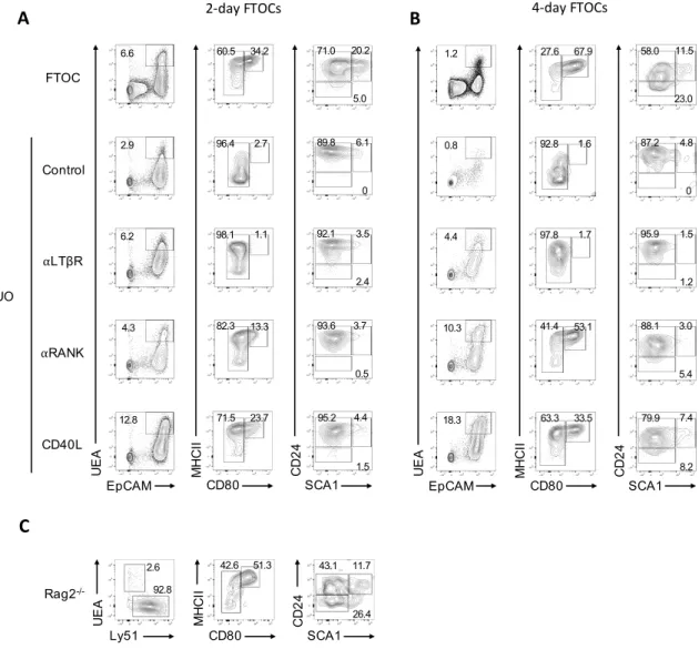
E14 thymic lobes were dissected and used to establish fetal thymus organ cultures (FTOCs). Three to four lobes were used per condition to normalize intrathymic variations. Lobes were cultured in 6-well plates on a 0.8 µm Isopore membrane filter (Milipore) resting on a foam sponge placed in 1 mL DMEM medium supplemented with 10% FCS, 1% L-glutamine 200mM (GIBCO), and with or without 360mg/L of 2-deoxyguanosine (dGuo) (Sigma). After 4 days, the dGuo-treated FTOCs were cultured in either medium alone or with 5μg/ml of recombinant CD40L (CD40L) (R&D Systems), 2μg/ml of agonist anti-RANK (αRANK) (R&D Systems) or 10μg/ml of agonist anti-LTβR (αLTβR) mAB AC.H6 (provided by Jeff Browning, Biogenidec, US). After 2 or 4 days in culture, the lobes were digested and analysed by flow cytometry.
Reaggregate thymus organ culture (RTOC)
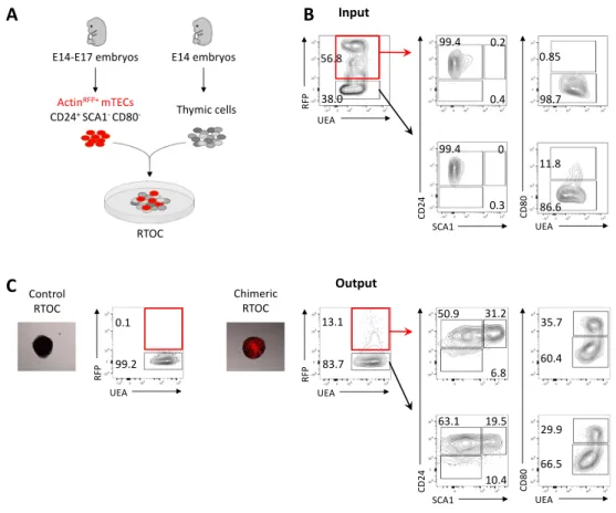
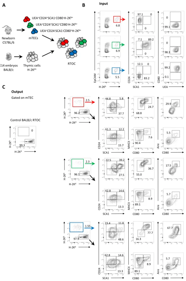
Thymic single-cell suspensions from embryonic (E14 to E17) ActinRFP mice or newborn (P1 to P3) C57BL/6 mice were stained as previously described and acquired in a FACSAria II (BD Biosciences) for the sorting of the following mTEC (CD45-EpCAM1+UEA+) populations: CD24+SCA1-CD80-, CD24+SCA1+CD80- and CD24-SCA1-CD80+. Isolated mTEC populations were mixed with single-cell suspensions from E14 C57BL/6 or BALB/c thymic lobes and reaggregates were performed as previously described [130] under the organ culture conditions described above. Chimeric reaggregate thymus organ cultures (RTOCs) were established by mixing 25.000 ActinRFP mTECs with 5x10⁷ E14 C57BL/6 thymic cells or 20.000-30.000 of the different sorted populations with 5x10⁷ E14 BALB/c thymic cells. After 7 days in culture, ActinRFP RTOCs were assessed for fluorescence in a IN Cell Analyzer 2000 (GE Healthcare). All RTOCs were subsequently dissociated and analysed by flow cytometry.
The differential expression of CD24 and SCA1 defines new mTEC
subsets
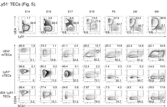
An in depth characterization of mTEC differentiation has been prevented due to the limited number of traits to define distinct developmental and functional states [131]. Our initial approach was to search for additional mTEC markers to further resolve the maturation program of mTECs. Using multicolour flow cytometry, we integrated the analysis of the expression of CD24 and SCA1 in the prototypical study of cTECs and mTECs. These two markers have been associated with stem/progenitor cell potential in other tissues [132, 133]. For this purpose, thymi were isolated from C57BL/6 mice ranging from embryonic day 14 (E14) to 6 weeks-old (6W). TECs were defined as CD45- EpCAM+ (supplemental Fig. 1) and subdivided into UEA+Ly51- mTECs, UEA-Ly51+ cTECs and UEA-Ly51- TECs [41]. At E14, all TECs were uniformly CD24+SCA1-. In both cTECs and UEA-Ly51- TECs, the expression of CD24 was reduced throughout embryonic life and these cells were mostly CD24-SCA1- at the neonatal period. However, at a later point of development, CD24-SCA1+ cells, as well as rare CD24+SCA1- and CD24+SCA1+ cells, appeared in cTECs and UEA -Ly51- TECs (Fig. 5).
Figure 5 - Differential expression CD24 and SCA1 defines novel subsets of mTECs. Thymi from C57BL/6 mice were
isolated at the indicated time points and total TECs (defined hereafter as CD45-EpCAM+) were analysed for expression of
Ly51 and UEA binding by flow cytometry (FC). UEA+Ly51- mTECs, UEA-Ly51- TECs and UEA-Ly51+ cTECs were analysed
for CD24 and SCA1 expression. Numbers in plots indicate the frequency of cells found within each gate. E – Embryonic day; P – Postnatal day; W – Weeks. Plots presented are representative of at least three analysis per time point.
E15 E17 E19 2W
E14 P5 1.7 39.8 3.8 23.3 6.9 6.8 13.0 6.4 37.9 6.6 54.8 69.5 80.8 77.4 52.7 Ly51 UE A 53.2 2.7 40.7 6W 89.8 4.9 3.1 UEA-Ly51 -TECs Ly51+ cTECs UEA+ mTECs SCA1 CD2 4 22.4 14.9 40.2 41.2 25.3 14.8 40.2 26.0 15.7 42.4 41.1 2.4 15.9 74.3 1.1 0.1 95.9 1.9 22.1 32.8 28.0 18.0 17.6 34.8 17.2 14.5 15.0 35.8 23.5 9.5 4.3 69.3 1.0 0.2 95.7 7.0 4.3 74.7 1.4 0.3 92.1 7.87 1.6 79.3 12.4 2.2 72.1 34.6 0.8 51.3 49.4 0.1 32.3 77.4 0.9 13.1 80.2 0.2 11.2 99.4 0.1 0.2 99.8 0.1 0
Interestingly, only a fraction of mTECs became CD24 during embryonic development, and a distinct CD24+SCA1+ subset appeared in the perinatal period (E19) (Fig. 5). Consequently, three mTEC subpopulations could be defined as CD24+SCA1-, CD24+SCA1+ and CD24-SCA1-, hereafter referred to as mTECI, mTECII and mTECIII. The diversity within the mTEC compartment defined by the expression pattern of CD24 and SCA1 was maintained in young adult mice (6W) (Fig. 5). Overall, these results reveal that the differential expression of CD24 and SCA1 allows the identification of novel mTEC subpopulations, whose developmental and functional characterization became the main focus of this work.
mTEC
IIand mTEC
IIIsubsets are respectively enriched for mTEC
lowand
mTEC
highWithin mTECs, two major subpopulations can be defined according to the co-expression of MHCII and CD80. On one hand, MHCIIlowCD80low mTECs, also known as mTEClow, include a mixture of immature precursors and terminally differentiated mTECs [102]. On the other hand, MHCIIhighCD80high mTECs, known as mTEChigh, include mostly mature cells, including Aire+ cells [100]. Thus, we sought to investigate how mTECI, mTECII and mTECIII were phenotypically related with mTEClow and mTEChigh. Throughout time, mTECI included both mTEClow and mTEChigh (Fig. 6, red). Notably, the mTECII encompassed mostly mTEClow, while the mTECIII were consistently enriched for mTEChigh (Fig. 6, green and blue).
mTEChigh present a further degree of heterogeneity, as a large fraction of these cells expresses the Aire protein and represents one of the later stages of mTEC maturation [100]. We next analysed how Aire+ cells were related with mTECI, mTECII and mTECIII populations. Throughout development, while Aire- mTEChigh distributed equally throughout the three subsets, Aire+ mTEChigh were located predominantly in mTECIII (Fig. 7). As such, the lack of SCA1 and CD24 expression is correlated with a mature Aire+ mTEC stage.
Figure 6 - Correlation between the mTECI-III subsets and mTEClow and mTEChigh. Thymi from C57BL/6 mice were isolated
at the indicated time points and UEA+ mTECs were analysed for CD80, MHCII, CD24 and SCA1 expression by FC. The
subsets defined by the coloured gates, mTECI (red), mTECII (green) and mTECIII (blue), were analysed for the expression of
CD80 and MHCII. Plots are representative of at least three analysis per time point. Pie charts represent the average proportion of mTEClow (light grey) and mTEChigh (dark grey) within each of the colour-coded mTEC subsets. E – Embryonic day; P –
Postnatal day; W – Weeks.
E15 E17 E19 2W
E14 P5 CD80 MH C II SCA1 CD2 4 32.0 57.9 16.8 74.1 53.7 33.2 44.1 46.3 18.3 73.6 58.8 28.5 56.9 36.0 21.1 74.4 60.8 31.0 78.6 17.6 38.9 55.8 70.7 18.0 94.2 4.5 80.3 15.6 57.8 37.4 39.3 55.1 33.3 57.7 29.4 61.2 22.4 14.9 40.2 41.2 25.3 14.8 40.2 26.0 15.7 42.4 41.1 2.4 15.9 74.3 1.1 0.1 95.9 1.9 6W 52.8 35.0 22.1 32.8 28.0 52.4 34.8 30.2 58.8 73.7 11.2 CD80 MH C II mTEChigh mTEClow mTECI mTECII mTECIII