D
ISINFECTION OF
H
EALTHCARE
W
ASTE
(HCW)
BY
A
LKALINE
H
YDROLYSIS
,
ITS
E
FFICIENCY AND
E
MISSIONS
D
ISINFECTION OF
H
EALTHCARE
W
ASTE
(HCW)
BY
A
LKALINE
H
YDROLYSIS
,
I
TS
E
FFICIENCY AND
E
MISSIONS
Dissertation presented by
S
ÍLVIA
C
ARDINAL
P
INHO
For the degree of
D
OCTOR OFP
HILOSOPHY INE
NVIRONMENTALE
NGINEERING to the Faculty of Engineering, University of PortoThesis supervised by
Professor Manuel Afonso Magalhães Fonseca Almeida
(Metallurgical and Materials Engineering Department)
Professora Olga Cristina Pastor Nunes
My sincere thanks to my Supervisors, for making this study possible, providing scientific and
tech-nical guidance during all work. To Professor Manuel Afonso Magalhães Fonseca Almeida I thank
for the personal support, help and advice for nearly two decades. To Professora Olga Cristina
Pas-tor Nunes thank you for accepting the supervision of this work and always be available to help at
all times.
I would like to thank the Fundação para a Ciência e Tecnologia for the fellowship
SFRD/BD/48956/2008 and the Project PTDC/SAU-SAP/114855/2009.
Thanks to the Metallurgical and Materials Engineering Department of the Faculty of Engineering of
Porto University (FEUP) and Laboratory for Processes Engineering, Environment, Biotechnology
and Energy (Lepabe) for accepting me as PhD student.
A very special thanks to Maria José from CTC for the support provided, by encouraging and, above
all for your friendship.
I would like to thanks to Joana Dias my colleague office and “Congresses” for support provided,
help and encouragement.
Thanks to all people who helped me throughout this work: Liliana Pereira, Carmen Deus, Sílvia,
Paula, “all girls of microbiology laboratory” especially at Luisa and Vera“, Sr. Ramiro and Anthony
Danko.
I wish to express my deepest thanks to my parents, Eduardo and Augusta, for proving my
educa-tion, for your care, support and love, for everything.
To my brothers, Helena, Anabela and André, thank you for your care and love.
To my husband, Rui, for whole patience, support and help my sincere thanks and love.
To my children, João Tomás and Mariana, my unconditional love and sorry by the time which I “stole” you during the last months.
vii
The present work aimed at providing additional knowledge about the application of wet treatments to healthcare waste (HCW), namely alkaline hydrolysis, through the study of transformations of some components usually present in HCW and characterization of effluents produced.
Samples of components usually present in HCW were subjected to autoclaving or alkaline hydrolysis under the same conditions of temperature and time. Alkaline hydrolysis caused appreciable degradation of most of the components, particularly in adhesives and diapers. The autoclaving treatment degraded the components in a much lesser extent than alkaline hydrolysis. The effluents obtained showed an appreciable organic load. Nevertheless, the effluents produced by autoclaving showed a lower organic load and were less biodegradable than the ones resulting from the alkaline hydrolysis treatment. The minimum conditions of temperature, time and concentration of NaOH to achieve the total destruction of animal tissues (pork and beef) were studied. The bone and meat containing samples were completely destroyed when subjected to temperatures higher than 95 °C with 1 M NaOH solution in less than 60 minutes. The effluents generated, although with very high pH and organic load, were biodegradable after neutralization.
Geobacillus stearothermophilus spores were used to assess the disinfection efficiency of alkaline hydrolysis. The survival curves and the D–values (decimal reduction time) were determined. The complete inactivation of spores (6 log10 reduction) was achieved at a temperature of 110 °C with 1 M NaOH in less than 5 minutes.
Based on the conditions obtained for the total destruction of animal tissues as well as for the inactivation of Geobacillus stearothermophilus, i.e. temperature of 110 °C, time 35 minutes and 1 M NaOH, animal tissues and discarded medical components usually present in healthcare waste were hydrolyzed and the effluents were characterized according to Portuguese legislation laying down emission limit values for discharges of waste water, and their aerobic and anaerobic biodegradability was evaluated. The effluents showed values lower than the discharge limit values for almost all the parameters, except pH, total nitrogen, TOC, COD and BOD5. The effluents showed a coefficient of total aerobic biological degradation of 50.5 % and 52.9 % and an anaerobic biodegradability of 22.3 % and 42.2 %, for discarded medical components and animal tissues, respectively.
A real sample of infectious HCW was subjected to alkaline hydrolysis at a pilot scale, using the conditions of the treatment defined in the laboratorial study. The characteristics of the resultant effluent corroborate with studies carried out in the laboratory.
ix
O presente trabalho pretendeu acrescentar conhecimento adicional sobre a aplicação dos tratamentos de calor húmido, isto é, hidrólise alcalina, aos resíduos hospitalares, através do estudo das transformações de alguns componentes usualmente presentes nos resíduos hospitalares e da caracterização dos efluentes produzidos.
Amostras de componentes normalmente presentes nos resíduos de hospitalares foram submetidas a autoclavagem e a hidrólise alcalina nas mesmas condições de temperatura e tempo usadas no actual processo de autoclavagem. A hidrólise alcalina causou uma degradação apreciável da maior parte dos componentes, sendo esta mais expressiva para o adesivo e a fralda. Embora na autoclavagem a degradação dos componentes tenha sido bastante inferior à hidrólise alcalina, os efluentes obtidos apresentaram uma carga orgânica elevada, mas menor biodegradabilidade do que os efluentes resultantes do tratamento por hidrólise alcalina.
Nos ensaios de hidrólise alcalina realizados com tecidos de origem animal (carne de porco e carne de boi) foram avaliadas as condições mínimas de temperatura, tempo e concentração de NaOH necessárias para a destruição total dos tecidos animais. Estes foram completamente destruídos, quando sujeitos a temperaturas superiores a 95 °C e NaOH 1 M em menos de 60 minutos. Os efluentes gerados embora com elevados valores de pH e carga orgânica são biodegradáveis após neutralização.
Nos ensaios efectuados para avaliar a eficiência de desinfecção da hidrólise alcalina foi usada a estirpe do microrganismo Geobacillus stearothermophilus. A eficiência do tratamento alcalino foi determinada pelas curvas de sobrevivência e pelos valores D (tempo de redução decimal). A completa inactivação dos esporos (redução 6 log10) foi conseguida à temperatura de 110 °C e a uma concentração de NaOH 1 M em menos de 5 minutos.
Com base nas condições obtidas para a destruição total dos tecidos de origem animal bem como para inactivação do Geobacillus stearothermophilus, isto é temperatura de 110 °C, tempo de 35 minutos e concentração de NaOH 1 M, as amostras foram hidrolisadas e os efluentes produzidos foram caracterizados de acordo com a legislação portuguesa que estabelece os valores limites de emissão de descarga de águas residuais. A biodegradabilidade aeróbia e anaeróbia dos efluentes também foi avaliada. Os efluentes, na maioria dos parâmetros excepção para o pH, azoto total, COT, CQO e CBO5, apresentaram valores inferiores aos valores limites de emissão. Os efluentes apresentaram um coeficiente de degradação biológica aeróbia total entre 50,5 % e 52,9 %, e uma biodegradabilidade anaeróbica entre 22,3 % e 42,2 %
Uma amostra de resíduos hospitalares do grupo III foi submetida ao tratamento por hidrólise alcalina, à escala piloto, nas condições de tratamento definidas nos ensaios laboratoriais. As características do efluente resultante do tratamento corroboraram os resultados obtidos nos ensaios laboratoriais.
xi
ACKNOWLEDGEMENTS v
ABSTRACT vii
RESUMO ix
LIST OF FIGURES xv
LIST OF TABLES xvii
LIST OF ACRONYMS AND SYMBOLS xix
CHAPTER 1 – INTRODUCTION 1
1.1 FRAMEWORK 1
1.1.1 RELEVANCE OF THE STUDY 1
1.1.2 OBJECTIVES OF THE WORK 4
1.1.3 OUTLINE 6
1.2 HEALTHCARE WASTE CHARACTERISTICS 7
1.2.1 DEFINITION AND CLASSIFICATION 7
1.2.2 GENERATION 16
1.2.3 COMPOSITION 22
1.2.4 OTHER CHARACTERISTICS 25
1.3 HEALTHCARE WASTE MANAGEMENT 28
1.3.1 SEGREGATION, COLLECTION AND STORAGE 33
1.3.2 TRANSPORT, TREATMENT AND DISPOSAL 36
1.4 HEALTHCARE MANAGEMENT IN PORTUGAL 37
1.5 REFERENCES 39
CHAPTER 2 – HEALTHCARE WASTE TREATMENT TECHNOLOGIES 45
2.1 INTRODUCTION 45
2.2 THERMAL PROCESSES 51
2.2.1 AUTOCLAVING 51
2.2.2 DRY HEAT 55
2.2.3 MICROWAVE AND MACROWAVE 57
2.2.4 INCINERATION 59 2.2.5 PYROLYSIS 67 2.2.6 GASIFICATION 68 2.2.7 PLASMA 70 2.3 CHEMICAL PROCESSES 71 2.4 IRRADIATION PROCESSES 73 2.5 BIOLOGICAL PROCESSES 73
xii
2.8 ACKNOWLEDGMENTS 76
2.9 REFERENCES 77
CHAPTER 3 – ALKALINE HYDROLYSIS 81
3.1 INTRODUCTION 81
3.2 CHEMICAL FUNDAMENTALS 84
3.3 DESTRUCTION/INACTIVATION OF DISEASE AGENTS 87
3.4 PRINCIPLE OF OPERATION 88
3.5 REFERENCES 90
CHAPTER 4 – EFFECTS OF ALKALINE HYDROLYSIS AND AUTOCLAVING ON DISCARDED MEDICAL COMPONENTS PRESENT IN HEALTHCARE WASTE
93
ABSTRACT 93
4.1 INTRODUCTION 94
4.2 MATERIALS AND METHODS 95
4.2.1 MATERIALS 95
4.2.2 METHODS 96
4.3 RESULTS AND DISCUSSION 99
4.3.1 HCW COMPONENTS TRANSFORMATIONS AND EFFLUENTS
PRODUCED 99
4.3.2 TG AND DSC ANALYSES 103
4.3.3 PRINCIPAL COMPONENTS ANALYSIS 107
4.4 CONCLUSIONS 109
4.5 REFERENCES 110
CHAPTER 5 – APPLICABILITY OF ALKALINE HYDROLYSIS TO DESTROY ANIMAL TISSUES PRESENT IN HEALTHCARE WASTE
113
ABSTRACT 113
5.1 INTRODUCTION 114
5.2 MATERIALSANDMETHODS 115
5.3 RESULTSANDDISCUSSION 116
5.4 CONCLUSIONS 121
5.5 REFERENCES 121
CHAPTER 6 – INACTIVATION OF Geobacillus stearothermophilus SPORES BY ALKALINE TREATMENT
xiii
6.2 MATERIALS AND METHODS 126
6.2.1 PREPARATION OF Geobacillus stearothermophilus SPORES 126
6.2.2 ALKALINE TREATMENT 127
6.2.3 INCUBATION AND SURVIVAL COUNTS 128
6.2.4 FLUORIMETRIC DETECTION OF DPA 128
6.2.5 TRANSMISSION ELECTRON MICROCOPY 129
6.3 RESULTS AND DISCUSSION 129
6.4 CONCLUSIONS 136
6.5 ACKNOWLEDGMENTS 137
6.6 REFERENCES 137
CHAPTER 7 – CHARACTERIZATION OF EFFLUENTS RESULTANT FROM ALKALINE HYDROLYSIS TREATMENT
141
ABSTRACT 141
7.1 INTRODUCTION 142
7.2 MATERIALS AND METHODS 143
7.2.1 MATERIALS 143
7.2.2 ALKALINE HYDROLYSIS TESTS 143
7.2.3 EFFLUENTS CARACTERIZATION METHODS 144
7.2.4 AEROBIC BIODEGRADATION TESTS OF EFFLUENTS 145
7.2.5 ANAEROBIC BIODEGRADATION TESTS OF EFFLUENTS 145
7.3 RESULTS AND DISCUSSION 145
7.3.1 EFFLUENTS CHARACTERIZATION 145
7.3.2 AEROBIC BIODEGRADATION OF EFFLUENTS 148
7.3.3 ANAEROBIC BIODEGRADATION OF EFFLUENTS 148
7.4 CONCLUSIONS 148
7.5 REFERENCES 149
CHAPTER 8 – SCALE UP EXPERIMENTS 151
ABSTRACT 151
8.1 INTRODUCTION 152
8.2 MATERIALS AND METHODS 152
8.2.1 PILOT SCALE REACTOR 152
8.2.2 MATERIALS 153
xiv
8.4 CONCLUSIONS 157
8.5 ACKNOWLEDGMENTS 157
CHAPTER 9 – CONCLUSIONS AND FUTURE WORK 159
9.1 CONCLUSIONS 159
9.2 FUTURE WORK 160
ANNEX 1 – CHEMICAL TEST METHODS DESCRIPTION 163
A.1.1 – TOTAL ORGANIC CARBON (TOC) 163
A.1.2 – CHEMICAL OXYGEN DEMAND (COD) 164
A.1.3 – BIOCHEMICAL OXYGEN DEMAND (BOD) 165
A.1.4 – TOTAL NITROGEN AND AMMONIA 167
ANNEX 2 – AEROBIC RESPIROMETRIC TEST 169
A.2.1 – INTRODUCTION 169
A.2.2 – PRINCIPLE 170
A.2.3 – DETERMINATION OF AEROBIC BIODEGRADATION 170
ANNEX 3 – ANAEROBIC BIODEGRADABILITY TEST 172
A.3.1 – INTRODUCTION 172
A.3.2 – PRINCIPLE 174
A.3.3 – REAGENTS 175
A.3.4 – TEST PROCEDURE 176
A.3.5 – DETERMINATION OF ANAEROBIC BIODEGRADATION 177
xv Figure 1.1 – Hazardous healthcare waste generated in Portugal. 2
Figure 1.2 – Healthcare waste classification. 15
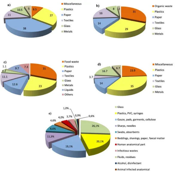
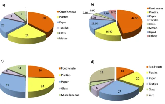
Figure 1.3 – The medical waste composition reported by four studies: a) Abdulla et al. (2008); b) Dehghani et al. (2008); c) Taghipour and Mosaferi (2008); d) Fraiwan et al.
(2013); Coker et al. (2009). 23
Figure 1.4 – The non-hazardous waste composition reported by: a) Sawalem et al. (2009); b) Taghipour and Mosaferi (2008); c) Alhumoud and Alhumoud (2007); d)
Mohee (2005). 24
Figure 1.5 – The hazardous waste composition reported by Taghipour and Mosaferi
(2008). 25
Figure 1.6 – a) colour plastics bags for waste collection; b) container for sharps
collection; c) waste containers. 35
Figure 2.1 – Incinerator used to treat HCW in a developing country. 47
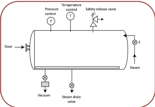
Figure 2.2 – Typical autoclave. 54
Figure 2.3 – Diagram of a mobile microwave unit: 1. Feeding hopper, 2. Feeding crank, 3.Shredder, 4. Connecting hopper, 5. Level sensors, 7. Microwave generators, 8. Temperature holding section, 9. Discharge conveyor auger, 10. Temperature sensors, 11. Filter system, 12. Water tank with pump and spraying connection, 13. Steam generator, 14.Steam connection, 15. Hydraulic aggregate, 16. Room heater, 17. Container.
58
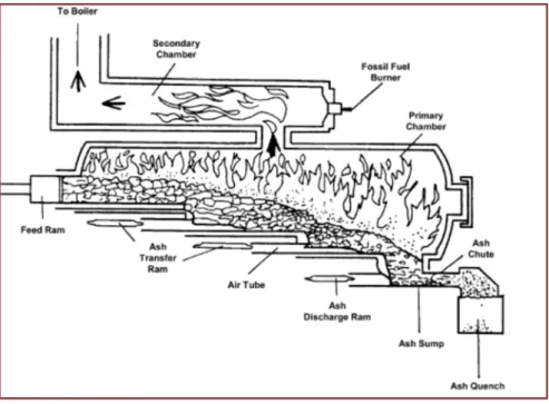
Figure 2.4 – Controlled air incinerator. 60
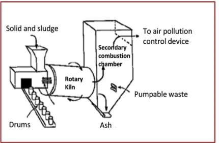
Figure 2.5 – Rotary kiln incinerator. 61
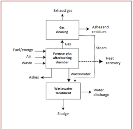
Figure 2.6 – Simplified flow scheme of the incineration process. 62 Figure 2.7 – Schematic diagram of cement encapsulation. 75
Figure 3.1 – Alkaline hydrolysis digester. 89
Figure 4.1 – Diagram of the alkaline process. 97
Figure 4.2 – TG profile of HCW components; (─) original; (---) hydrolysed; (∙∙∙∙) autoclaved.
104
Figure 4.3. – DSC profile of HCW components, (─) original; (---) hydrolysed; (∙∙∙∙) autoclaved. The DSC profiles of untreated and hydrolysed bag collector for urine coincide with DSC profiles of examination glove.
106
Figure 4.4 – Principal components analysis of the HCW treatments results and spatial distribution of the samples; arrows refer the parameters considered in the analysis 108
xvi
Figure 5.1 – Reactor and temperature control. 116
Figure 5.2 – Alkaline hydrolysis of pork ((□) 1 M NaOH; (■) 2 M NaOH) and bovine ((○) 1 M (●); 2 M NaOH) meat at 90 °C over time.
118
Figure 5.3 – Hydrolyzed bone e hydrolyzate. 119
Figure 5.4 – TG (a) and DSC (b) profiles of bone; (─) untreated; (─··─) alkaline treated at 100 °C (---) alkaline treated at 90 °C; (····) alkaline treated at 80 °C. 120
Figure 6.1 – TEM images of Geobacillus stearothermophilus. Vegetative cells suspension (A), spores suspension after addition of lysozyme (B), after addition of SDS (C) and spore debris after alkaline treatment (D). sc, spore coat; co, cortex; cr, core. 130
Figure 6.2 – Survival curves for the Geobacillus stearothermophilus exposed to alkaline treatment at 100 °C with various NaOH concentrations; 0 M NaOH (□); 0.1 M NaOH(■); 0.25 M NaOH(∆); 0.5 M NaOH (●); 0.75 M NaOH(○); 1 M NaOH(▲) and predicted by the Cerf model (― ). Vertical bars represent standard deviations of the means.
132
Figure 6.3 – Survival curves for the Geobacillus stearothermophilus exposed to alkaline treatment at 110 °C with various NaOH concentrations; 0.1 M NaOH (■); 0.25 M NaOH (∆); 0.5 M NaOH (●); and predicted data by the Cerf model in the absence of components ( ― ); discarded medical components (----) and animal tissues (····). Vertical bars represent standard deviations of the means.
134
Figure 8.1– Pilot reactor and real infectious HCW used in the alkaline hydrolysis tests. 153 Figure 8.2 – Effluent from the scaled-up alkaline hydrolysis treatment of real infectious
HCW. 155
Figure 8.3 – Real infectious HCW after alkaline hydrolysis treatment. 156 Figure A.1.1 – TC analyser used in TOC and TC determination. 164 Figure A.1.2 – Digester and spectrophotometer used in COD determination. 165 Figure A.1.3 – DO meter used in BOD5 determination. 167 Figure A.2.1 – Respirometer used in the aerobic tests. 170 Figure A.2.2 – Dissolved oxygen versus time for sample and acetate. 171 Figure A.3.1 – The samples, blank and controls vessels used in anaerobic tests. 177 Figure A.3.2 – Cumulative methane production (at 0 °C and 1 atm) of samples and
controls.
xvii
Table 1.1 – Outline of the work. 6
Table 1.2 – Healthcare waste classification according to WHO. 9 Table 1.3 – Healthcare waste classification according U. S. Environmental Protection Agency.
10
Table 1.4 – Healthcare waste classification in Greece according to the Hellenic legislation (HMWC, 2003).
10
Table 1.5 – Healthcare waste classification in Turkey according to the Medical Waste Control Regulation (MWCR, 2005).
11
Table 1.6 – Healthcare waste classification according to the China Ministry of healthcare, 2003.
11
Table 1.7 – Healthcare waste classification in Brazil according to federal resolutions (ANVISA, 2004 and CONAMA, 2005).
12
Table 1.8 – Healthcare waste classification in Korea according to Waste Management Act in 1999.
12
Table 1.9 – Healthcare waste classification in India according to the Biomedical Waste Rules, 1998.
13
Table 1.10 – Healthcare waste classification in Spanish Autonomous Community of Madrid
13
Table 1.11 – Healthcare waste classification according Portuguese legislation (Order No 242/96).
14
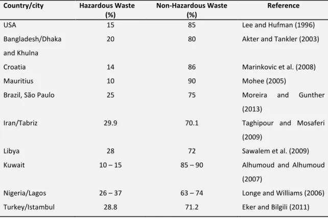
Table 1.12 – Healthcare waste classification according to the European Waste List. 16 Table 1.13 – Healthcare waste generation in different countries. 19 Table 1.14 – Hazardous and non-hazardous wastes in different countries. 22 Table 1.15 – Specific weight of healthcare waste in different studies. 26 Table 1.16 – Specific weight, moisture content and heating value of various components
of healthcare waste in Ecuador, India and USA. 27
Table 1.17 – WHO – Recommended segregation scheme. 34
Table 2.1 – Levels of microbial inactivation 46
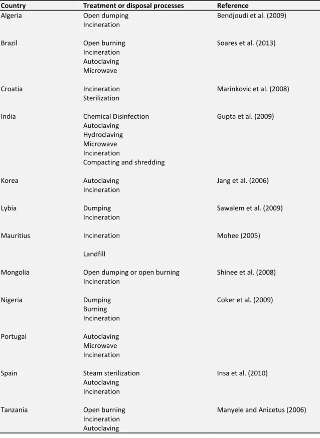
Table 2.2 – Treatment and disposal processes of HCW in different countries 48 Table 2.3 – Advantages and disadvantages of autoclaving process. 53 Table 2.4 – Advantages and disadvantages of dry heat process. 56 Table 2.5 – Advantages and disadvantages of microwave process. 59
xviii Table 2.7 – Advantages and disadvantages of pyrolysis process. 67 Table 2.8 – Advantages and disadvantages of chemical process. 72 Table 2.9 – Advantages and disadvantages of irradiation process. 73 Table 2.10 – Advantages and disadvantages of encapsulation process. 76 Table 3.1 – Advantages and disadvantages of alkaline hydrolysis. 83 Table 4.1 – Components of the HCW used in the experimental work, their materials and
carbon content. 96
Table 4.2 – Weight losses (WL) in the samples of HCW components subjected to autoclaving and alkaline hydrolysis tests at 135 °C.
99
Table 4.3 – Carbon losses (CL) in the samples of HCW components subjected to autoclaving and alkaline hydrolysis tests at 135 °C.
100
Table 4.4 – TOC, in mg C/L, and pH in the effluents resulting from autoclaving and alkaline hydrolysis of individual samples of HCW components.
101
Table 4.5 – COD in the effluents from autoclaving and alkaline hydrolysis tests of samples of individual components.
102
Table 4.6 – TOC, as mg C/L, and COD and BOD5, as mg O2/L, in the effluents from
autoclaving and alkaline hydrolysis tests of mixtures with samples of the components.
103
Table 5.1 – Alkaline hydrolysis conditions of samples composed by pork meat including bones versus hydrolysis time, residue amount, TOC, COD and BOD5 of the effluent
produced.
117
Table 5.2 – Alkaline hydrolysis conditions versus hydrolysis time and residue amount of the meat and bone.
119
Table 6.1 – Decimal time reduction (D–value), estimates of the model parameters and standard derivation values for the Cerf model. Data presented are the mean of three independent experiences with standard deviation.
131
Table 6.2 – Concentration of 10 nM standard DPA, and DPA released from endospores after autoclaving and alkaline treatment.
133
Table 6.3 – Decimal time reduction (D–value) described in literature for
G.stearothermophilus spores under different treatments.
136
Table 7.1 – Chemical methods to characterize effluents. 144 Table 7.2 – Characterization of effluent from alkaline hydrolysis of discarded medical
components 146
Table 7.3 – Characterization of effluent from alkaline hydrolysis of animal tissues. 147 Table 8.1 – Composition of infectious HCW sample used in the tests. 151 Table 8.2 – Characterization of the effluent from the scaled up alkaline hydrolysis
treatment of real infectious HCW.
xix BAT – Best Available Techniques
BEP – Best Environmental Practices
BOD – Biochemical oxygen demand
CDJ – Creutzfeldt–Jajob disease
CL – Carbon loss
COD – Chemical oxygen demand
DGS – Direção Geral Saúde
DO – Dissolved oxygen
DPA – Dipicolinic acid
DSC – Differential Scanning Calorimetric
EPA – Environmental Protection Agency
EU – European Union
EWL – European Waste List
GEF – Global Environment Facility
HBV – Hepatitis viruses B
HCV –Hepatitis viruses C
HCW – Healthcare waste
HHW – Household hazardous waste
HIV – Immunodeficiency virus
KOH – Potassium hydroxide
NaOH – Sodium hydroxide
PAHs – Polycyclic aromatic hydrocarbons
PCA – Principal Components Analysis
PCDDs – Polychlorinated dibenzodioxins
xx POPs – Persistent organic pollutants
PVC – Polyvinyl chloride
SDS – Dodecyl sulphate
STAATT – State and Territorial Association on Alternate Treatment Technologies
TC – Total carbon
TG – Thermogravimetric
TOC – Total organic carbon
TSE – Transmissible Spongiform Encephalopathy
UK – United Kingdom
USA – United States of America
USEPA – United States Environmental Protection Agency
VOCs – Volatile organic compounds
WHO – World Health Organization
1
CHAPTER 1
– INTRODUCTION
1.1
F
RAMEWORK1.1.1 RELEVANCE OF THE STUDY
Waste management is a matter of concern in developed countries due to environmental
consequences, public health and valuable resources waste, many of them strategic. In the
European Union, 2.5 billion tonnes of waste are produced annually out of which 100 million
tonnes are hazardous waste (Eurostat, 2010).
Healthcare waste (HCW) is a small fraction of solid waste produced, since it represents only
0.3 % of total generated in Europe (Eurostat, 2010), but it requires special attention due to the
danger it represents to public health and environment. Nevertheless, the amount of HCW is
increasing compared to other types of waste, mainly due to the use of medical disposable
2
HCW includes all the waste generated by healthcare establishments, research facilities and
laboratories (Prüss et al., 1999). It is a heterogeneous mixture of waste with different
components produced in variable amounts, differing among countries according to their
particular medical management practices (Prüss et al., 1999).
Characterisation, composition and quantities of HCW generated per year and per country or
region can be found in the literature (Lee and Huffman, 1996; Diaz et al., 2008). The majority
of HCW, of about 75 – 90 % is not infectious, thus can be treated as municipal waste posing no
additional risk to health or the environment. The remaining 10 – 25 % is classified as hazardous
waste, and it represents the fraction that requires special attention and specific treatments
(WHO, 2005). In Portugal, in recent years, the quantity of hazardous HCW generated was
about 30 000 ton (Figure 1.1).
Figure 1.1 – Hazardous healthcare waste generated in Portugal (source: INE, 2014).
The implementation of efficient and environmentally friendly technologies for HCW waste
treatment requires considerable technical and funding resources and a legal framework, all
which are mostly lacking in developing countries. For these reasons, in these countries, is
common to watch an inappropriate treatment and uncontrolled deposition of HCW that, due
24000 25000 26000 27000 28000 29000 30000 2008 2009 2010 2011 2012 H az ar d o u s H C W ( to n ) Year
3 to the infectious nature of the waste, may contaminate surface and groundwater, exposing the
population to diseases and parasites and increasing the epidemics risk (Hossain et al., 2011).
There are several HCW treatment processes, including incineration, pyrolysis, autoclaving,
chemical disinfection, encapsulation, microwave disinfection, irradiation and gasification.
Autoclaving and incineration are the main processes used for treating HCW (Sukandar et al.,
2006), the last being the oldest and, until now, the most used (Lee and Huffman, 1996).
However, the incineration process presents a drawback, and may emit a wide range of
hazardous pollutants including dioxins, furans, heavy metals, fine dust particles and other
pollutants resultant of incomplete combustion (Alvim-Ferraz and Afonso, 2005; Sukandar et
al., 2006; Singh and Prakash, 2007).
In 2004, the Stockholm Convention on Persistent Organic Pollutants put a focus on eliminating
or reducing releases of 12 persistent organic pollutants (POPs) including dioxins and furans.
Accordingly, stricter emissions limits for incinerators were introduced in the European Union
(EU). Additionally, World Health Organization (WHO) has been promoting the use of
non-incineration technologies to treat medical waste, eliminating the risk of dioxins and furans
emissions that may be generated by uncontrolled medical incinerators.
Also, the small producers of HCW, due to issues of current technique as the storage areas
needed for the HCW and the high transportation costs of untreated waste to the treatment
unit, are seeking for viable alternatives technologies.
Some of the treatments applied to HCW, as autoclaving and microwave, require a mechanical
process to reduce the waste volume, which increases the operating costs. Additionally, due to
the low mass reduction achieved in these treatments, there is a considerable mass of waste
that is disposed of in landfills; and, in some countries as Korea, there was reluctance to accept
4
Under such guidelines, alkaline hydrolysis may be an alternative treatment process for HCW in
some countries (Health Care Without Harm, 2004). This treatment has been shown to have
significant advantages compared to other HCW treatments, because it sterilizes and destroys
at once, and it also reduces the total waste volume. Also, alkaline hydrolysis may have a range
of application larger than autoclaving, since it can also accept organic tissues.
However, the effects of various materials present in HCW during the autoclaving process as
well as the final effluent composition and its treatability are not well known. This lack of
knowledge makes authorities to have some reluctance in licensing autoclaving plants and in
permitting their effluents to be discharged into domestic sewage treatment systems.
Thus, once the consequences of application of alkaline hydrolysis are well known, it may
advantageously substitute autoclaving and incineration to treat some types of HCW and offer
an interesting basis of decentralised treatment for reducing the risks of infection from handling
and transporting HCW.
1.1.2 OBJECTIVES OF THE WORK
The present work aimed at providing some additional light on the application of wet
treatments to HCW, namely autoclaving and alkaline hydrolysis. It intends to help waste
treatment operators, regulatory authorities, general public concerned with wastes and as well
as researchers to clarify some particularities of those two processes through carefully obtained
experimental data. Some of these aspects include answers to questions as the following: how
common materials in HCW behave under these treatments, and, in particular, which are the
minimum conditions of alkaline hydrolysis to destroy animal tissues? Which conditions
guarantee disinfection when HCW contain very resilient infectious microorganisms? What kind
of effluents is produced and are they compatible with discharge in public sewage network?
5 1 – Study the transformations of some components generally present in HCW and characterize
the effluents produced, at laboratory scale using similar conditions to autoclaving.
2 – Study the transformations of some components generally present in HCW and characterize
the effluents produced by alkaline hydrolysis, i.e. using similar conditions of temperature and
time to those used in autoclaving but with different NaOH solution concentrations, at
laboratory scale.
3 – Make the alkaline hydrolysis process an alternative treatment for animal tissues and
discarded medical components present on HCW by determining in laboratory:
i. milder conditions than those described in the literature for destroying animal tissues
with bones and make them unrecognizable.
ii. effective minimum sterilization conditions using a very resilient biological indicator.
iii. the characteristics of the effluents from the treatment.
iv. the aerobic and anaerobic biodegradability of the effluents obtained.
4 – Test the alkaline hydrolysis process with a real HCW sample under pilot scale conditions:
i. design and build a reactor to scale up the laboratorial process to the range of tenths of
liters not far from 100 L.
ii. make at least one test with a real sample of hazardous HCW using the conditions of
treatment defined in the laboratorial study and determine the characteristics of the
6
1.1.3 OUTLINE
The outline of the work is shown in Table 1.1, including the synthetic description of the
content approached in each chapter.
Table 1.1 – Outline of the work
CHAPTERS CONTENT
CHAPTER 1
INTRODUCTION Introduces the subject relevance, objectives and outline of
the work.
Presents an overview of HCW as: definition; classification; generation; composition and management.
CHAPTER 2
HEALTHCARE WASTE TREATMENT TECHNOLOGIES
Presents and describes the technologies used on the treatment of HCW.
CHAPTER 3
ALKALINE HYDROLYSIS Describes the alkaline hydrolysis process.
CHAPTER 4
EFFECTS OF ALKALINE HYDROLYSIS AND AUTOCLAVING ON DISCARDED MEDICAL COMPONENTS PRESENT IN HEALTHCARE WASTES
Studies the behaviour of some discarded medical components usually present in healthcare waste when subjected to treatments of alkaline hydrolysis or autoclaving. CHAPTER 5
APPLICABILITY OF ALKALINE HYDROLYSIS TO DESTROY ANIMAL TISSUES PRESENT IN HEALTHCARE WASTES
Studies the behaviour of animal tissues usually present in HCW when subjected to alkaline hydrolysis treatment.
CHAPTER 6
INACTIVATION OF Geobacillus
stearothermophilus SPORESBY ALKALINE TREATMENT
Studies the effects of NaOH concentration and temperature on the inactivation of Geobacillus stearothermophilus spores.
CHAPTER 7
CHARACTERIZATION OF EFFLUENTS
RESULTANT FROM ALKALINE HYDROLYSIS TREATMENT
Characterize the effluents produced when alkaline hydrolysis is applied to animal tissues and discarded medical components common in HCW and assess their aerobic and anaerobic biodegradability.
CHAPTER 8
SCALE-UP EXPERIMENTS Alkaline hydrolysis tests with a real infectious HCW sample
using a designed reactor with 70 L of capacity. CHAPTER 9
7
1.2
H
EALTHCARE WASTE CHARACTERISTICS1.2.1 DEFINITION AND CLASSIFICATION
The definition of healthcare waste is not universal and is relatively variable worldwide. Many
terms as “medical waste”, “clinical waste”, “hospital waste”, “biomedical waste” and
“infectious waste” have often been used in the literature. These terms can have different
meanings according to its designation; the severe differences between them often depend on
which legal or regulatory authority is defining the term. Indeed, in some countries the
definition of medical waste is often different due to the existing regional legislation. In Spain,
most of regional laws use the common term “medical waste” and only one region, of
Cantabria, uses the term “hospital waste”.
Medical waste is defined, according to the WHO, as “all the waste generated within healthcare
facilities, research centers and laboratories related to medical procedures. In addition, it
includes the same types of waste originating from minor and scattered sources, including
waste produced in the course of healthcare undertaken in the home (e.g. home dialysis,
self-administration of insulin, recuperative care)”. In the United States of America (USA), the
Medical Waste Tracking Act of 1988 (MWTA, 1988) defines medical waste as "any solid waste
that is generated in the diagnosis, treatment, or immunization of human beings or animals, in
research pertaining thereto, or in the production or testing of biologicals”. This definition is
commonly used in some countries, such as Greece, (Hellenic legislation, Ministerial Decision
37591/2031) or India, although in the later the term used is “biomedical waste” (Biomedical
Waste Rules, 1998). According to the Chinese legislation the medical waste is defined “as
waste characterized by infectious, toxic and other hazardous properties deriving directly or
indirectly from medical treatment, prevention, health protection, and other related activities
8
waste in the United Kingdom (UK) is taken from The Controlled Waste Regulations 1992,
issued under the Environmental Protection Act 1990. It has remained unchanged since it was
first issued under the Collection and Disposal of Waste Regulations 1988, issued pursuant to
the Control of Pollution Act 1974. Clinical waste is defined as: (a)“...any waste which consists
wholly or partly of human or animal tissue, blood or other bodily fluids, excretions, drugs or
other pharmaceutical products, swabs or dressings, syringes, needles or other sharp
instruments, being waste which unless rendered safe may prove hazardous to any person
coming into contact with it; and (b)“…any other waste arising from medical, nursing, dental,
veterinary, pharmaceutical or similar practice, investigation, treatment, care, teaching or
research, or the collection of blood for transfusion, being waste which may cause infection to
any person coming into contact with it.”
The Portuguese legislation defines hospital waste as “waste resultant from healthcare
activities to humans or animals, in the areas of prevention, diagnosis, treatment, rehabilitation
or research and education, as well as other activities involving invasive procedures such as
acupuncture, piercings and tattoos” (Decree Law No 73/2011).
Medical waste is often considered as a subcategory of healthcare waste or hospital waste
which contains the waste with infectious character. WHO defined infectious waste as “the
waste type suspected to contain pathogens (viruses, bacteria, parasites or fungi) in enough
concentration or quantities to cause disease in susceptible hosts” (Prüss et al., 1999).
The classification of healthcare waste is not uniform worldwide and brings some confusion
and disagreement between countries, regions and institutions. Several classifications have
been used in legislation and in the literature. Healthcare waste may be classified into different
types according to the source of production, chemical and physical composition, type and risk
9 suggested a simple classification of HCW into eight categories; currently there are seven
categories as shown in Table 1.2 (Chartier et al., 2014).
Table 1.2 – Healthcare waste classification according to WHO.
Waste type Description
General Waste
Infectious to potentially infectious waste
Wastes that have similar properties to the household solid waste. Waste contaminated with blood and its by-products, cultures and stocks of infectious agents, waste from patients in isolation wards, discarded diagnostic samples containing blood and body fluids, infected animals from laboratories, and contaminated materials (swabs, bandages) and equipment (such as disposable medical devices).
Pathological waste Human tissues or fluids, e.g. body parts, blood and other body fluids, fetuses.
Sharps waste Syringes, needles, disposable scalpels and blades, etc.
Chemicals waste Waste containing chemical substances, e.g. laboratory reagents, film developer, disinfectants that are expired or no longer needed, solvents.
Pharmaceuticals waste Expired, unused and contaminated drugs; vaccines and sera.
Radioactive waste Such as glassware contaminated with radioactive diagnostic material or radiotherapeutic materials.
According to United States Environmental Protection Agency (EPA 40 CFR Part 60.51c), the
wastes are divided into six categories (Table 1.3). In Greece, according to the Hellenic
legislation (HMWC, 2003), the wastes are classified into three categories; in Turkey, the
Medical Waste Control Regulation (MWCR, 2005) categorized wastes in four categories;
Chinese national legislation (PR China MOH, SEPA, 2003) and the federal resolutions of Brazil
(ANVISA, 2004 and CONAMA, 2005) classify wastes in five categories; in Korea, under Waste
Management Act in 1999, exist six categories, and India define ten categories in accordance
with Biomedical Waste Rules, 1998 (Table 1.4 to Table 1.9). In the case of Spain, wastes
classification varies from region to region in the range of three to seven categories, according
10
Table 1.3 – Healthcare waste classification according U. S. Environmental Protection Agency.
Waste type Description
Cultures and stocks Cultures and stocks of infectious agents and associated biologicals, including cultures from medical and pathological laboratories; cultures and stocks of infectious agents from research and industrial laboratories; wastes from the production of biologicals; discarded live and attenuated vaccines; and culture dishes and devices used to transfer, inoculate, and mix cultures.
Pathological and chemo wastes
Human pathological wastes, including tissues, organs, and body parts and body fluids that are removed during surgery or autopsy or other medical procedures and specimens of body fluids and their containers.
Human blood and blood product
Liquid waste human blood; products of blood; items saturated and/or ripping with human blood; or items that were saturated and/or dripping with human blood including serum, plasma, and other blood components.
Sharps Sharps that have been used in animal or human patient care or treatment or in medical, research, or industrial laboratories, including hypodermic needles, syringes (with or without the attached needle), Pasteur pipettes, scalpel blades, blood vials, needles with attached tubing, and culture dishes (regardless of presence of infectious agents).
Animal wastes Contaminated animal carcasses, body parts, and bedding of animals that were known to have been exposed to infectious agents during research (including research in veterinary hospitals), production of biologicals, or testing of pharmaceuticals.
Isolation wastes Biological waste and discarded materials contaminated with blood, excretion, exudates, or secretions from humans who are isolated to protect others from certain highly communicable diseases or from isolated animals known to be infected with highly communicable diseases.
Table 1.4 – Healthcare waste classification in Greece according to the Hellenic legislation (HMWC, 2003).
Waste type Description
Household type medical wastes Wastes that have similar properties to the household solid waste.
Hazardous medical wastes Waste with solely infectious properties, such as body tissues, blood, fecal and urine samples from patients with an infectious disease, needles etc.
Waste with both infectious and toxic properties, such as chemotherapy related waste, waste from bio-pathology and histology laboratories, etc.
Waste with solely toxic properties, such as waste that contains mercury, hazardous organic waste, expired drugs, filters, etc. Other types of medical waste Radioactive waste, batteries, pressurized cans, etc.
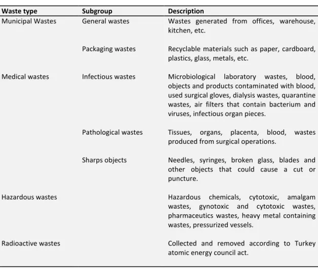
11 Table 1.5 – Healthcare waste classification in Turkey according to the Medical Waste Control Regulation (MWCR, 2005).
Waste type Subgroup Description
Municipal Wastes General wastes
Packaging wastes
Wastes generated from offices, warehouse, kitchen, etc.
Recyclable materials such as paper, cardboard, plastics, glass, metals, etc.
Medical wastes Infectious wastes
Pathological wastes
Sharps objects
Microbiological laboratory wastes, blood, objects and products contaminated with blood, used surgical gloves, dialysis wastes, quarantine wastes, air filters that contain bacterium and viruses, infectious organ pieces.
Tissues, organs, placenta, blood, wastes produced from surgical operations.
Needles, syringes, broken glass, blades and other objects that could cause a cut or puncture.
Hazardous wastes Hazardous chemicals, cytotoxic, amalgam
wastes, gynotoxic and cytotoxic wastes, pharmaceutics wastes, heavy metal containing wastes, pressurized vessels.
Radioactive wastes Collected and removed according to Turkey atomic energy council act.
Table 1.6 – Healthcare waste classification according to the China Ministry of healthcare, 2003.
Waste type Description
Infectious waste Hazardous medical wastes carrying pathogenic microorganisms which can cause the infectious disease transmission.
Pathological waste Medical waste generated in the human body and medical laboratory animal carcasses during the diagnosis and treatment, including tissues, organs, blood and body parts and fluids.
Sharp objects Needles, syringes, broken glass, blades and other items that could cause a cut or puncture.
Pharmaceutical waste Overdue, eliminated or contaminated waste drugs.
Chemical waste Hazardous chemicals, heavy metal containing wastes, toxic, corrosive, flammable and explosive waste drugs.
12
Table 1.7 – Healthcare waste classification in Brazil according to federal resolutions (ANVISA, 2004 and CONAMA, 2005).
Waste type Description
A Waste representing risk to public health and the environment due to presence of biological agents such as: blood, bodily fluids, drainage fluids or excreta (ex. surgical gloves, gauze, cotton, bandages; laboratory plates and blades; microorganism cultures; vaccines discarded and devices used for transference, inoculation or stir cultures).
B Substances representing risk to public health and the environment, depending on their characteristics of inflammability, corrosiveness, reactivity and toxicity (ex. some chemical products and pharmacological substances).
C Radioactive waste.
D General waste represented by recyclable materials (e.g. paper, cardboard, plastic, metals, and glass) and non-recyclable (e.g. organic substances, food leftover, and toilet paper).
E Sharp devices (ex. needles, syringes, lancets and similar tools).
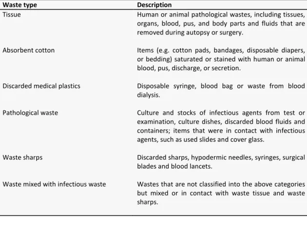
Table 1.8 – Healthcare waste classification in Korea according to Waste Management Act in 1999.
Waste type Description
Tissue Human or animal pathological wastes, including tissues,
organs, blood, pus, and body parts and fluids that are removed during autopsy or surgery.
Absorbent cotton Items (e.g. cotton pads, bandages, disposable diapers, or bedding) saturated or stained with human or animal blood, pus, discharge, or secretion.
Discarded medical plastics Disposable syringe, blood bag or waste from blood dialysis.
Pathological waste Culture and stocks of infectious agents from test or examination, culture dishes, discarded blood fluids and containers; items that were in contact with infectious agents, such as used slides and cover glass.
Waste sharps Discarded sharps, hypodermic needles, syringes, surgical blades and blood lancets.
Waste mixed with infectious waste Wastes that are not classified into the above categories but mixed or in contact with waste tissue and waste sharps.
13
Table 1.9 – Healthcare waste classification in India according to the Biomedical Waste Rules, 1998.
Waste type Description
Human anatomical waste Human tissues, organs and body parts. Animal waste
Microbiology and biotechnology waste
Animal tissues, organs, body parts, carcasses, bleeding parts, blood and experimental animals used in research. Waste from lab culture, specimens from microorganisms, vaccines, cell cultures, toxins, dishes, devices used to transfer cultures.
Waste sharps Syringes, needles, disposable scalpels and blades, glass. Discarded medicines and cytotoxic drugs Contaminated, expired, discarded drugs.
Soiled waste Waste contaminated with blood and body fluids including cotton, dressings, and soiled plasters.
Solid waste Tubes and catheters.
Liquid waste
Incineration waste Chemical waste
Waste generated from laboratory and washing, cleaning, disinfection.
Ash from incineration of any medical wastes
Chemicals used in production of biologicals, disinfection
Table 1.10 – Healthcare waste classification in Spanish Autonomous Community of Madrid.
Groups Waste type Description
I Municipal waste Waste comes from no polluted and infectious risk areas of the hospital (ex. papers, glass, plastic, metals, wood or food remains).
II Non-specific waste Includes waste from medical activities without infectious or toxic risk, such as dialysis filters, gloves, probes, and bandages, which have not been in contact with patients.
III Anatomical substances These substances belong to category 1 of the hazardous waste listed by Directive 91/689/EEC and Royal Decree 833/1988.
IV Large pieces of human remains
V Chemical products Includes chemical substances, pharmaceuticals, medicines and veterinary compounds, biocides and phyto-pharmaceutical substances.
VI Cytostatic waste Waste is only generated by medical activities related to cancer treatment as carcinogenic, mutagenic and teratogenic drugs.
VII Radioactive waste This waste refers to radioactive chemical elements that do not have a practical purpose, as well as those products that have come in contact with them, including solid and liquid substances.
14
Portuguese legislation classifies HCW in four categories (Table 1.11), the two first are
considered non-hazardous waste and the third and fourth ones are hazardous waste (Order No
242/96).
Table 1.11 – Healthcare waste classification according Portuguese legislation (Order No 242/96).
Groups Waste type Description
I Wastes similar to municipal ones Wastes that do not present special requirements in their treatment such as: wastes from general services, support services, kitchen and packaging (paper, cardboard, etc).
II Non-hazardous hospital wastes Wastes that do not require specific treatment and can be considered similar to municipal wastes. Orthopedic supplies, diapers and disposable guards, personal protective equipment from general and support services without contamination and no traces of blood. Serum bottles without contamination except those from group IV. Empty containers of drugs except those included in group III and group IV.
III Hospital wastes with biological risk Waste contaminated or suspected of contamination susceptible of incineration or other effective pre-treatment before elimination as municipal wastes, such as waste from rooms and wards for infectious patients or suspected of infectious, materials contaminated with blood or traces of blood, anatomical not identified pieces, materials used in dialysis, etc.
IV Specific hospital wastes Wastes with compulsory incineration, such as animal tissues, organs, body parts, carcasses of animals used in research, sharps materials, chemical substances, pharmaceuticals and cytostatic waste.
In some countries of European Union, as United Kingdom, the healthcare classification is based
in the European Waste List (EWL). In order to standardize the waste classification, in December
1993, the European Union publishes the EWL adopted by Commission Decision 2000/532/EC,
as amended by Commission Decisions 2001/118/EC, 2001/119/EC and 2001/573/EC. In the
15 to the EWL chapter, the second two digits relate to sub-grouping within the chapter, and the
final two digits are specific to the waste (Table 1.12). HCW are identified with code 18 in the
EWL, which includes various categories. Wherein, the first sub-grouping corresponds to the
waste generated in diagnosis, treatment or prevention of disease in humans and the second
sub-grouping corresponds to the waste generated from research, diagnosis, treatment or
prevention of disease involving animals. In general, the healthcare wastes are categorized in
two types: general waste and special waste or non-infectious and infectious waste, thus
defined as non-hazardous and hazardous wastes. Therefore, the EWL considers both types of
waste and defines a separate category for cytotoxic agents and chemicals. Indeed, the HCW
can be further characterized to a more universal form as shown in Figure 1.2 (Chartier et al.,
2014).
Figure 1.2 – Healthcare waste classification. HCW General waste Hazardous waste Infectious waste Chemical and pharmaceutical waste Radioactive waste Genotoxic waste Pathological waste
16
Table 1.12 – Healthcare waste classification according to the European Waste List.
18 01 Wastes from natal care, diagnosis, treatment or prevention of disease in humans 18 01 01 Sharps (except 18 01 03)
18 01 02 Body parts and organs including blood bags and blood preserves (except 18 01 03) 18 01 03* Wastes whose collection and disposal is subject to special requirements in order to
prevent infection
18 01 04 Wastes whose collection and disposal is not subject to special requirements in order to prevent infection (e.g. dressings, plaster casts, linen, disposable clothing, diapers, 'Controlled Waste' as defined in NZS 4304:2002 Management of Healthcare Waste) 18 01 06* Chemicals consisting of or containing hazardous substances
18 01 07 Chemicals other than those mentioned in 18 01 06 18 01 08* Cytotoxic and cytostatic medicines
18 01 09 Medicines other than those mentioned in 18 01 08 18 01 10* Amalgam waste from dental care
18 02 Wastes from research, diagnosis, treatment or prevention of disease involving animals
18 02 01 Sharps (except 18 02 02)
18 02 02* Wastes whose collection and disposal is subject to special requirements in order to prevent infection
18 02 03 Wastes whose collection and disposal is not subject to special requirements in order to prevent infection
18 02 05* Chemicals consisting of or containing hazardous substances 18 02 06 Chemicals other than those mentioned in 18 02 05
18 02 07* Cytotoxic and cytostatic medicines
18 02 08 Medicines other than those mentioned in 18 02 07
1.2.2 GENERATION
There has been an increase in the quantities of HCW compared to municipal waste mainly due
to changes in healthcare policies, higher number of healthcare facilities, medical services and
to the use of medical disposable products. In USA more than 3.5 million tons of HCW are
produced each year (Lee et al., 2004); India generates about 3 million ton/year (Mohankumar
17 Portugal, near 90 000 ton are generated every year (DGS, 2006). Thus, the quantities of HCW
generated do not depend only on the population but also on several factors such as the
medical situation of each country and its waste management systems.
The major sources of HCW are hospitals, but other sources as clinics, healthcare
establishments, laboratories and research facilities, mortuary and autopsy centers, animal
research, blood banks, veterinary surgeons, dentists, nursing homes are also generating
extensive quantities of HCW (Prüss et al., 1999). In Portugal, about 94 % of the total HCW
production has been generated by hospitals (DGS, 2006).
In recent years, there have been an increasing number of patients receiving a home healthcare
and medical services. The home-based healthcare services also produce substantial quantities
of HCW with a variety of waste materials. Some researchers (Ojeda-Benítez et al., 2013;
Yasuda and Tanaka, 2006) have studied the characteristics, generation and composition of
household hazardous waste (HHW). Gu et al. (2014) found that approximately 17.7 % of the
HHW stream was medicines in Suzhou, China. These wastes are still included in general
household waste even though they are infectious (Blenkharn, 2008). Nevertheless, there is no
management guideline and legislation for this type of waste even in developed countries as
Japan (Miyazaki et al., 2007).
The quantities of HCW generation differ among countries by their waste medical management
practices allied on the level of economic development. The developing countries generate
lower quantities of HCW than the developed countries (Prüss et al., 1999). The quantities of
HCW generated are usually depicted as a function of the bed capacity of healthcare services
(kg/bed/day); however, in healthcare facilities where the beds are not available the production
is expressed as kg/patient/day. For the low-income countries, WHO estimated a HCW
generation range of 0.5 – 3 kg/bed/day and for the high income countries a HCW generation
18
average up to 0.5 kg/bed/day of hazardous waste, while low-income countries generate on
average 0.2 kg/bed/day of hazardous waste. However, the real quantity of hazardous waste
can be much higher in low-income countries because the HCW are often not separated into
hazardous or non-hazardous wastes (WHO, 2014). According to WHO, the North America
generates 7 – 10 kg/bed/day of HCW, while South America produces just 3 kg/bed/day. In Asia,
richer countries generate 2.5 – 4.0 kg/bed/day and poorer countries produce
1.8 – 2.0 kg/bed/day of HCW. The same happens in Europe, on Western the production rate is
about 3 – 6 kg/bed/day while on Eastern the production rate is 1.4 – 2.0 kg/bed/day of HCW
(Prüss et al., 1999). Diaz et al. (2008) reported that the quantities of HCW in various types of
facilities located in developing countries are lower than in some industrialized countries. They
described that the range of HCW and infectious wastes in developing countries varies from
0.016 to 3.23 kg/bed/day and 0.01 to 0.65 kg/bed/day, respectively. This wide variation is due
to the fact that some facilities, used in their study, provide very basic services and thus
quantities of the waste generated are relatively small. In fact, the quantities of HCW generated
depend on the level of economic development, and the developed countries generate more
quantities of HCW mainly due to the the use of medical disposable products, higher number of
healthcare facilities and medical services and greater consumption of disposable instruments
and packing materials. Moreover, in developing countries the HCW are still handled, stored
and disposed together with municipal wastes for which reason the amount of HCW is smaller
than really produced (Rudraswamy et al., 2013; Hossain et al., 2011; Bendjoudi et al., 2009;
Sawalem et al., 2009).
The quantities of HCW generated per country in kg/bed/day can be found in the literature and
Table 1.13 shows the values found in some references.
The quantity of HCW generated varies not only from country to country, but also within the
19 methods adopted by healthcare services managers, hospitals departments or services, and the
use of medical disposable products as a result of developing healthcare technology. Prüss et al.
(1999) estimated HCW generation according to source size of healthcare establishment,
namely university hospital, general hospital, district hospital and primary healthcare center.
Table 1.13 – Healthcare waste generation in different countries.
Country/city Waste generation
(kg/bed/day)
Reference
Algeria 0.7 – 1.22 Bendjoudi et al. (2009)
Bangladesh/Dhaka and Khulna 0.55 – 1.10 Akter and Tankler (2003)
China 0.5 – 3.0 Changping et al. (2012)
Croatia 1.2 Marinkovic et al. (2008)
El Salvador/San Salvador 0.37 Johnson et al. (2013)
Greece/Attica 0.24 – 0.27 Komilis et al. (2011)
Iran/Tabriz 3.48 Taghipour and Mosaferi (2009)
Italy 1.3 – 2.87 Giacchetta and Marchetti (2013)
Jordan (northern) Jordan (southern) 0.83 0.73 Abdulla et al. (2008) Fraiwan et al. (2013)
Korea 0.48 Jang et al. (2006)
Kuwait 3.87 – 7.44 Alhumoud and Alhumoud (2007)
Mongolia/Ulaanbaatar 0.78 Shinee et al. (2008)
Nigeria/Lagos 0.43 – 0.67 Longe and Williams (2006)
Portugal 3.3 Botelho and Pinto (2010)
Sudan/Khartoun 0.80 – 1.71 Saad (2013)
Taiwan 2.41 – 3.26 Cheng et al. (2009)
Tanzania 0.75 Manyele and Anicetus (2006)
Turkey/Istambul USA
2.11 5 – 7
Eker and Bilgili (2011)
Medical Waste Committee (1994)*
*Taken from Lee et al. (2004).
The highest generation rate was in university hospital with 4.1 – 8.7 kg/bed/day followed by
general hospital, 2.1 – 4.2 kg/bed/day, district hospital and primary healthcare center with a
20
al. (2008) reported medical waste generation rate (kg/patient/day) in the inpatient services of
public healthcare facilities was 1.4 to 3.0 times higher than in the outpatient services a sample
of 56 healthcare facilities at Ulaanbaatar, capital of Mongolia. Sawalem et al. (2009) studied
the amounts of HCW generated in fourteen different healthcare facilities, in three cities with
different size and population, in Libya. These cities were: Tripoli a large city, Misurata a
medium-sized city and Sirt a small city. They have showed that the highest generation rate was
found in Tripoli and the lowest rate was found in the clinics and rural health centers. This fact
was due to the medical centers in Tripoli were more developed general public facilities and
serve a higher number of patients. Abdulla et al. (2008) and Fraiwan et al. (2013) studied the
medical waste management practices in northern and southern Jordan. The average amount
of medical waste generated in both regions is not the same, the northern produced
0.83 kg/bed/day and southern produced 0.73 kg/bed/day.
In the Portuguese hospitals, according to DGS (2006), the region of Lisbon and Tejo Valley
produced the highest generation rate of 3.8 kg/bed/day followed by the northern region with
2.6 kg/bed/day. Those two regions are the ones with higher population density. Coker et al.
(2009) characterized and quantified the medical wastes generated in Ibadan, Nigeria. They
selected 52 healthcare facilities based in size and function. Their results indicated that large
hospitals and clinics generated the greatest amounts of medical wastes followed by the small
healthcare units, which have facilities to treat only outpatients, and diagnostic service
laboratories. Different results were obtained by Cheng et al. (2010) when investigated the
quantities of medical waste generated in small clinical facilities (private clinics, medical
laboratories, blood centers and public clinics) in Taiwan. They reported that the production
rate of medical wastes was higher at the small clinics (3.97 kg/bed/day) than at the large
hospitals (2.41 – 3.26 kg/bed/day). The highest quantities of infectious wastes generated were
21 (0.053 kg/bed/day). Graikos et al. (2010) evaluated the production rate of HCW generated in
two laboratories and in two departments of the healthcare facility of social insurance institute
of Xanthi, Greece. The injection therapy showed the highest HCW production rate of
0.145 kg/patient/day while the surgery department had a production rate of
0.041 kg/patient/day. The clinical pathology laboratory showed higher HCW production rate
(0.071 kg/patient/day) than X-ray laboratory (0.023 kg/patient/day). Taghipour and Mosaferi
(2009) determined the quantity and generation rate of medical waste generated in 10
hospitals at the city of Tabriz, Iran. The maximum quantities produced of medical waste and
hazardous–infectious waste were associated with non-governmental organization and private
hospitals. A similar conclusion was reported by Eker and Bilgili (2011) when studied the
amounts of HCW generated in 375 healthcare services in Istanbul, Turkey. Their study
indicates that the major producers of medical waste were the private hospitals, while the
amount of hazardous waste generation is much higher at state hospitals. Komilis et al. (2011),
in the study conducted in 95 public and private medical facilities in the Attica region, Greece,
concluded that no correlation among the number of beds and the unit medical waste
generation rate could be established. Each hospital should be studied separately due to the
differences in the type of hospitals, the number of occupied beds, number of external patients
and the types of departments or laboratories.
The implementation of standardized and optimized management practices leads to the
reduction of HCW quantities, mainly a decrease of generation rate of hazardous waste.
According to Chartier et al. (2014), 75 – 90 % of HCW is classified as general waste and
10 – 25 % is classified as hazardous waste, of which 1 % is made of cutting and perforating
materials, 3 % are discarded chemicals and pharmaceuticals and less than 1 % is radioactive