Ana Gomes de Lacerda
Acant
hamoeba and Cont
act Lenses
Ana Gomes de Lacerda
Acanthamoeba and Contact Lenses
Universidade do Minho
Escola de Ciências
Ana Gomes de Lacerda
Acanthamoeba and Contact Lenses
Dissertação de Mestrado
Mestrado em Optometria Avançada
Trabalho efetuado sob a orientação de:
Professora Doutora Maria Madalena Cunha Faria Lira
Universidade do Minho
Escola de Ciências
Acknowledgements
As palavras nunca serão suficientes para agradecer a todos os estiveram presentes e me incentivaram para que eu chegasse a este ponto da minha vida pessoal e académica.
Aos meus pais, que não há palavras para descrever, e aos quais devo tudo o que sou e tenho. Obrigada por nunca desistirem de mim, por perceberem o que quero e por fazerem de tudo para que cumpra todos os meus sonhos. Por tudo aquilo que são e fazem! Tenho um orgulho infinito em vocês!
Aos meus irmãos Filipe e João, que me ajudaram a crescer e estão sempre presentes! Ao meu namorado, que está sempre presente e me incentiva sempre a fazer mais e melhor. Obrigada por acreditares tanto em mim!
A todos os membros da minha família que me incentivaram e acompanharam nesta caminhada: o meu avô Armando, por todas as mensagens e carinho, os meus tios Palmira e Jorge, por toda a ajuda, a minha cunhada Mina e ao meu sobrinho Pedro, à minha Guida e ao meu Zé, aos meus primos Jorge, Andreia e Salvador por todo o carinho e incentivo, à minha madrinha Joana, por estar presente desde sempre e à minha afilhada Maria.
À minha Kiara!
À minha melhor amiga Lena, pela amizade que nunca muda, e a todos os meus amigos. Aos meus amigos do coração, a Bruna, a Cláudia e o Alexandre, que trouxeram alegria e companheirismo a esta caminhada.
Às minhas afilhadas, Andreia, Cristina e Diana! Aos meus doutores e aos meus caloiros!
Um agradecimento especial à minha orientadora a Professora Doutora Madalena Lira pelo apoio incansável durante todo o processo de desenvolvimento e escrita.
À Universidade do Minho e a todo corpo docente da Licenciatura em Optometria e Ciências da Visão e do Mestrado em Optometria Avançada.
“Cada um tem a vista da montanha que subir” e todos vós me ajudaram nesta escalada! A todos o meu Muito Obrigada!
Statement of Integrity
I hereby declare having conducted this academic work with integrity. I confirm that I have not used plagiarism or any form of undue use of information or falsification of results along the process leading to its elaboration.
I further declare that I have fully acknowledged the Code of Ethical Conduct of the University of Minho.
Resumo
Acanthamoeba e Lentes de Contacto
A Queratite por Acanthamoeba é uma infeção causada por um protozoário unicelular do género Acanthamoeba que está universalmente distribuído. Desta forma, e porque o contacto com o ser humano acontece diariamente, o risco de exposição a esta infeção pode ser elevado. Até aos dias de hoje, a maioria dos casos são reportados em usuários de lentes de contacto, porém é também uma realidade em não usuários, mais associados ao trauma corneal. Para além desta diferença entre usuários e não usuários verifica-se também uma variação na incidência entre países desenvolvidos e em desenvolvimento, no que toca à etiologia da infeção.
O trauma corneal, que pode ter variadas causas tal como o uso de lentes de contacto, facilita a invasão deixando uma porta aberta para os micro-organismos penetrarem na córnea. Para além do trauma, os fatores de risco estão maioritariamente associados aos comportamentos do usuário, como o contacto da lente com água contaminada no banho, na piscina, na praia, etc., a falta de cuidados de higiene e de limpeza das lentes de contacto e dos estojos, e o uso de soluções de manutenção ineficazes contra a Acanthamoeba. O facto de uma solução de manutenção não ser completamente eficaz contra quistos e/ou trofozoitos, as duas formas do ciclo de vida da Acanthamoeba, pode fazer com que haja um reaparecimento da infeção, pois apenas um quisto leva ao aparecimento de uma nova população de Acanthamoeba.
Este trabalho baseia-se numa revisão bibliográfica e tem como principal objetivo aprofundar o conhecimento sobre a Queratite por Acanthamoeba, apresentando os principais fatores de risco e salientar as medidas de prevenção para este tipo de infeções corneais, uma vez que os tratamentos nem sempre são eficazes. Tem como público alvo os especialistas na área da saúde visual, reforçando os seus conhecimentos nesta área, para que possam informar melhor os seus pacientes sobre cuidados de higiene, medidas de desinfeção apropriadas e formas de minimizar o risco de infeção. Nesta fase, é importante realçar o papel essencial que os especialistas têm no momento da adaptação, monitorização e acompanhamento dos pacientes para minimizar o perigo de infeção.
“An ounce of prevention is worth a pound of cure” (Benjamin Franklin)
Palavras-chave: Acanthamoeba, Queratite por Acanthamoeba, lentes de contacto, soluções de manutenção e prevenção.
Abstract
Acanthamoeba and Contact Lenses
Acanthamoeba keratitis is an infection caused by a unicellular protozoan of the genus Acanthamoeba which is universally widespread. Thus, and because contact with humans happens daily, the risk of exposure to this infection is very high. Until now, the majority of cases are reported in contact lens wearers although it’s also a reality in non-wearers, mostly connected with corneal trauma. In addition to this difference between wearers and non-wearers, there was also a variation in incidence, regarding the etiology of the, between develop and developing countries.
Corneal trauma, which can be caused by varied causes and the use of contact lenses, facilitates invasion by leaving an open door to microorganisms to penetrate the cornea. In addition to trauma, risk factors are mostly associated with patients’ behaviors, such as contact of lenses with contaminated water in the shower, swimming pools and beach, etc., lack of hygiene habits with contact lenses and respective cases and the use of ineffective disinfecting solutions against Acanthamoeba. The fact that a disinfecting solution isn’t completely effective against trophozoites and/or cysts, both forms of Acanthamoeba’s lifecycle, can cause the infection to reappear as only one cyst leads to the emergence of a new population of Acanthamoeba.
This work is based on a literature review and the main goal is to deepen the knowledge about Acanthamoeba keratitis, presenting the main risk factors and focusing on prevention actions for this type of corneal infection, since the treatments are not always effective. It targets specialists in visual health to strengthening their knowledge in this area as well as that they can better inform their patients about hygiene care, appropriate measures of disinfection and ways to minimize the risk of infection. At this stage it’s important to highlight the essential role that specialists play in fitting, monitoring and follow-up of patients to minimize the danger of infection.
“An ounce of prevention is worth a pound of cure” (Benjamin Franklin)
Key words: Acanthamoeba, Acanthamoeba keratitis, contact lenses, maintenance solutions and prevention.
Index
Direitos de Autor e Condições de Utilização do Trabalho por Terceiros ... ii
Acknowledgements ... iii
Statement of Integrity ... iv
Resumo ... v
Abstract ... vi
Index. ... vii
Acronyms and Abbreviations ... ix
Index of figures ... xi
Index of tables ... xii
1. Introduction ... 13
2. Methodology ... 14
3. Acanthamoeba ... 15
Definition ... 15
Distribution of Acanthamoeba in nature ... 15
Effect of Acanthamoeba in nature ... 16
Live cycle of Acanthamoeba ... 16
Biology of Acanthamoeba ... 18
Reproduction of Acanthamoeba ... 20
Classification of Acanthamoeba ... 21
Genotyping of Acanthamoeba ... 22
Alimentation ... 23
Relationship between Acanthamoeba and P. aeruginosa ... 24
What Acanthamoeba can cause ... 24
4. Acanthamoeba Keratitis... 25
Definition ... 25
Incidence of AK ... 26
Immune response to AK ... 30 Symptoms of AK ... 31 Signs of AK ... 32 Risk factors of AK ... 34 Diagnostic of AK ... 36 Treatment of AK ... 39 Visual Prognosis ... 45
5. Acanthamoeba and Contact Lenses ... 47
Effect of Contact Lenses in Eye Structures ... 47
Contact Lens Types ... 48
Adhesion of Acanthamoeba to Contact Lenses ... 49
Disinfection methods of CL ... 51
6. Conclusion ... 57
Acronyms and Abbreviations
AK: Acanthamoeba keratitisBAK: Benzalkonium chloride CL: Contact lens
DALK: Deep Anterior Lamellar Keratoplasty DMSO: Dimethyl sulfoxide
E. Coli: Escherichia coli
EDTA: Ethylenediaminetetraacetic acid FDA: Food and Drug Administration
GAE: Granulomatous amoebic encephalitis IgA: Immunoglobulin A
IgG: Immunoglobulin G
ISO: International Organization for Standardization LC: Langerhans cell
MAPD: Myristamidopropyl dimethylamine MBP: Mannose-binding protein
MPS: Multipurpose solution m: Micrometers
PAPB: Polyaminopropyl biguanide PCR: Polymerase Chain Reaction PHMB: Polyhexamethylene Biguanide PK: Penetrating keratoplasty
R: Riboflavin
RGP: Rigid gas permeable
SEM: Scanning Electron Microscope sIgA: Secretory immunoglobulin A TEM: Transmission Electron Microscope UV: Ultraviolet radiation
Index of figures
Chapter 3 - Acanthamoeba
Figure 3.1 - Trophozoite form of Acanthamoeba (Magnification x1000) (Reproduced from Walochnik et al., 2015). . 17 Figure 3.2 - Schematic representation of the Acanthamoeba life cycle. The trophozoites, which are able to reproduce and produce infection, when put under harsh conditions transforms into cyst (encystation). The cyst, more resistant under unfavorable conditions, it's able to transform back into the trophozoite form when the conditions become favorable (excystment) (Adapted from Siddiqui and Khan, 2012). ... 17 Figure 3.3 - Cyst form of Acanthamoeba (Reproduced from Siddiqui and Khan, 2012) ... 18 Figure 3.4 - Trophozoite of Acanthamoeba with identification of the cell nucleus, spinous process and air vacuole (Magnification x1000) (Reproduced from Sun and Wang, 2018). ... 19 Figure 3.5 - Cyst of Acanthamoeba with identification of the cell nucleus (Magnification x1000) (Reproduced from Sun and Wang, 2018). ... 19 Figure 3.6 - Acanthamoeba cysts in interference contrast microscopy (A) morphological group I, (B) morphological group II, (C) morphological group III. Scale bar: 10 m. (Reproduced from Lorenzo-Morales et al., 20157 ... 22
Chapter 4 - Acanthamoeba Keratitis
Figure 4.1 - A) Normal human eye; B) Eye with AK. The ring-like stromal infiltrate (pointed with the arrow) is a characteristic feature of AK. (Reproduced from Neelam and Niederkorn, 2017). ... 25 Figure 4.2 – Schematic representation of Acanthamoeba life cycle in humans. Trophozoites and cysts are present in contaminated air, water and soil and can infect the human body through the eye, nasal passages and broken skin which can result in AK and GAE, respectively (Adapted from Maycock and Jayaswal, 2016). ... 28 Figure 4.3 - Pseudodendrite from an AK case (Reproduced from Maycock and Jayaswal, 2016). ... 32 Figure 4.4 - Cornea with subepithelial infiltrates, pointed by the white arrow (Reproduced from Wong et al.,2007) .. 33 Figure 4.5 - Ring infiltrate from a late advanced stage of AK (Reproduced from Garg et al., 2017). ... 33 Figure 4.6 - Microscopic features of Acanthamoeba cyst in corneal scraping – Gram stain (a) and calcofluor‑white (b). Note hexagonal inner wall of the cysts (Reproduced from Garg et al., 2017). ... 37 Figure 4.7 - Final outcome after penetrating keratoplasty. Diffuse observation at 12× magnification (Reproduced from Arance-Gil et al., 2014). ... 44
Chapter 5 - Acanthamoeba and Contact Lenses
Figure 5.1 - SEM image from a cosmetic lens surface. C represents colored areas and N represents non-colored areas of contact lenses (Original magnification x 1000) (Reproduced from Lee et al., 2018). ... 50
Index of tables
Chapter 3 - Acanthamoeba
1.
Introduction
The continued growth of contact lens (CL) wearers associated with a lack of information on hygiene habits, risk factors and handling procedures can lead to an increase risk of infections, in which Acanthamoeba keratitis is inserted.
This revision work intends to minimize this information gap through gathering as much information as possible about Acanthamoeba keratitis in order to understand the disease mechanism, risk behaviors, diagnostic and treatment procedures so that eye care professionals can be better informed about this condition when a patient recurs to them with the characteristic signs and symptoms. If the eye specialist is well informed about this condition, he will be able to convey a correct message to their patients.
The main target of this work are professionals in the vision science area, to make them better understand which clinical signs to look for, the risk factors to what patients are exposed in order to prevent these incidents and what message is important to their patients to minimize these risks.
2.
Methodology
The methodology adopted for this review work involved an intensive research in several databases as PubMed, Google Scholar, B-On, ScienceDirect and Web of Science for scientific articles and books related with the topic, both used in this paper.
The minimum goal of this paper included the analyzes of 150 articles related with the theme. Overall, 170 articles were analyzed.
Initial search method in online databases involved specific keywords and their variations, from the most inclusive to the most restricted, in the order in which they are referred:
1. Acanthamoeba
2. (Acanthamoeba and contact lens) and (Contact lens and Acanthamoeba) 3. (Acanthamoeba and contact lenses) and (Contact lenses and Acanthamoeba)
4. (Acanthamoeba and contact lens and review) and (Contact lens and Acanthamoeba and review)
5. (Acanthamoeba and contact lenses and review) and (Contact lenses and Acanthamoeba and review)
6. Acanthamoeba keratitis
7. (Acanthamoeba keratitis and contact lens) and (Contact lens and Acanthamoeba keratitis)
8. (Acanthamoeba keratitis and contact lenses) and (Contact lenses and Acanthamoeba keratitis)
9. (Acanthamoeba keratitis and contact lens and review) and (Contact lens and Acanthamoeba keratitis and review)
10. (Acanthamoeba keratitis and contact lenses and review) and (Contact lenses and Acanthamoeba keratitis and review)
11. (Acanthamoeba keratitis and disinfecting solutions) and (disinfecting solutions and Acanthamoeba keratitis)
Then, in addition to the articles withdrawn first, were analyzed the available articles proposed by first ones.
3.
Acanthamoeba
Definition
Acanthamoeba is an opportunistic unicellular1 free-living protozoan2–10 of genus Acanthamoeba11,12 which is environmental widespread13 and can affect the eye.14 Acanthamoeba comes from term “Acanth” who means “spikes” or “peaks”15 and “amoeba” that refers to the surface “spin-like” structures.2 Has two different strains: pathogenic and nonpathogenic.13
Sometimes Acanthamoeba is called parasite but the term can’t be applied because the amoeba can survive and reproduce on their own, it doesn’t need a host.16
As it’s the most common protozoa17 and has an ubiquitous distribution in the environment, human exposure is inevitable2 through inhalation or airborne cysts1 and is frequent to find specific IgA1 antibodies against Acanthamoeba in healthy patients.18,19
Distribution of
Acanthamoeba
in nature
Acanthamoeba lives in soil, dust, air and water.2,13,16,20–22 Because of its ubiquitous presence in the environment it’s common to have contact with the amoeba frequently.2
Acanthamoeba can be found in many water sources and supplies such as, tap and bottle water, distilled water bottles, jacuzzi tubs, spas, sea water, pond water, beaches, ocean sediments, fresh and salt water lakes, swimming pools,2,18 rivers, springs, irrigation water for crops, aquariums dental treatment units,13 shower heads and sewage.2,23
It has also been isolated from water-air interface, air, soil, mud,24 vegetation and animals like mammals, amphibia, fish and reptiles.17 Some hospitals, surgical instruments, sterile intravenous saline,25 dialysis machines,2,13 filters of ventilation systems8 and air-conditioning units, eyewash stations, CL, lens cases2,11,18,23,26–28 and CL solutions2,11,18,23,27,28 has been identified with varied Acanthamoeba strains.2
This amoeba was recovered from human tissues such as corneal biopsies, human nasal cavities, skin lesions, brain necropsies and cerebrospinal fluid, lung tissues, throat, human faeces,2 kidneys18 and intestines of healthy people.29
Human contamination is more likely to occur through CL, cases and solutions infected with this free-living amoeba.30,31 When Acanthamoeba is under certain conditions can be a considered a pathogenic microorganism.32
Acanthamoeba is sometimes retrieved from CL surfaces and storage cases not only in symptomatic but also in asymptomatic patients.33
Effect of
Acanthamoeba
in nature
Acanthamoeba is an organism present in soil that promotes the presence and regulation of bacterial populations and maintenance of ecosystems. This combination improve the mineralization of nitrogen, phosphorus and carbon.2
Live cycle of
Acanthamoeba
Acanthamoeba it’s a resistant dimorphic organism that presents two distinct forms: an active form – trophozoite – and a dormant stage – cyst.2,17,27,34,35
Trophozoites are the active form of Acanthamoeba and presents vegetative growth. Is characterized by having an irregular, oval, round29 or pear-shaped,7 acanthopodia (protoplasmic projections) that emerge from the clear hyaline ectoplasm laterally and anteriorly,36 the size varies depending on the author and the genotypes but is comprehend between 15-50 micrometers (m),4,15,29 a large numbers of mithocondria,2 and different kind of vacuoles: small digestive vacuoles and a large and refractive contractile vacuole responsible of osmotic regulation (Figure 3.1).18 In this stage, trophozoites are able to replicate, move, feed, adhere to surfaces and prey through acanthopodia on the surface.12,18
The movement of trophozoites is due to the formation of hyaline pseudopodium.4,17 They’re able to connect to lesions on corneal surface created by trauma with CL or other injuries through specific mannosylated proteins.13
In periods of environmental adversity, trophozoite is put under unfavorable conditions of pH, temperature of -20 to 56ºC,25 dryness, dessication,30,33 ultraviolet radiation (UV), presence of chemicals, accumulation of metabolic products and toxins, changes in osmolarity,37 nutrient limitation and has the ability of differentiate into a double-cyst wall developing two walls, exocyst (exterior wall) and endocyst (interior wall).38 In the cystic form, Acanthamoeba is more resistant to disinfection11,16 and can survive under this conditions during extended periods of time.1 Cellular dehydration among other causes led to a great loss of volume from trophozoite to cysts.18 It’s possible to revert into the trophozoite stage when the situation is favorable, called excystment (Figure 3.2).17,38
Figure 3.2 - Schematic representation of the Acanthamoeba life cycle. The trophozoites, which are able to reproduce and produce infection, when put under harsh conditions transforms into cyst (encystation). The cyst, more resistant under unfavorable conditions, it's able to transform back into the trophozoite form when the conditions become favorable (excystment) (Adapted from Siddiqui and Khan, 2012).2
Figure 3.1 - Trophozoite form of Acanthamoeba (Magnification x1000) (Reproduced from Walochnik et al., 2015).19
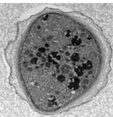
Cysts are the inactive form of Acanthamoeba that presents minimal metabolic activity2 and is the cause of persistent infections (Figure 3.3).15 They can be found in two different forms: mature and immature.13 The size varies depending on the author but is comprehended between 5-30 m,15,18,29 stellate form with 6-8 rays, round, ovoid, triangular, hexagonal or polygonal bodies,27,28 that varies morphologically between isolates. The structure of the cyst contains a cellulose double wall with a fibrous laminar exocyst and a granular smooth endocyst. The composition of cyst wall has 1,4 b-glucan, also known as cellulose39, and other polysaccharides that makes the wall more robust for protection.40 The endocyst is mainly constituted by proteins and cellulose and the wrinkled exocyst is composed of polysaccharides41 and proteins.2 Between this two walls exists a blank space that is thinner around a series of pores, known as ostioles. Possesses an operculum where the trophozoite exits the cyst stage when extern conditions allow.18 The cyst cytoplasm is composed by dense granules of food particles, mitochondria, lipid droplets and glycogen.29
After decades of storage, cysts can remain pathogenic and viable.42 According to Niszl et al. “cyst walls become thicker as the cyst ages”43 and mature cysts are more resistant than immature cysts.25 As they are very resistant to CL chemical and disinfection solutions containing antimicrobial agents, are hard to treat and are able to contaminate lens storage cases.44,45
Biology of
Acanthamoeba
Acanthamoeba species “belong to the phylum Amoebozoa and the family Acanthamoebidae”.46
Acanthamoeba is constituted by the following main structures: nucleus, cytoplasm, vacuoles, membrane, endoplasm, ectoplasm and pseudopods (Figure 3.4 and 3.5).2,15
Figure 3.4 - Trophozoite of Acanthamoeba with identification of the cell nucleus, spinous process and air vacuole (Magnification x1000) (Reproduced from Sun and Wang, 2018).29
It’s usually uninucleate, local where possesses the genetic information, that control the activities of the cell and the expression of different genes.2,15
The membrane define the limit of Acanthamoeba15 and presents contractile vacuoles that maintain osmotic regulation by the control of water quantity inside the cell.2,15
Under Transmission Electron Microscope (TEM) observation can be perceived the division of the trophozoite membrane in three coats and the cyst wall is marked by two layers. The internal and external layers are separated by a transparent space that split the tenuous fibrils inner coat of wrinkled and fibrinous outer coat. These layers may merge and form spinous foramens established in the equatorial zone.29
Figure 3.5 - Cyst of Acanthamoeba with identification of the cell nucleus (Magnification x1000) (Reproduced from Sun and Wang, 2018).29
Plasma membrane is a trilaminar membrane that enclose the cytoplasm and it’s composed by proteins (33%), lipophosphonoglycan (29%), phospholipids (25%) and sterols (13%).2 The main phospholipids found in Acanthamoeba are phosphatidycholine (45%), phosphatidylethanolamine (33%), phosphatidylserine (10%), phosphoinositide (6%) and diphosphatidyglycerol (4%).2 There are also chains of fatty acids like “oleic acids (40-50%) and longer polyunsaturated fatty acids (20-30%)”.2
Cytoplasm has glycogen, lipid and food vacuoles. The last ones regulate digestion15 and are also present in trophozoites lysosomes,2 abundant mitochondria, Golgi complex, free ribosomes, microtubules and smooth and rough endoplasmic reticulum.17,29
Endoplasm includes the central portion of the cell while the ectoplasm represents the outer superficial film.15
The pathogenicity and virulence of the ameba can be define by the conditions in which the specie of Acanthamoeba develop and grow.47 Pathogenicity was also determined by the observation in Scanning Electron Microscope (SEM) of an elevated quantity of acanthopodia on the surface.48
Reproduction of
Acanthamoeba
Acanthamoeba reproduction occurs on the trophozoite stage by binary fission17,28 and mitotically division41 when the conditions are favorable.2 The binary fission is divided into different stages: prophase, metaphase, interphase, anaphase and telophase.29
The prophase consists on the split and disappearance of the nucleolus. Next, in the metaphase, the division in two parts starts in the equatorial zone with a banding shape of chromatin. After metaphase, interphase which englobes the adherence of the dense long column of centrosome with nucleus cells to tubular structures. The next phase, anaphase, refers to the arrival of the fine fiber’s chromatin to the two poles of the cell. In the final phase – telophase - exists the reformation of the nuclear membrane and circle the new formed cells, completing the formation of the two new trophozoites.29
Classification of
Acanthamoeba
According to Marciano-Cabral and Cabral, Volkonsky proposed a classification system for Acanthamoeba in 1931 based on the presence of acanthopodia.17
Both Bouheraoua et al. and Sun refer the classification system of Pussard and Pons presented in 1977 that establish a division of 18 species in three groups based on the cyst morphology (size and shape of cysts).15,29 Assigning one of three groups depend on the average diameter and characteristics of the outer and inner layer:29
• Group I: englobe the cysts with average sizes of eighteen micrometers or more. The inner layer presents a star shape, the outer layer can be wrinkled or smooth, ostioles present at tips of the rays,36 and the space between both layers is large.29
• Group II: englobe the cysts with average sizes of less than eighteen micrometers. The inner layer presents different forms like round, oval, triangle, polygon or star shape; the outer layer is wrinkled17 and/or wavy, ostioles present at angles of the rays,36 and the space between both layers differ. Most of pathogenic species was attributed to this group.29 Human pathologies are mostly implicated by A. polyphaga and A. castelanii, species of this group.15
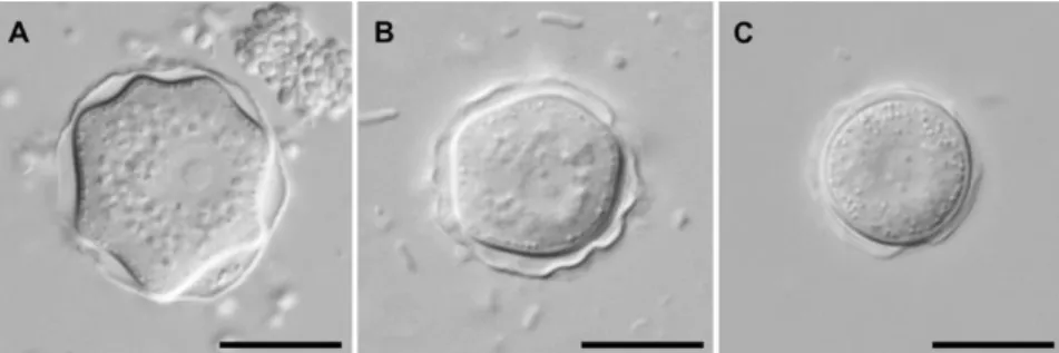
• Group III: englobe the cysts with average sizes of less than eighteen micrometers. The inner layer soft presenting three to five angles, the outer layer may or may not present wrinkles and ostioles are single or obscure. Although most of pathogenic species belong to group II, A. culbertsoni belongs to group III (Table 3.1 and Figure 3.6).29
Group I Group II Group III
A. astronyxis A. castellanii A. palestinensis
A. comandoni A. mauritaniensis A. culbertsoni
A. echinulata A. polyphaga A. lenticulata
A. tubiashi A. lugdunensis A. pustulosa
A. quina A. royreba A. rhysodes A. divionensis A. paradivionensis A. graffini A. tringularis A. hatchetti
It was identified that group II and III, particularly, have more virulent characteristics when related with human keratitis.45
In the year of 1991, some species of Acanthamoeba like A. tubiashi and A. hatchetti was added to group I and II, respectively, based on their characteristics.29
In 1999 was created a new system of classification by Stothard et al. based on the analysis of “18S nuclear subunit ribosomal RNA gene” that allow the discover of 53 isolates17 and the division in 12 groups (T1-T12).15,49 It’s possible to relate both classification system of Stothard and Pussard and Pons establishing the next groups:
• Group I: T7, T8 and T9 • Group II: T3, T4 and T11
• Group III: T1, T2, T5, T6, T10 and T12.17,19
Genotyping of
Acanthamoeba
The benefits of genotyping are allowing us to understand what the epidemiological links are so that it’s possible to established drug susceptibility, virulence factors, infectious isolates, and the most relevant clinically, the clinical outcome.50
The division by genotype of the specie Acanthamoeba is based on the rRNA gene sequences and has to have a difference of 5%, at least, so that they are considered different
Figure 3.6 - Acanthamoeba cysts in interference contrast microscopy (A) morphological group I, (B) morphological group II, (C) morphological group III. Scale bar: 10 m. (Reproduced from Lorenzo-Morales et al., 2015).47
sequences2 Exists both nonpathogenic and pathogenic strains in Acanthamoeba genotypes but the pathogenic ones happens equally in nature.13
To date are recognized 25 species29 and 20 different genotypes of Acanthamoeba (T1-T20).30,48,51–54 The genotype T4 is identified as the most common clinical isolate28–30,38,52,55 of the majority of human infections and in 90% of cases is linked to Acanthamoeba keratitis (AK)2,19,32,50,53,56,57 and 79,3% of cases in brain, skin and lung infections.13 Unlike the genotype T4 that is abundant in nature,54 the genotypes T3 and T11 are rare in the environment.15
Genotypes related to corneal infections as AK are: “T2, T3, T4; T5, T6, T10, T11, T13, T15 and T16”.2,29,48 The genotype T4 is related with both CL and non-CL related keratitis58 particularly the species A. culbertsonii, A. castelanii and A. polyphaga1,4 that have higher pathogenic potential. This pathogenic potential is related with the capacity of binding to corneal tissues, extracellular protease production, specific enzymatic activity and reproduction speed.29,50
According to Ledee et al. the genotype T5 is believe to be not pathogenic to the human species but with the increase of CL use is possible to start finding this isolate more often.50
Recently, in a 2018 report of Orosz, E. et al was identified for the first time the presence of genotype T8 as a cause for AK related with the use of CL.51
The species of Acanthamoeba that are recognized for causing Acanthamoeba keratitis are A. culbertsoni, A. castelanii, A. polyphaga,1 A. hatchetti, A. griffin, A. rhysodes, A. lugdunensis, A. quina.17,27,36,38,41
Alimentation
Acanthamoeba trophozoites feeds on algae, bacteria,20 fungus,9,16 cells, yeast,17 other protozoans, organic matter,2,41 deskerocytes15 and keratocytes in the cornea.4 Between the bacterial selection, they show a preference of coliforms and non-fermentative Gram negatives bacteria.8,59
The feeding procedure occurs by two distinct methods: phagocytosis and formation of food cup sucking.17,29 The ameba forms temporary structures on its surface like food cups to help ingesting food and nutrients.17
According to Qureshi et al. species as Pseudomonas aeruginosa can be lethal to Acanthamoeba trophozoites or cysts60 and Neff et al. refers that the growth of the ameba species is favored by death cells of Saccharomyces cerevisiae and live or death Escherichia coli and Aerobacter aerogenes.61 In Qureshi et al. was refered that beyond the species refered anteriorly, also species of bacteria like Pseudomonas paucimobilis, Xanthomonas maltophilia and Flavobacterium breve enhance growth of Acanthamoeba.60
Relationship between
Acanthamoeba
and
P. aeruginosa
Acanthamoeba and P. aeruginosa have lethal relation when exist contact between P. aeruginosa and the cells on the surface of Acanthamoeba. Despite this relationship, they are both similar eye pathogens. Both are capable of penetrate in damaged corneal tissue through “tissue destructive” enzymes, contaminate CL cases and care systems.60
The production of different exoenzymes as elastase (destroy epithelial cells in humans), blue-green pigment pyocyanin (able to affect human nasal epithelial cells by inhibiting the cells function) and phospholipase C are capable of injure Acanthamoeba’s membrane.47,60
What
Acanthamoeba
can cause
The ameba can cause rare human infections36 like AK62, potentially blinding infection of the cornea;1,63 granulomatous amoebic encephalitis (GAE), fatal disease that affects the central nervous system;17 sinus infections, cutaneous lesions2,16 and, depending on the strain, serve as a host for other microorganisms.1 This infections are related with immunocompromised individuals and all users of CL.2,11
Acanthamoeba is contracted by contact with contaminated water, soil, dust, air or the inhalation of cysts20 that invades the human body through several ways.18
4.
Acanthamoeba
Keratitis
Definition
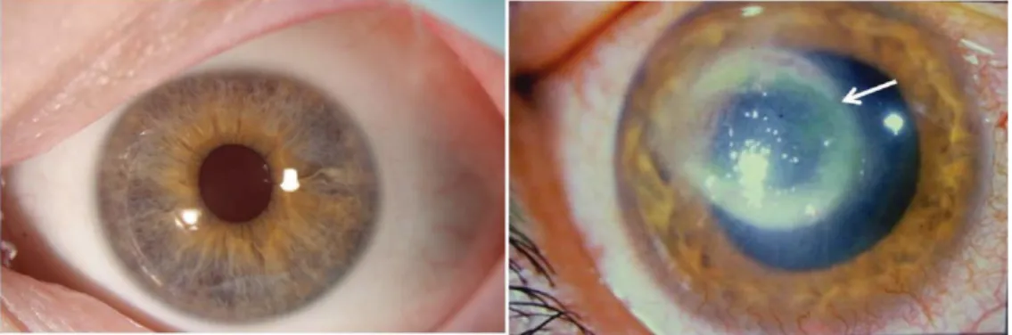
AK is a rare but severe corneal infection15,64 caused by Acanthamoeba species,5,25,65–70 painful and difficult to diagnose and treat1,7 that can lead to blindness or even enucleation.18,23,30,33,35,40,58,71–74 The devastating risk of vision loss is connected with scarring and corneal ulceration75 causing massive ocular injury.52 Sight-threatening AK is mostly found in soft CL wearers, young ages and agricultural workers even though it can be present in non-CL wearers (Figure 4.1).76
When AK isn’t associated with the use of CL usually presents in an advance stage with worse diagnostic and poor visual outcome with acute vision damage.1,77
Cases of AK are normally connected with corneal trauma, lack of personal hygiene, biofilm formation, poor cleaning of CL and contact with contaminated water, materials or solutions.20 There are also reported cases of AK after LASIK and radial keratoplasty.18
Infection by Acanthamoeba starts on the epithelium and progress slowly into the stroma. When treatment isn’t adequate, AK can lead to perforation or corneal melting.78 This process of infection is complex and requires both host and pathogens factors.47
Figure 4.1 - A) Normal human eye; B) Eye with AK. The ring-like stromal infiltrate (pointed with the arrow) is a characteristic feature of AK. (Reproduced from Neelam and Niederkorn, 2017).41
In most of the cases AK is unilateral but can also affect both eyes18,35,55,79,80 like 7.5% in UK.15 AK can also be found in animals like monkeys, fish, reptiles, bulls, dogs, kangaroos, buffalo and echinoderms.18
Patients with AK may lose stereopsis due to central corneal inflammation and presents difficulties on the daily tasks like reading.16
Incidence of AK
As state by several authors in scientific literature18,22,55,81–84 the first report of AK was described in 1974 by Nagington.85 In the following years, AK was mainly related with ocular trauma.34 Although the ubiquitous presence of Acanthamoeba species in nature and the appearance from all over the world,76 the infection caused by this ameba still is rare.13
After the first recognition of the disease was verified an increase in the incidence of AK in parallel with growing of CL users.76 The incidence of AK differs between develop and developing countries, geographical areas and within the countries.25
As reported by Jain et al. in 2015 in a developing country the incidence of AK is lower when compared with a develop country. In this last one, the majority of the cases are liked with the use of contact lenses86, more specifically soft CL76,87 in 85-88% of cases15,40,52 even though it can be present with other kind of CL like rigid lens34 and orthokeratology.78 In developing countries, as India, where the use of CL is not considerable, most cases are reported in non-CL wearers76 and associated with trauma.16 In cases of non-CL wearers the diagnostic of AK is, most of the time, late due to the low incidence around 3-15% in UK and USA15 and are associated with trauma in corneal tissue with contaminated water, soil, vegetal material18 and surgical trauma. Often found in farmers or artisans.15
According to Seal et al. in 1995 the incidence of AK in USA was low about 1 in 250.000.11 A study realized in the US around the decade of 1990 pointed numbers of 1.65-2.01 cases of AK per million of CL wearers in the US and numbers of 17.54-21.14 per million of CL in the UK.6,31 This contrast is thought to be due to the use of water storage tanks in house lofts in UK.37 In 1988, was announced the first incidence of this disease in Japan81 and in the southeast and southern
regions of Brazil.53 In countries like Japan where the prevalence of myopia is high and orthokeratology lenses are popular, leads to the increase of infection78 by fifteen more times than United States numbers.7,83 In China the first case of AK occurred in 1991,70and it’s similar to Japan since presents a high adherence to orthokeratology for myopic control and high rates of AK when compared with other countries like Nepal and India.88 Other than orthokeratology, AK in China is also very evident in cases of soil-contaminated matter and corneal trauma.66
As reported by Lorenzo-Morales et al. in 2013, in the years of 1980 to 1990 the incidence of AK was relatively low of 1-2 cases per million of CL wearers.18 The relationship between the use of CL and AK was first established in 1984.34 With the increase of wearers was observed a direct increase of reported cases. The recognition of risk factors like the incorrect use of non-sterile homemade saline prepared from salt tablets and tap water for rinsing and storing CL34 lead to a decrease in AK numbers until the year 2000.18 Even though the introduction of silicone-hydrogel lenses in the late 1990s, which was expected to reduce the incidence of AK, the numbers never back down.58
The inefficacy of certain CL care systems like Complete MoisturePlus (Abbott Medical Optics, Santa Ana, CA, USA) and ReNu MoistureLoc (Bausch&Lomb, Rochester, NY, USA) were associated with outbreaks of AK and Fusarium, respectively. After the recall of this products from the market, the numbers in the US remained higher than the anterior baseline levels pointing to other risk factors.42
The difference of incidence of AK between countries varies depending on diverse factors as the prevalence of CL use, virulence of Acanthamoeba strains, diagnostic techniques used to detect AK, risk factors, contact with contaminated water in domestic or public environments, climate, amoebicidal efficacy of CL care system15 and the lack of care with the maintenance of CL and cases.65 This lack of maintenance of CL cases can lead to the formation of biofilm that acts like a food source to Acanthamoeba and makes disinfection difficult due to the resistance of polysaccharide conglomerates of microorganism.1
Factors as younger age and gender also appears to have relevance in the incidence of AK. Even though several reports refer that the female gender present a greater risk for contracting AK because of lack of care and disregard for clinical evaluation30,74,88,89, there is one report that defend
that the male gender seems to be at higher risk of contracting AK due to less compliance to hygiene behaviors.68
As it was referred by Bouheraoua et al. the increase incidence of AK in warmer periods like summer and early autumn is associated with the increase in the number of amoeba in nature and the number of water activities.15
The increasing number of CL wearers all over the world due to refractive errors or cosmetic reasons is leading to the increase of AK cases every year.32 This increase in the develop countries is often caused by contamination with Acanthamoeba during cleaning procedures.53
Pathogenesis of AK
In most cases pathogenesis of AK involves the contaminated CL as a transmitting vector of Acanthamoeba.11,90,91 The principal ways by which the protozoa comes in contact with CL is through water and air (Figure 4.2).12,19
CL wear can induce hypoxia in corneal epithelial cells that decreases metabolic rates and debilitate the capacity of resistance of the cornea, particularly in overnight wear. Other than Figure 4.2 – Schematic representation of Acanthamoeba life cycle in humans. Trophozoites and cysts are present in contaminated air, water and soil and can infect the human body through the eye, nasal passages and broken skin which can result in AK and GAE, respectively (Adapted from Maycock and Jayaswal, 2016).4
hypoxia, soft CL can also cause decrease of in tear exchange and minor mechanical trauma in eyelids causing alterations in corneal cells.72,92
Some theories point CL storage cases as one of the principal source of infection and where Acanthamoeba can adhere to all kinds of CL material and then transferred to the cornea.11,93
Most of the time, AK is caused by T4 genotype because it seems to attach to host cells more easily and therefore it’s the genotype more commonly found AK cases.1
First, Acanthamoeba is enclosed in the epithelium. The proliferation to the stroma occurs with the progression of the disease that causes inflammation and severe damage.17 A number of steps occur before keratitis happen like adhesion of the amoebae to CL, transference to the cornea, adhesion to the corneal surface,94,95 disintegration of the epithelial barrier, stromal invasion and final stromal necrosis with blinding consequences.2,15 This invasion mainly occurs when corneal surface is damage, otherwise the amoebae is rapidly removed through tears, blinking1 and host immune system.41 During the use of CL the posterior surface of the CL is relatively immobile so the microorganisms can adhere and proliferate more easily to the corneal surface.1
Adhesion to the corneal surface, more specifically to the epithelium surface, depends mainly on the number of acanthopodia on the trophozoite surface and mannose-binding protein (MBP).18 Both this characteristics are influenced by the degree of pathogenicity once non-pathogenic trophozoites present low capacity of binding to the host cell.18 Pathogenic trophozoites presents over than a hundred acanthopodia per cell and higher levels of the adhesion MBP protein, non-pathogenic trophozoites has a maximum of twenty acanthopodia per cell and low levels of MBP.18
Breakdown of the epithelial barrier is facilitated by the abrasion of corneal surface through the use of soft CL and/or trauma and contact with the inoculum of Acanthamoeba. Once in the surface of the epithelium trophozoites destroys corneal integrity through the fabrication of an enzyme denominate cytotoxic serine protease. A 136-kDa MBP adheres to the exposed mannosylated glycoproteins in the surface of the corneal epithelium.17,18,45,53,58 This event is only possible because of the high quantity of both this proteins in injured corneal surfaces that are essential for the adherence of Acanthamoeba to corneal epithelium.18
Trophozoites releases MIP133, protease able to dissolute cells, when exposed to mannose.1 Acanthamoeba is also able to produce three different types of proteases, like cysteine proteases,
serine proteases and metalloproteases, that are enzymes capable of degrade protein substrates. Serine proteases ease are capable to corrupt collagen type 1, present in corneal stroma that regulates corneal integrity,17,18 secretory immunoglobulin A (sIgA), immunoglobulin G (IgG), laminin, fibrinogen, fibronectin, fibrin, lasminogen, hemoglobin and bovine serum albumin (BSA).18 Enzymes as elastases act on connective tissues degrading them and phospholipase A causes demyelination of brain nerves.18
Stromal invasion derives from the destruction of epithelial cells and perforation of Bowman’s membrane caused by the formation of an enzyme of serine protease and the secretion of collagenolytic factors from trophozoites lead to the dissolution of stromal matrix.13,53 This stromal degradation provides a deeper invasion of the cornea.4
During the process of invasion by Acanthamoeba the amoeba induces cytolytic mechanisms that cause changes in the osmotic pressure inside the cell as well as ionic disruption causing apoptosis of the host cell.18
The last stage involves death of corneal cells and corneal nerves94 due to the activation of the caspases-3 and -10 dependent apoptosis cascade.13,96 Absence of trophozoites in the anterior chamber is connected with the robust neutrophil response.66 In some cases, the protozoan destroys all layers of corneal tissue and can lead to perforation.67
In the cornea Acanthamoeba trophozoites are able to convert into cysts which may prolong the presence of the protozoa in the cornea during infection.
Immune response to AK
Although the contact with Acanthamoeba is common the infection is not. When healthy people get in touch with the amoeba triggers a humoral immune response and production of specific antibodies against the protozoa.29
Antibodies have an important role in the prevention or regulation of Acanthamoeba adhesion to the host18 as they restrain trophozoite movement and/or neutralize cytotoxic factors of the amoeba.17 Individuals affected with AK possesses low levels of IgA1 and serum IgG41 in tear fluid
when compared with non-affected individuals.1 As referred in the anterior chapter serine proteases are capable of destroying IgA and IgG antibodies present in human tears.9,17
Tears present natural defenses like anti-pathogenic substances capable of dissolve and kill pathogenic microorganism. IgA is the principal immunoglobulin in tears and function as the main obstacle for adhesion.18 IgA antibodies acts on Acanthamoeba MBP inhibiting the adhesion to corneal epithelium and IgA-independent factors restrain the activity of cytotoxic proteases protecting from cytopathic effect on the cornea.1
Other than antibodies exist a cellular response to the site of infection. In the first moment there is a flow of neutrophils97 followed by macrophages. These types of inflammatory cells are found around the tissue containing the amoeba. Langerhans cells (LC) are known for postpone AK progress by migrating from the peripheral to the central cornea and retarding hypersensitivity. This kind of cells might act by destroying the trophozoites or inhibiting the adhesion to the epithelium.29 T-cells appears around the final stage of infection. The affluence of neutrophils cause corneal infiltration and a significant humoral response result in corneal opacity or perforation.17
Corneal infection caused by Acanthamoeba doesn’t produce immunity so recurrence of this infection is frequent.2 This persistence is due to the capacity of Acanthamoeba to avoid human response to the infection and it’s able to persist in host tissues during extended periods of time.29
Symptoms of AK
Symptoms of AK are most of the time incapacitating driving from periods of improvement and setbacks16 where severe ocular pain out of proportion is a distinct characteristic.20 In the majority of times is unilateral,29 even though it can appear in both eyes.98
The symptoms may varied between discomfort,20 blepharospasm, conjunctival hyperemia, foreign body sensation, eyelid ptosis, edema, excessive tearing,36,99 blurred vision, decrease of visual acuity,34 photophobia and severe ocular pain disproportionate to clinical signs 18,30,41,47,68,81,82,100,101 even though it could not present some of these symptoms.1,4,15,16,31,102
Pain is related with the inflammatory process67 and the intensity of pain derives from nerve affectation.103,104 When AK is presented with no pain associated could be connected with the
retention of infection in the epithelium, treatment with steroids that decreases inflammation and cornea could be already neurotrophic.104
At an early stage visual loss is not significant and gets worse with the delay of diagnosis, as well as all other symptoms.29 The symptoms may persist during a long period of time and according to Jain et al. that may vary from 3-240 days.76
The symptoms reported above are common to other bacterial, viral and fungal keratitis what makes the diagnose difficult and facilitates misdiagnoses.18,54
Signs of AK
Some clinical signs aren’t specific and may be confused with other ocular disease15 and so it gets worse over time.15 The clinical sign characteristics of AK is the ring infiltration that results from the stromal invasion of Acanthamoeba.29
In the early stage of AK most of the signs are confined to the epithelium and presents like epithelial lesions as punctate epithelial erosions, pseudodendrites (Figure 4.3),1 subepithelial and perineural opacities (Figure 4.4),16,27 conjunctival hyperemia, eyelid ptosis17 and watering.40 These initial signs are often confused with herpetic keratitis.5,67,105
The progression of the disease through the stroma can produce neovascularization,83 anterior stromal infiltration,76 scarring,103 ring infiltrate (Figure 4.5),18,80 central inflammation which causes increase vision loss, corneal opacification, stromal thinning, ulcers with increase diameter and unclear boundary,29 hypopion15 and scleritis.1,16,34,106,107 In a late stage the patient can present severe headache and orbital pain.29
During the process of inflammation corneal sensitivity decreases.15 When trophozoites invade the corneal nerves they are able to cause neuritis and, consequently, necrosis.17
In some rare cases, AK can produce cataracts, glaucoma30,34,105 and dacryoadenitis.101 According to Tu et al. there are five classifications depending on the severity of disease: epithelitis, epithelitis with keratopathy, previous stromal involvement, deep stromal involvement and immune ring and scleritis, respectively, from stage one to five.108,109 Sometimes in more acute stages surgery may be required and also enucleation as a last resort.110
Figure 4.5 - Ring infiltrate from a late advanced stage of AK (Reproduced from Garg et al., 2017).80
Figure 4.4 - Cornea with subepithelial infiltrates, pointed by the white arrow (Reproduced from Wong et al.,2007).124
Risk factors of AK
As referred in chapter 3.2, in industrialized countries about 85% of AK cases are related with the use of CL.41,51,68,81,89,99,111 Trauma is the main cause of AK in developing countries, farmers70 and non-wearers of CL.16,22,70,100,112,113 Other factors are also pointed for the increased number of infections related with Acanthamoeba as CL multipurpose disinfecting solutions,77 CL materials,18 contact with contaminated water,77,114,115 use of contaminated storage cases,116 trauma with soil or vegetation,1,17 ocular surface disease, ocular surgery,84 systemic diseases117 and climate.29 The majority of the patients with AK are immunocompetent CL wearers.6,18,23
The following describes some risk factors associated with this ocular complication: Age: AK is more likely to appear in younger patients,77 with ages under 26 years, but can also be present in the oldest, with ages above 55 years.1,16 As orthokeratology is associated with younger ages and less hygiene, the risk of contracting AK is higher.118
Gender: female patients are at higher risk of contracting AK. Normally, women wear CL for cosmetic reasons that sometimes leads to cheaper choices without eye examinations. Furthermore, the application of makeup as mascara can induce the adhesion of pigments to the surface of CL facilitating the adhesion of microorganism.30
Systemic Risks: decreased immunity, genetic mutations, diabetes, thyroid disease and general health problems.117
CL wear: is considered the major risk factor of AK.4,17,18,30,33,42,59,72,74,77,94,119,120 The main risk behaviors are related with lack of hygiene habits with CL,17,36,52 absence of rubbing the lens during the process of disinfection, hand hygiene, overwear of CL,121 extended wear,1,16 non-compliance with replacement schedules,111,119,122 overnight wear75,105,123,124 and biofilm formation in both CL and their storage cases.110 The overwear issues are related with many characteristics as lens type, oxygen transmission and permeability.72
CL material: soft CL are mostly related with AK,74,125,126 although AK can appear in all types of lens materials1 as rigid gas permeable (RGP), hard CL,27 cosmetic lenses,127 hybrid lenses and orthokeratology lenses.75,77,78,128
CL disinfection methods: like Multipurpose solution (MPS) are easy to manipulate but they are not completely effective against AK.4,18,95,129,130 Solutions as “ReNu MoistureLoc”, “Advance Medical Optics Complete Moisture Plus”64 and “Moisture Plus Complete No-Rub” were withdrawn from the market because they were not effective against some species that cause keratitis, within which Acanthamoeba was included.18 CL maintenance solution and hygiene procedures of CL have an impact in the onset of the disease.1 Risk behaviors like topping off1,89,131 or reuse of the solution,119 expired CL solutions,40 failure to follow manufacturer’s protocols,9,37,132 the use of home-made saline solution from distilled water and salt tablets and chlorine based disinfection show increased risk to the disease.3,5,14,25,27,123,133 This subject will be explored in chapter 4.
CL lens storage cases are the source for many pathogenic agents.42,58,134 Some of the main risk behaviors are infrequent replacement of storage cases,12,121 wash and rinse the CL storage case with tap water21 and then keep the case wet when it should be air-dried.120 A contaminated storage case with bacteria, protozoa and fungus offer a useful environment to the developing of Acanthamoeba once the bacteria work as food supply to trophozoites.7
Contaminated water: This contact may occur in daily activities such as shower, hot tub, lakes,34 tap water,120,135 storage water tanks, swimming,14,22,57 fishing, snow and water sports.1,25,100,106 The exposure of CL to water during the process of disinfection such as rising lens,14 placement and cleaning the lens with tap water before placed them in the storage case1 is very prejudicial. Tap water despite being treated to be drinkable isn’t sterile and it’s not free of microorganisms that can cause keratitis.89 Hard water supplies are favorable environments for the development of Acanthamoeba species and can originate a decrease efficacy of some disinfection systems.79
Other risk behaviors: Sleeping with CL118,121 presume close lids and absence of lid movements, which leads to corneal hypoxia, edema, subepithelial infiltrations, central corneal thinning72 and can result in superficial punctate keratitis.30 More, rubbing the eyes with CL on causes corneal trauma facilitating the invasion by Acanthamoeba species.30 Sometimes, the purchase of CL online is seen as a risk factor once the seller doesn’t require a prescription and the user doesn’t see the purpose of a thorough visual examination and important procedures of disinfection.136
Climate: According to Sun et al. in the summer there is an increase of cases when compared with other seasons,29 which is associated with the increase in the number of amoeba in nature and the number of water activities.15
Diagnostic of AK
Diagnostic of AK is complicated due to unspecific signs and symptoms.31 AK is very often confused with herpes simplex keratitis34,53,115,137 and other types like viral, fungal or bacterial keratitis.2
Early diagnosis and treatment is essential to maintain visual capacity since the delay of diagnosis leads to worse prognosis.76,138 Suspicion is vital to obtain an early diagnosis and avoid delays.79 As state by Chynn et al. and Sharma et al. the time for the diagnosis is about half in CL wearers when compared with non-users of CL.100,126
In cases of ocular trauma and all types of CL wear,5 contact with contaminated water or soil associated with severe pain should alert to the suspicion of AK.2,15
Improvement of diagnostic tools and evaluation technics should allow a correct interpretation of signs and symptoms. The delay on the diagnosis or the wrong one can lead to permanent blindness or the need for corneal transplant.68
Since diagnosis based on clinical signs is difficult, diagnostic methods such as biomicroscopy, corneal scraping, histopathology, Polymerase Chain Reaction (PCR)16 and confocal microscopy76 allow faster and more accurate diagnosis66 with higher sensitivity.6
Initially, biomicroscopy remains an important diagnostic tool to detect modifications on corneal integrity and evaluation of clinical signs even though it doesn’t allow direct visualization of pathogens.29 Despite advances in other diagnostic techniques, PCR method doesn’t replace the biomicroscopic evaluation with fluorescein which allow the visualization of damage area.139
Corneal scraping is the gold standard method for AK diagnosis with a specificity of 60%. This procedure starts with the extraction of a small part of the already fragile epithelium and separated in two parts. After this division, half is cultivated with non-nutritive agar cover with E. Coli, that function as food for Acanthamoeba,16 or inoculates into liquid medium.17 Trophozoites appear first with slow and contractile moves and cysts just develop after a few days when the food is over.1,16 The other part of the tissue sample is submitted to microscopy with tension immunohistochemisthry.16
When corneal cultures results are negative should be elaborated cultures in CL, lens cases18 and lens-cleaning solutions of patients.
Histopathology includes the staining of tissue sample and visualization of microorganisms and antigens under fluorescent microscopy or light. During the biopsy procedure the extraction of trophozoites can provoke the damage in the amoeba cells.1 Stains normally used by this technic are Gram-stain,80 Calcofluor White (Figure 4.6) and Periodic Schiff.1 The different stains allow the visualization of structures in detail:
o Calcofluor White Stain: gives a pale blue tone to the cyst wall because of the chitin presence in the cysts wall.
o Acridine Orange Stain gives an orange-red tone to Acanthamoeba
o Lactophenol Cotton Blue Stain: gives a blue tone to cyst wall and thorn-like projections of trophozoites.
o Iodine Tincture Stain: gives a yellow tone to Acanthamoeba cysts and trophozoites because of the levels of glycoproteins in Acanthamoeba surface.
o Fuchsin Stain: gives a pale brown tone to Acanthamoeba’s nucleolus.29
PCR has a sensibility of 85%,16 specificity of 94-100% and sensitivity of 80-84%.15,76 This molecular method consists in the identification of microorganisms through the gene sequence of 18rRNA gene collected from corneal scrapping.1,16,106 This technique uses “oligonucleotide primers to an Acanthamoeba-specific target sequence”27 but doesn’t allow to distinguish living beings from the dead ones making it difficult to realize if the cornea is healthy or the infection is still active.1 It allows the identification of Acanthamoeba strains28 and others genotypic information.50
Figure 4.6 - Microscopic features of Acanthamoeba cyst in corneal scraping – Gram stain (a) and calcofluor‑white (b). Note hexagonal inner wall of the cysts (Reproduced from Garg et al., 2017).80
Garg et al. referred a new method of loop-mediated isothermal amplification where the DNA sequence is amplified under isothermal conditions in a quickly and efficient way. When compared with PCR technique this method takes two or three hours less.80
Confocal microscopy has a specificity and sensitivity of 90%.15,76 It’s rapid, non-invasive18 and in-vivo method36 using the scanning confocal microscope through the visualization of all layers of the cornea for identification of trophozoites and double wall cysts of Acanthamoeba.17 Cysts presents as hyperreflective, round or oval form and diameter of 15 to 30 m.15 Sometimes cysts are confused with macrophages, neutrophils and immune cells1,15 and trophozoites with keratocyte, leukocytes47 and inflammatory cells.80 For this technique is required a cooperative patient.27 For some authors like Zhong et al. this method is considered as the best diagnostic technique due to its characteristics such as the high levels of sensitivity and specificity.66 With this procedure it’s possible to repeat it during the evaluation of infection and the treatment,29 verifying if the cornea has any sign of infection with living cysts or trophozoites before it’s submitted to a corneal transplant, if necessary.22
According to Rodríguez-Martin et al. the Schirmer test showed a sensitivity of 100% and it can be used to acquire samples from the ocular surface in an aseptic and fast form which allows the study of the pathogens.140
To confirm the diagnostic of AK is necessary to be fulfilled with one or more of the following criteria:
• Positive result from a culture of corneal scraping, corneal biopsy or keratoplasty specimen;
• Histologic evidence from corneal biopsy or keratoplasty specimen and detection of cysts and/or trophozoites;
• Clinical manifestation of at least one of the symptoms presented in the chapter 2.6.14
Treatment of AK
Good visual prognosis derives from an early diagnosis and correct treatment,2 but the risk of recurrence is high.40 Treatment should act on trophozoites, cysts and inflammation.141 Its aim is to removal of all pathogens from corneal tissue with therapy with anti-amoeba agents and elimination of inflammatory response by the use of anti-inflammatory drugs.1,4,80,106
In the course of diagnosis with epithelial debridement it’s thought that in early stage of disease this procedure allow the removal of some of the pathogens from the corneal tissue and helps in the penetration of medication.1,4,17,34,67,137 Before diagnostic confirmation of AK, the treatment with corticosteroids, antiviral, antibacterial or antifungal may cause some improvement in the condition but is followed by periods of deteoration.17
Therapeutic principles depend on the gravity of the disease and stage of infection, the combination of drug therapies for first line of treatment, duration of treatment longer than six months and a surgical option must be placed when the drug therapy isn’t effective.29
Treatment outcome is mostly related with the initial stage of the disease in the time of diagnosis.70 When early diagnosis is performed with organisms still limited to the most superficial layers of the cornea there is a probability of minimal scarring.103 On the contrary, late diagnosis usually leads to risk of corneal transplants and/or permanent blinding.142 Thus, with the monitorization of DNA copy number the treatment can be conducted or modified accordingly the stage of disease.143
The first response to a successful treatment includes a combination therapy which is more effective than a monotherapy69 because one agent is not enough to act against all genotypes of Acanthamoeba.2,27,47,56,58,82,140 While trophozoites are sensitive to different therapies as antiprotozoals, antiseptics, antibiotics and antifungals, cysts are highly resistant to treatment1,16,20,80 and responsible for the persistent infection since they can remain in corneal surface for a long period of time.4,20 There is no single agent that proved to be effective for both cysts and trophozoites.17
There is no specific treatment directed to AK but there is evidence that exist effective substances to treat this disease. In cases where treatment is delayed, the progression of the infection through the corneal layers and treatment can be continued for periods of 1 year or more.17 The effectiveness of treatment also depends on the Acanthamoeba strain,16,48 since they present
different drug susceptibilities during the different growing phases,29 and virulence of the strain,20,144 health of corneal tissue, degree of the disease and the experience of eye health professional.58 Due to the factors mentioned before is difficult to create a link between in vivo and in vitro efficacy.18,47
Recommended effective topical treatments for AK comprehend the use of aromatic diamidines, biguanides, aminoglycosides and imidazoles. Successful therapy englobe the use of biguanides together with diamidines,56,69,74,84,106,140 although the biguanides can be used without the use of diamidines.15 This comprehends multiple agents as polyhexamethylene biguanide (PHMB), chlorhexidine, propamidine isethionate, dibromopropamidine and hexamidine.129
o Biguanides
o First choice in clinical treatment of AK.29
o These cationic surfactants interact with phospholipids on the cytoplasmic membrane, damaging both respiratory enzymes and cell components with modification of configuration and membrane fluidity.4,106,145 After, the interaction with the biguanides, there is a contraction of cyst’s contents and swollen of cyst wall.
o They are often combined with aromatic diamidines to enhance treatment of AK.140 o The most used biguanides are:
o 0.02%-0.06% PHMB1–4,15,16,27,34,36,47,54,56,67,99,118,140,146– effective in low concentrations against both trophozoites and cysts105 of different strains of Acanthamoeba however causes toxic reactions in corneal cells18,47,107,129 when combined with other substances.17
o 0.02%-0.2% chlorhexidine1,2,4,15,16,27,36,54,56,67,140 - effective against both trophozoites and cysts and in low concentrations doesn’t cause toxic reactions in corneal cells.18,47
o Use of biguanides with concentrations above 0.02% are used in cases of resistance to treatment.15 In this cases, PHMB and chlorhexidine concentrations may be increased to 0.06% and 0.2%, respectively.4,29
o Diamidines
o The cationic surface effect produced by the bipolar molecule in diamidines can modify cell membrane structure and increased permeability,106 leading to the escape of ions and biomolecules from the interior of the cell and, consequently,
the cell death.29 After penetrate into the cytoplasm they cause denaturation and coagulation of cytoplasmatic enzymes and structural proteins, acting effectively against cysts and trophozoites,4 by interfering with the synthesis of nucleic acids.29
o Present an additional anti-amoebic effect140 and effective cysticidal effect often used in combination with chlorhexidine.4,15
o This kind of treatment is tolerated by ocular tissue but the extension of treatment can lead to toxic corneal reactions.4,15
o The most used diamidines are:
o 0.1% propamidine isethionate1,2,56,118,4,15,17,18,27,34,36,47 – active substance against trophozoites of Acanthamoeba but not suitable for prolonged used due to the possibility of causing signs of corneal toxicity.
o 0.15% dibromopropamidine17,18,29,47
o 0.1% hexamidine1,2,4,15,47,56,67 – has been proven to present a faster effect against both forms of Acanthamoeba4
o Pentamidine isethionate29 o Diminazene aceturate29
o Hydroxystilbamidine isethionate29
o Combination of 0.1% propamidine isethionate and 0.1% hexamidine proved clinically effective against both forms of Acanthamoeba even though hexamidine present a faster amoebicidal response when compared with propamidine isethionate.15
o According to Marciano-Cabral et al. combination of chlorhexidine and propamidine isethionate is rapid and efficient in AK infection.17,36
o Diamidines can’t work as a monotherapy, always have to be combined with other kind of treatment.15
o Triazoles – has successful results when combined with biguanides and diamidines, in cases with resistance to therapy and limiting the contamination of scleral tissue from corneal tissue. Voriconazole appears effective against clinical strains of Acanthamoeba140 and also proved effective in both topical and systemic application.18 It’s sometimes used in cases where the patient doesn’t respond to the conventional treatment plan.80
o Imidazoles
o Broad-spectrum antifungals.29
o Should never be used alone, usually combined with other therapies147 like biguanides.29
o This compound influenced the permeability of the cell membrane and leads to death of the microorganisms, but the concentration used in medical treatment isn’t enough to kill them, they only inhibit their activity.29
o The most used imidazoles are: o Miconazole18,20,29 o Ketoconazole29
o Clotrimazole29 – only effective against cysts34
o Fluconazole29 – used during keratoplasty to irrigate the anterior chamber impeding the contamination with ameba in the chamber.66 o Aminoglucosides
o Main capacity of modifying the bacterial membrane permeability and inhibiting of the synthesis of bacterial protein.29
o Clinically used in combination with biguanides.29
o The most used imidazole in AK is neomycin, with a concentration of 0.5% in clinical use.29
Treatment is usually applied every hour during the first 48 to 72 hours followed by an hourly application only during daytime80 with a minimum recommended of nine applications per day, from 3 weeks to many months.54 This decrease in treatment dose is due epithelial toxicity derived from the excessive application of drugs.1,4,45
Guidelines suggested by Holland et al. (1987) in an example of treatment guide involves: 1 drop of propamidine isethionate every hour, miconazole and solution of neomycin-polymyxin-gramicidin every two hours when the patient is awake and propamidine isethionate before bed.137
Most ocular antibiotics aren’t effective in this pathogens.20 As refer by some authors, it was successfully used neomycin-polymyxin,16,18 miconazole and propamidine isethionate.18,20 Siddiqui et al. also recommended the use of chloramphenicol.2 When AK is associated with bacterial infection is also necessary the use of antibiotics as neomycin or chloramphenicol.2 Agents as clotrimazole and neomycin are only effective against trophozoites not cysts.