i
UNIVERSIDADE DA BEIRA INTERIOR
Ciências da Saúde
Disruption of the Choroid Plexus Circadian
Rhythm’s in Alzheimer’s Disease
(Versão final após defesa)
André Filipe Lino Furtado
Dissertação para obtenção do Grau de Mestre em
Ciências Biomédicas
(2º ciclo de estudos)
Orientador: Prof. Doutora Telma Alexandra Quintela Paixão
Co-orientador: Prof. Doutora Cecília Reis Alves dos Santos
iii
Dedication
I would like to dedicate this work to my family. Without them this wouldn’t be possible, and because of them, this was all worth it.
v
Acknowledgements
First of all, I would like to thank PhD professor Telma Quintela for accepting me as her pupil, allowing me to work in a field of my interest, and for all the knowledge, guidance, support and most important for her availability when needed.
To PhD professor Cecilia Santos for the advices, scientific knowledge and guidance that allowed for a better outcome of this work.
I would like to thank MSc Ana Catarina Duarte, MSc Ana Raquel Costa, MSc Ana Raquel Brito and all my other group colleagues for the advices and laboratory work teachings and support along this year.
To my friends, who supported me during this year and made me smile in the toughest moments, I thank you all.
vii
Resumo
A doença de Alzheimer (AD) é uma doença neurodegenerativa caracterizada pela deposição do péptido beta-amilóide (Abeta) e proteína Tau no parênquima e vasos sanguíneos do cérebro. A acumulação destes, quando ocorre em áreas do cérebro responsáveis pelo controlo de ritmos circadianos, pode conduzir a alterações nos ritmos de atividade. O ritmo circadiano é coordenado por um marcador de atividade circadiana central que está localizado no núcleo supraquiasmático (SCN) do hipotálamo. No plexo coroide (CP), um oscilador extra-SCN recentemente caraterizado como tal, também se verificam alterações morfológicas na AD que são representadas pela acumulação de Abeta nas suas células epiteliais . A melatonina é uma hormona secretada pela glândula pineal, cujos níveis se encontram diminuídos na AD. Em condições normais, a melatonina tem uma função neuroprotetora na AD, e mesmo quando a doença já está instalada, esta ação continua presente, embora ainda se desconheça parcialmente os mecanismos responsáveis por esta neuroproteção. A melatonina funciona também como “Zeitgeber”, sincronizando os ritmos de expressão dos diferentes genes relógio, regulando desta forma os diversos relógios circadianos distribuídos pelo organismo. Neste estudo, foi utilizado um modelo de murganhos da AD (APP/PS1), de ambos os sexos e com idades de 6 e 12 meses, para determinar alterações na expressão dos genes relógio no CP, em diferentes momentos ao longo do tempo. Para os estudos in vitro foi utilizada a linha celular Z310 tratada com Abeta e melatonina de modo a avaliar se a melatonina tinha um efeito modulador na ritmicidade do Bmal1. Demonstramos que apenas o Bmal1 sofreu alterações na expressão circadiana e que esta ocorre em modelos APP/PS1 com 12 meses de idade. Contrariamente, a expressão do Per2 e Cry2 não é afetada no modelo APP/PS1. Também foi observado que a melatonina tem a capacidade de modular diversos parâmetros na ritmicidade do Bmal1. Estes resultados sugerem uma desregulação do ritmo circadiano da expressão do
Bmal1 num modelo da AD, bem como um efeito modulador da melatonina na expressão dos
genes relógio do CP.
Palavras-chave
ix
Resumo Alargado
Em mamíferos, diversos processos fisiológicos, como ciclos de sono e vigília, secreção hormonal, entre outros, ocorrem associados a uma ritmicidade circadiana. Estes processos com ritmo circadiano associado apresentam dimorfismo entre sexos em várias espécies.
O processo responsável pelo controlo temporal envolve diversos genes denominados de genes relógio. Estes incluem genes como Clock, Bmal1, Per1, Per2, Cry1e Cry2 que interagem entre si de forma a criar sistemas de retroação transcricionais autorregulatórios que constituem o relógio molecular circadiano. Um sistema de retroação positivo é acionado pela formação do heterodimero CLOCK/BMAL1 e promove a transcrição do PER e CRY. Outro sistema de retroação negativo termina quando o complexo PER/CRY, após se deslocar para o núcleo da célula, se liga ao CLOCK/BMAL1 inibindo a sua própria transcrição. O principal relógio em mamíferos é o núcleo supraquiasmático (SCN)do hipotálamo. No entanto, existem outros relógios extra-SCN, tanto no sistema nervoso central como no periférico. Um relógio recentemente descoberto foi o existente no plexo coroide (CP). Os CPs são estruturas vascularizadas localizadas no sistema ventricular do cérebro. O CP é formado por uma camada de células epiteliais cuboides que se encontram interconectadas por junções apertadas. O lado basal está em contacto com uma vasta rede de capilares sanguíneos e o lado apical está em contacto com o líquido cefalorraquidiano (CSF). O CP, entre outras funções, atua como barreira à passagem de substâncias nocivas do sangue para o CSF, e é ainda responsável pela produção deste mesmo líquido. O CP também destoxifica o CSF de produtos tóxicos resultantes do metabolismo cerebral.
A demência é descrita pela organização mundial de saúde como sendo “uma síndrome no qual existe uma deterioração da memória, pensamento e na capacidade para realizar tarefas comuns do dia-a-dia”, sendo que 50 milhões de pessoas sofrem com esta condição em todo o mundo. A demência pode ser causada por diversas patologias sendo que a mais comum é a doença de Alzheimer (AD). A acumulação de beta amiloide (Abeta) e de formas anormais de proteína tau estão descritas como sendo duas das principais alterações que contribuem para o desenvolvimento da AD. Na AD estão descritas diversas alterações que ocorrem no CP, tanto a nível morfológico como fisiológico. Umas dessas alterações é a deposição de Abeta que ocorre nas células epiteliais do CP, descrita como sendo tóxica para as mesmas. A acumulação de Abeta nas células epiteliais do CP ocorre tanto em indivíduos com AD como em indivíduos saudáveis. Isto ocorre porque o CP é responsável por retirar o Abeta do CSF e o transportar para a corrente sanguínea de modo a ser eliminado. Este mecanismo de limpeza do Abeta do CSF parece estar associado a um ritmo circadiano, uma vez que os níveis de Abeta aumentam durante o ciclo de vigília e diminuem durante o sono. A este processo está também associado um maior ritmo de limpeza do Abeta durante o sono. Como se sabe, os ciclos de vigília e sono estão associados ao SCN. Os genes relógio, quando mutados no SCN levam a alterações do sono
x
e na AD está provado que a expressão dos genes relógio está afetada, levando consequentemente a alterações do sono. Uma vez que a limpeza de Abeta está associada a uma ritmicidade circadiana, poderá a AD estar a provocar alterações na expressão dos genes relógio no CP?
A melatonina é uma hormona secretada pela glândula pineal. A sua produção está associada a uma ritmicidade circadiana, atingindo níveis máximos no plasma durante a noite. A melatonina não é a “hormona do sono” sendo que atua mais como um sincronizador endógeno dos ritmos circadianos dos diferentes relógios existentes no corpo. O CP é uma das estruturas descritas como contendo recetores para a melatonina. Níveis diminuídos de produção desta hormona estão diretamente relacionados com o avançar da idade sendo que este pode ser um dos fatores que contribui para o desenvolvimento de doenças neurodegenerativas como a AD. A suplementação com melatonina provou ser eficaz em indivíduos com AD, aumentado a quantidade de sono e melhorando a sua qualidade. Ainda possui diversas capacidades relacionadas com a melhoria da cognição, antiamiloidogénicas, antioxidantes, antidepressivas, entre outras.
De modo a verificar os efeitos da AD sobre a expressão dos genes relógio no CP, utilizamos murganhos APP/PS1 que são um modelo duplamente transgénico da AD. Estes murganhos, de ambos os sexos tinham 6 e 12 meses de idade. Como controlo foram utilizados animais wild-type (WT) que variavam de igual forma em idade e sexo. Com o objetivo de verificar se a melatonina exercia algum efeito modulador na expressão de Bmal1 no CP, utilizaram-se 4 grupos constituídos por uma linha celular de CP à qual foram feitos diferentes tratamentos de melatonina previamente à adição de Abeta.
Para analisar a expressão circadiana dos genes relógio no CP dos modelos animais, os dados de
Real-Time reverse transcriptase polymerase chain reaction (RT-PCR) foram analisados com
recurso ao software CircWave. A análise de Circwave revelou que em machos WT, apenas o
Bmal1 apresentava ritmicidade de expressão, enquanto que em fêmeas WT, para além do Bmal1, também o Per2 apresentava ritmicidade. Estes dados estão de acordo com outros
estudos que também verificaram a expressão diferencial dos genes relógio entre sexos, reforçando a ideia de uma expressão dependente da ação das hormonas sexuais. Em animais APP/PS1 de ambos os sexos, o Bmal1 é expresso de forma rítmica aos 6 meses de idade, mas perde essa ritmicidade em animais APP/PS1 de ambos os sexos com 12 meses de idade. Estes resultados claramente sugerem um efeito da AD sobre a expressão rítmica dos genes relógio no CP. De acordo com a literatura este efeito pode ser explicado pela desregulação dos ciclos de metilação do Bmal1 provocada pelo Abeta. Enquanto em machos APP/PS1 o Per2 nunca apresentou expressão rítmica, em fêmeas o Per2 apresentou expressão rítmica tanto em animais APP/PS1 com 6 meses, como em animais APP/PS1 com 12 meses. A literatura sugere que este resultado se deve a um efeito modulatório do estrogénio sobre a expressão do Per2. O Cry2 não apresentou ritmicidade de expressão em nenhuma das condições testadas.
Tendo a confirmação que a AD altera a expressão rítmica do gene relógio Bmal1 no CP, fomos testar se a melatonina tinha capacidade modulatória sobre a expressão deste mesmo gene na
xi presença de Abeta numa linha celular de CP. O grupo com tratamento contínuo de melatonina e o grupo com tratamento descontínuo de melatonina, ou seja na presença da mesma aquando da adição do Abeta apresentaram alterações nos parâmetros de expressão rítmica do Bmal1 em comparação com um grupo controlo que não teve tratamento com melatonina e que apenas teve exposição ao Abeta. Estes resultados estão de acordo com as funções sincronizadoras que já foram descritas para a melatonina. Também se verificou que o grupo tratado apenas com Abeta não perdeu a ritmicidade na expressão do Bmal1, o que sugere que a concentração de Abeta utilizada neste estudo não tem capacidade para desregular a expressão dos genes relógio nas células utilizadas. O grupo tratado com estímulos descontínuos de melatonina, mas que não tinha melatonina aquando da adição do Abeta, perdeu completamente a ritmicidade na expressão do Bmal1. Admitimos que possivelmente a melatonina estará a influenciar o equilíbrio redox existente nas células e que a sua remoção aquando da exposição ao Abeta facilita a ação do Abeta sobre a ritmicidade das células.
Estes resultados demonstram que estes modelos de murganhos duplamente transgénicos para AD apresentam desregulação da expressão dos genes relógio no CP e que a melatonina apresenta uma possível capacidade modulatória sobre a expressão dos genes relógios no CP.
Palavras-chave
xiii
Abstract
Alzheimer´s disease (AD) is a neurodegenerative disorder, characterized by amyloid beta (Abeta) and tau protein deposition in the brain parenchyma and blood vessels. Abeta accumulation in areas of the brain controlling circadian rhythms can delay or shift activity rhythms. The circadian rhythm is coordinated by the master circadian pacemaker located in the suprachiasmatic nucleus (SCN) of the hypothalamus. The choroid plexus (CP), a recent characterized extra-SCN circadian oscillator, is also known to exhibit morphological changes in AD which are exacerbated by the presence of Abeta deposits in CP epithelial cells. Melatonin is a hormone secreted by the pineal gland and there’s some evidence that, in AD patients, the circulating levels of this hormone are diminished. Under normal circumstances, melatonin acts as a neuroprotector against AD, but how this protection occurs is still to be fully comprehended. It also acts as a Zeitgeber, synchronizing the rhythms of the circadian genes, regulating the body’s circadian clocks. In this study we addressed the question whether Abeta contributes to CP’s circadian clock disruption and if melatonin modulates circadian clock genes expression therein. Using an AD mouse model (APP/PS1), we investigated changes in the expression of CP clock genes at different time points, in female and male animals, aged 6 and 12 months old. In addition, in vitro studies using Z310 cell line treated with Abeta and melatonin were used to examine if melatonin modulated Bmal1 circadian expression. We demonstrated that only Bmal1 circadian expression is altered in AD mice model 12 months old. Contrarily, Cry2 and Per2 expression were not affected in the APP/PS1 model. In addition, we found that melatonin modulated several parameters in the circadian expression of Bmal1. These results indicate that Abeta deposition on the CP disrupted the rhythmic circadian expression of Bmal1 and that melatonin modulates CP clock gene circadian rhythms in the presence of Abeta.
Keywords
xv
Index
I. Introduction 1
1. Circadian Rhythms 1
1.1 Molecular Circadian Clock 1
1.2 Suprachiasmatic Nucleus 3
1.3 Extra-SCN Clocks 3
1.3.1 The CP as a New Extra-SCN Clock 3
2. Alzheimer’s Disease 5
2.1 AD and Circadian Rhythmicity 6
3. Melatonin 7
3.1 Biosynthesis 7
3.2 Distribution and Action 7
3.3 AD and Melatonin Action 8
II. Aim 11
III. Material and Methods 13
1. Animal Samples 13
2. Cell Culture 13
2.1 Cells Synchronization 14
2.2 Treatment of the Z310 cell line with Abeta and Melatonin 14
3. tRNA Collection 16
3.1 In vivo Samples: tRNA extraction 16
3.2 In vitro Assays: tRNA extraction 16
4. cDNA Synthesis 16
5. Conventional PCR 17
6. Real-Time PCR 18
7. Statistical Analysis 20
IV. Results 21
1. Changes in CP Clock Genes Expression in AD 21
2. In vitro Assays 23
2.1 Dexamethasone is a Synchronizer Agent for the Z310 cell line 23
2.2 Melatonin Treatment Modulates Bmal1 Expression in the Presence of Abeta 25
V. Discussion 29
VI. Conclusion and Future Perspectives 33
VII. Bibliography 35
xvii
Figures List
Figure 1. The mammalian molecular clock. p.2
Figure 2. The CP localization in the CNS. p.4
Figure 3. The CP structure. p.5
Figure 4. Melatonin’s protective role in AD. p.9
Figure 5. CircWave curves of Bmal1 expression in the CP of WT animals. p.22
Figure 6. CircWave curves of Bmal1 expression in the CP of APP/PS1 animals. p.22
Figure 7. CircWave curves of Per2 expression in the CP of females. p.23
Figure 8.
One-way ANOVA analysis of the clock genes treated with
dexamethasone and vehicle, and CircWave curves of Bmal1, Per2 and
Cry2 expression in the Z310 cell line treated with dexamethasone. p.24
xix
Tables List
Table 1. Number of samples collected for each condition used during the experiment. p.13
Table 2. Scheme of stimulus done to the different groups of the assay. p.15
Table 3. Primers and respective annealing temperatures used on the Conventional PCR and for Real-Time RT-PCR. p.18
Table 4. Each assay’s specific Real-Time RT-PCR specifications and reaction mixes. p.19
xxi
Abbreviations and Acronyms list
AD Alzheimer’s disease
Abeta Amyloid beta
APP Amyloid precursor protein
AANAT Aryalkylamine-N-acetyl transferase
Bmal1 Brain and muscle ARNTL-1
CNS Central nervous system
CSF Cerebrospinal fluid
CP Choroid plexus
Clock Circadian locomotor output cycles kaput
cDNA Complementary deoxyribonucleic acid
Cry Cryptochrome
DNA Deoxyribonucleic acid
DEPC Diethylpyrocarbonate
DMEM Dulbecco’s modified Eagle medium
Pgp Glycoprotein-P
HIOMT Hydroxyindole-O-methyl transferase
LRP Lipoprotein receptor-related protein
mRNA Messenger ribonucleic acid
miRNA Micro ribonucleic acid
ROR Nuclear retinoic acid receptor-related orphan receptor
Per Period
PBS Phosphate buffered saline
PCR Polymerase chain reaction
TRI-reagent Reagent for the isolation of high quality total ribonucleic acid
RT-PCR Reverse transcriptase polymerase chain reaction
RNA Ribonucleic acid
SIRT1 Sirtuin 1
SCN Suprachiasmatic nucleus
tRNA Total ribonucleic acid
TTR Transthyretin
WT Wild-type
1
I. Introduction
1. Circadian Rhythms
An organism behavior, physiology, and biochemistry changes during the day, exhibiting
circadian rhythmicity 1,2. Robust circadian rhythms can be found governing diverse physiological
processes in mammals, from sleep-wake cycles, to hormone secretion and many other functions
3. The alignment of the circadian rhythm to the environmental stimuli or “Zeitgebers” to
maintain periodicity is named “entrainment” 4. Light is the strongest Zeitgeber, and knowing
that a day on earth has a duration of 24 hours, the circadian clock entrains to a 24 hour
light-dark cycle resulting in diurnal, nocturnal or crepuscular behaviors 4,5. Maintaining both
biochemical and behavioral rhythms aligned with the circadian time is essential for survival since it enables all species to match their physiological and behavioral processes in synchrony
6. Even without external temporal cues, circadian rhythms maintain their period of 24 hours,
approximately, if the conditions remain constant 7. This supports the hypothesis that circadian
rhythms are generated by internal clocks instead of external cues such as light or feeding 7.
Differences in the circadian rhythms between sexes are evident and seen across a multitude of
species like mice, rats, hamsters and even humans 8,9. Sexually dimorphic differences are seen
in the distribution of daily activity 10, time spent sleeping 11, phase responses to light pulses
12,13, rates of re-entrainment 14, susceptibility to splitting 15 and free-running period 8,11,12,16,17.
A sexually dimorphic circadian period in adults, is only achieved in post-gonadal puberty
completion, highlighting a significant impact of gonadal hormones on circadian period 18. More
recently, it was described that during the perimenopausal period, changes in the circulating hormones occur, and there is evidence that lower levels of estradiol and increasing follicular
stimulating hormone levels are related to poorer sleep quality 19. This shows the importance of
understanding the connection between hormones and sleep, improving sleep quality and well-being.
1.1 Molecular Circadian Clock
The process of timekeeping involves a set of circadian clock regulated genes, which include
circadian locomotor output cycles kaput (Clock), brain and muscle ARNTL-1 (Bmal1), Period (Per1 and Per2) and Cryptochrome (Cry1 and Cry2) that interact between them, forming
transcriptional autoregulatory feedback loops that constitute the molecular circadian clock
(Figure 1) 20–23.
The positive feedback loop is powered by the heterodimerization of CLOCK protein with BMAL1. In certain tissues or cells where CLOCK is not expressed, neuronal PAS domain-containing
protein 2 can take its place in the heterodimer 24. CLOCK/BMAL1 promotes the transcription
of clock controlled genes which include the ones encoding for PER and CRY proteins20,21,23. The
2
with CLOCK/BMAL1, inhibiting their own gene transcription 20,21,23. Nuclear retinoic acid
receptor-related orphan receptor (ROR) and REV-ERB, a nuclear receptor that acts as a
ligand-dependent suppressor of gene transcription 25, are all involved in the auxiliary loop, with
CLOCK/BMAL1 mediating activation through E-boxes in their promoters 26. REV-ERB represses 27
while ROR regulates positively Bmal1 expression. 22,28.
Figure 1. The mammalian molecular clock. The mammalian molecular clock oscillates within a 24 hour cycle, in a series of transcriptional and translational feedback loops. It can be divided in different loops: the positive, the negative and the auxiliary feedback loop. Adapted from Logan and McClung, 2019 22.
3
1.2 Suprachiasmatic Nucleus
The suprachiasmatic nucleus (SCN) in the hypothalamus is the primary circadian pacemaker in
mammals 29–33. Grafts of SCN tissue restore circadian rhythmicity in arrhythmic animals that had
their own nucleus destroyed 34–37. In mammals, the SCN exerts direct control over many
physiological and neuroendocrine rhythms 38,39.
The SCN is essential for synchronization of the internal circadian time with the external environmental time that surrounds the organism, integrating different stimuli from internal and
external sources 6,39. Synchronization (“entrainment”) of the SCN to the light/dark cycle is
essential for the synchronization of SCN clock genes expression 23. This entrainment involves
the retinohypothalamic tract which sends time-of-day specific signals from the retina to the
SCN using different combinations of peptides and thus modulating the SCN activity 40,41. The
sensitivity of the SCN to photic inputs can be modulated by extra visual stimuli 42. The core of
the SCN is essential for the production of the rhythmic output signal for the central nervous
system (CNS) 43,44. Within the SCN core there are small-world neuronal networks which are a
robust and redundant way of maintaining circadian synchronization 45.
The SCN is not the only circadian clock present in the organism. Several extra-SCN clocks have been described in the CNS as well as in extra-CNS tissues, receiving not only SNC originating inputs, but also sending their own outputs (neurotransmitters or other modulators like steroid
hormones) to the SCN, modulating it 46. Extra-CNS oscillators, denominated as peripheral
oscillators, are present in the retina, lung, liver, ovary and even fibroblasts 47–51. This peripheral
oscillators are all synchronized by the SCN 52 but they are also entrained by different non-SCN
cues like food intake, one of the strongest entrainment cues in these tissues 49,53,54. Other cues
such as temperature 55,56, oxygen 57 and glucocorticoids 58 also have influence over the
entrainment of these peripheral oscillators circadian clocks.
1.3 Extra-SCN Clocks
Desynchrony between SCN and extra-SCN clocks is suggested as a precipitating factor leading
to disease 59. It is a SCN responsibility to synchronize all the clocks present in the organism.
Other synchronizing cues exist, and their continuous action over the body’s circadian clocks might overrule SCN’s synchronizing action leading to internal conflicts between different clocks
and the associated functions 59. Cellular oscillators located centrally at the CNS include the
pineal and pituitary glands, the arcuate nucleus, median eminence and the retrochiasmatic and
supraoptic nucleus of the hypothalamus 60. In this list we also have the olfactory bulb 61,
considered for many years the most robust extra-SCN circadian clock and the recently
discovered choroid plexus (CP) 62–64. More details concerning this new clock will be given in the
next section.
1.3.1 The CP as a New Extra-SCN Clock
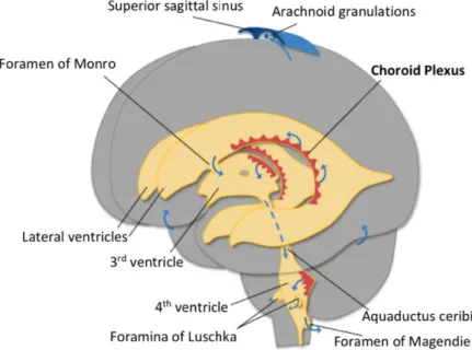
The CPs are vascularized structures located in the cerebroventricular system: third, fourth and
4
CP is a modified version of the ependyma that coats the inside of those same ventricles 66.
Thus, the CP is constituted by a single layer of cuboidal epithelial cells connected to each other
by tight junctions (Figure 3) 66,67. On the basal side of the CP there’s a dense network of capillary
which are fenestrated 68,69. This allows for molecules and fluids to exit the blood stream and
integrate the interstitial fluid 70, with the CP acting as a barrier to the passage of polar
compounds from the blood to the cerebrospinal fluid (CSF), 71. Together with the blood-brain
barrier, the blood-CSF barrier are essential in protecting the CNS from pathogens, toxins and
inflammatory molecules circulating in the blood stream 72
Figure 2. The CP localization in the CNS. The CPs are located in the third, fourth and lateral ventricles which are all interconnected to enable the circulation of the CSF. Adapted from Damkier, Brown and
Praetorius, 2013 73.
Besides other functions, the CPs are responsible for the production of CSF 71,73. The side of the
CP in contact with the CSF presents numerous microvilli which are thought to mix the CSF
maintaining and blending the different compounds present in this fluid 71. The CP is also
responsible for detoxifying the CSF from toxic products resulting from brain metabolism 74 and
producing numerous proteins that have nutritional and neuroprotective properties in the brain
75. It may use olfactory-like chemosensing systems to detect alterations to the CSF chemistry
in order to respond to those alterations maintaining brain homeostasis 76–78. Therefore, the CP
has a crucial role in the brain’s metabolism, neuronal function, neuro-signaling, immunological
and inflammatory processes, neuroprotection and neurodegeneration 79. As reported
previously, the CP is now considered a strong circadian clock. The presence in CP of clock genes (Bmal1, Clock, Cry1, Cry2, Per1, Per2 and Per3) messenger ribonucleic acid (mRNA) expression
and corresponding proteins was first reported in 2015 64. The existence of a functional CP
5
Bmal1-ELuc mice 62,63, showing that the CP is able to generate circadian rhythmicity with a
mean period around 24 hours.
Figure 3. The CP structure. The CP is constituted by a single layer of cuboidal epithelial cells connected to each other by tight junctions. Adapted from Nakada and Kwee, 2019 80.
Moreover, it was demonstrated that clock genes are under circadian regulation in a sex
dependent way in the rat CP 64. Bmal1, Cry2 and Per2 are rhythmically expressed in the CP of
female rats while only Cry2 and Per2 express rhythmic expression along the day in males 64.
Clock seems to be constitutively expressed throughout the day in both sexes 64. In the males’
CP, Bmal1 is downregulated when compared to females’, whereas Per2 and Per3 are
upregulated, suggesting that sex hormones control the CP clock gene expression 81. Thus, the
male CP clock has a less pronounced circadian variation of the clock genes than females’
evidencing the sex hormones influence and their regulatory effect 64. The CP possesses estrogen
receptors 82 but these are scarce in the SCN 83. Thus, the CP may act as an extra-SCN site of
action for estrogen, communicating with the SCN and adjusting the SCN circadian rhythmicity
84 The variations that occur on the expression of the CP clock genes , resulting from sex hormone
action, can be part of that regulatory mechanism. This regulatory effect might be supported by the fact that the circadian rhythms of expression of CP clock genes are more robust than
the ones exhibited by the SCN 62.
2. Alzheimer’s disease
Dementia is described as being “a syndrome in which there is a deterioration in memory, thinking, behavior and in the ability to perform everyday activities” by the World Health Organization (14, May 2019) , that also reports that, worldwide, 50 million people suffer from this condition. Dementia can be caused by several pathologies but none has the impact of
Alzheimer’s disease (AD). Between 75% to 80% of dementia cases are caused by AD 85.
AD was first reported in 1907 86 and today it’s considered by the world health organization as a
6
Accumulation of both Amyloid beta (Abeta) and of an abnormal form of tau protein are thought to be two of the major alterations occurring in the brain tissue contributing for AD development
88. It can also encompass dystrophic neurites, microglial activation, neuropil threads and
associated astrogliosis and also cerebral amyloid angiopathy 89. All these pathological processes
have consequences, culminating in neurodegeneration characterized by synaptic and neural
loss, to the point where macroscopic atrophy is visible 87 being that the first pathological signs
can appear approximately 15 years earlier than the onset of cognitive impairment 90. In AD, the
CP suffers some anatomical changes that are an exacerbation of the ones occurring with normal
ageing 91. Height of the epithelial cells can be reduced in 22% in relation to age matched
controls 92. The basement membrane becomes thicker and irregular and the villi stroma
becomes fibrotic with extensive vascular thickening 92,93. There is a greater intracellular
distribution of pathological entities like lipofuchsin vacuoles and Biondi Ring Tangles, and these
are present in a larger number of epithelial cells 94,95. CSF production is also reduced in AD
patients when compared to age matched controls 96. Deposition of Abeta is neurotoxic for CP
epithelial cells 69, which might be one of the factors contributing to the alterations that occur
in the CP in AD and in normal ageing.
2.1. AD and Circadian Rhythms
The CP epithelia presents Abeta accumulation not only in AD patients 97 but also in healthy
subjects 98. The CP is responsible for the uptake of Abeta, and this mainly occurs from the CSF
to the blood stream 98,99. Several transporters are involved in this clearance process like the
low-density lipoprotein receptor-related protein (LRP), megalin mediated transcytosis and
glycoprotein P (Pgp) 99. In normal aging, there’s a decline in megalin mediated transcytosis
100,101, which is balanced with an increase in LRP and Pgp 101.
The CP synthesizes several Abeta scavenger proteins like transthyretin (TTR) and clusterin 102.
TTR is a protein known to bind Abeta 103 preventing the formation of Abeta plaques 104, while
clusterin after binding to Abeta facilitates its transport across the blood-CSF barrier into the
blood stream 102,105. The Abeta clearance mechanism seems to be associated with a circadian
rhythmicity. In amyloid precursor protein (APP) transgenic mice and in young healthy men
volunteers, Abeta levels increase during wakefulness and decrease during sleep 106,107. The mice
model was also subjected to chronic sleep deprivation and this lead to greater Abeta plaques
deposition when compared to their age-matched littermate controls 106. Abeta plaques
formation was reported to disrupt the sleep/wake cycle in a mouse model of AD amyloidosis
107. Abeta clearance from the interstitial fluid to the CSF and from the CSF to the blood stream
is greatly enhanced during sleep time 108. This highlights the tight relation between sleep and
Abeta formation and clearance.
As described above, circadian rhythms of sleep-wake cycles are driven by the master circadian
clock 109. The SCN core clock genes when mutated, lead to circadian rhythm sleep disorders in
humans 110. Changes in the clock genes expression can have different outcomes since, for
7
hour period 111. Per1 expression shortens the period and Per2 lengthens it 111. AD was found to
have an effect over the clock genes, disrupting their expression in the SCN and in other brain
regions such as the hippocampus, frontal cortex and the brainstem 112. These changes,
eventually result in desynchrony between various brain circadian oscillators which can also
explain the sleep-awake cycle disruptions 113 associated with Abeta plaques formation 107.
Increasing severity of dementia in AD was also associated with disorganization and decreased
amplitude of the daily pattern of activity 114.
If the clearance of Abeta is linked with the circadian rhythm mechanism, and the development of AD is associated with the disruption of the clock genes expression, the disruption of the CP circadian clock might lead to a deficient Abeta clearance and an aggravation of the disease and patient’s condition. In addition, sex hormones also have a significant impact on the prevalence of the disease. The Framingham study found that there is a higher risk of developing the disease
in women 115. At the age of 65 years, the risk of developing AD is almost two times higher in
women 115. A faster decline and greater deterioration of cognition occurs in elderly women
compared to elderly men suffering from the disease 116. We believe that understanding the link
between the dysregulation of the CP circadian clock and AD, bearing in mind the influence of sex hormones on the process, might represent an interesting target to prevent AD.
3. Melatonin
3.1 Biosynthesis
Melatonin is a hormone secreted by the pineal gland. There, tryptophan is absorbed from the blood stream and converted into serotonin, which is then transformed into melatonin by a two
step process involving two enzymes that act in a sequential fashion 117: Arylalkylamine-N-acetyl
transferase (AANAT), which is the limiting enzyme, and hydroxyindole-O-methyl transferase
(HIOMT) 118. Melatonin rhythmic secretion has its origin in the SCN 119. The SCN sends neural
projections to the superior cervical ganglia 120 which then projects to the pineal gland.
Norepinephrine is the main neurotransmitter regulating the pineal gland activity 117. This
neurotransmitter is released during the night in response to signals coming from the SCN 117.
Norepinephrine activates adenylate cyclase which promotes the biosynthesis of AANAT 121. Light
inhibits the secretion of melatonin, and darkness enhances it 122.
The retina, bone marrow cells, platelets, skin, the gastrointestinal tract of vertebrate species, lymphocytes, Harderian gland, cerebellum and the CP are all extra pineal sites for melatonin
secretion 123–131.
3.2 Mechanisms of Action
Due to its high solubility in lipids 132, melatonin is able to reach the brain, getting through the
blood-brain barrier, as well as reaching other body tissues 133.
Melatonin has two membrane bound receptors: MT1 and MT2 134,135. Both of these receptors
8
are present in a variety of tissues: blood vessels, heart, gonads, liver, kidneys, adrenal cortex, pancreas, spleen, breast and mammillary glands, placenta, skin, immune system, pituitary
gland, adipose tissue, gastrointestinal tract and several brain structures 137. The CP is one of
the brain structures expressing melatonin receptors 138. Melatonin also exerts action over
nuclear receptors, one of which is ROR 139, an important piece of the circadian clock’s network
22, suppressing RORalpha’s transcriptional activity 140. Some investigators defend the idea that
melatonin binds directly to nuclear receptors 141 while others think that melatonin’s effect is
mediated by MT1 membrane receptor 140.
Melatonin is not the “hormone of sleep” but acts more as a night period indicator to the different circadian rhythmic physiological processes that occur in the body, like body
temperature variations 142. Melatonin may act as an endogenous synchronizer of the body’s
circadian rhythms, maintaining the different clocks working in synchrony 143.
3.3 AD and Melatonin Action
After birth, melatonin levels increase until puberty, where they reach a peak 144. Later in life,
melatonin synthesis 145 and consequent circulating levels, start to decline 146. Decreasing levels
of melatonin production in older individuals 147 can be a major point contributing to the
development of neurodegenerative diseases 148. Melatonin levels seem to be directly correlated
with AD. Decrease of melatonin levels in the CSF occur at the same time as AD neuropathology
progresses 149 and AD patients show lower levels of melatonin when compared to their healthy
counterparts 150,151. As described earlier, AD was found to disrupt the expression of circadian
clock genes in the SCN and in other brain regions such as the hippocampus, frontal cortex and
the brainstem 112. Some reports also propose that poor sleep quality and quantity is directly
related with an higher risk of developing dementia 152. So circadian rhythms disruption, as we
can see, can be interconnected to AD at the different levels of the development of the disease,
both as a risk factor and as a consequence 112,152.
Melatonin supplementation improves total sleep time at night in AD patients 153. Sirtuin 1
(SIRT1) is known as the “longevity protein” and in cellular and mouse models of AD it was
described to attenuate Abeta production 154. This protein expression is promoted by melatonin
signaling, and SIRT1 overexpression may be a protection factor in AD phenotypes 154. Melatonin
stimulates antioxidant defense systems in the brain 155,156 and is itself a free radical scavenger
157,158. Tau hyperphosphorylation is reduced in the presence of melatonin, this reduction can be
partially due to melatonin’s antioxidant properties preventing phosphorylation 159. It also
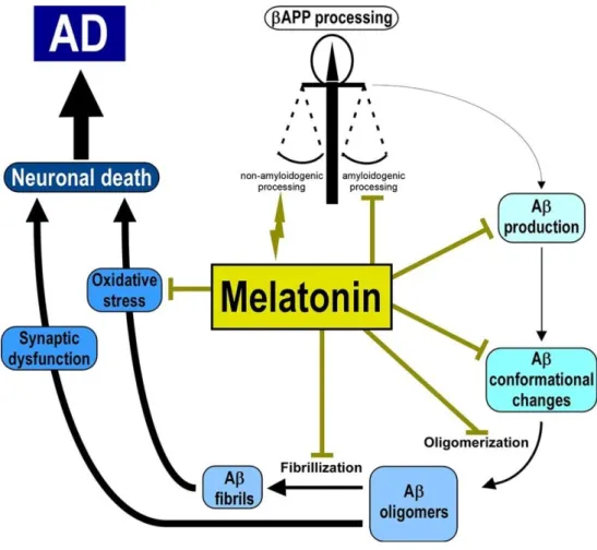
presents anti-amyloidogenic capabilities 159,160 (Figure 4). By binding to Abeta, melatonin
prevents Abeta aggregation 161 and could have a regulatory effect over APP expression,
diminishing beta-APP levels 160. This amyloidogenic properties might also present an
anti-inflammatory effect since Abeta plaque formation is responsible for neuroinflammation 162.
Melatonin also presents the ability to prevent the formation of Abeta fibrils 163 by forming
non-covalent complexes with Abeta itself 164. Cognitive impairment was reduced in a mouse model
9
antioxidant pathway 165. Again, using a mouse model of AD treated with melatonin and
subjected to behavioral tests, as for example elevated plus maze test and forced swimming
test, it was demonstrated that melatonin prevented anxiety and depression-like behaviors 166.
This effect was associated to an augment in glutathione S-transferase P-1 (an
anxiety-associated protein) and complexin-1 (a depression-anxiety-associated protein) in the hippocampus 166.
A commonly observed condition in AD patients is “Sundowning” 167. This condition is
characterized by disorganized thinking, reduced focus, wondering, agitation, perceptual and emotional disturbances, and is associated to a circadian rhythmicity, manifesting late in the
afternoon or in the early evening 167. Melatonin treatment is associated with improvements in
sundowning 167. Together, melatonin seems to have a very powerful protection effect against
AD development and the understanding of how this protection occurs might lead to better options regarding treatment of AD.
Figure 4. Melatonin’s protective role in AD. Melatonin inhibitory effect over the different pathological processes that occur during the development of AD. Vincent, 2018 168.
11
II. Aim
The clearance of Abeta from the CSF is tightly associated with the CP and this process occurs with a circadian rhythmicity. There is some evidence relating clock gene’s rhythmicity disruption to neurodegenerative diseases, particularly AD. There’s also proof that AD has a sex dependent prevalence suggesting an effect of sex hormones in the development of the disease. Melatonin is a hormone secreted by the pineal gland and there is some evidence that, in AD patients, the circulating levels of this hormone are diminished. Under normal circumstances, melatonin acts as a neuroprotector against and during AD, but how this protection occurs is still to be fully comprehended. It also acts as a Zeitgeber, synchronizing the rhythms of the circadian genes, regulating the body’s circadian clocks.
In this work we aim to evaluate AD effects on the rhythmicity and expression of several clock genes in the CP. We also plan to verify if melatonin is able to modulate the expression of CP’s clock genes in the presence of Abeta.
13
III. Materials and Methods
1. Animal Samples
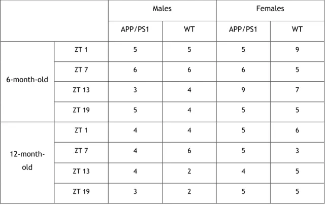
In this work, we used samples of mouse’s CP that were collected by our collaborators of the research group located in the Research Institute Hospital 12 de Octubre (i+12) in Madrid, Spain. The samples were obtained from female and male, 6- and 12-month-old APP/PS1 transgenic and wild-type (WT) mice, at different time points during a 24 hour period (Table 1). The APP/PS1 strain of double transgenic mice results from a cross between Tg2576 (overexpressing
human APP695) and mutant PS1 (M1462) 169. The time points at which the samples were
collected are denominated of Zeitgeber Time (ZT). Lights are turned on at ZT0 (7 a.m.) and off at ZT12 (7 p.m.).
Table 1. Number of samples collected for each condition used during the experiment.
Males Females APP/PS1 WT APP/PS1 WT 6-month-old ZT 1 5 5 5 9 ZT 7 6 6 6 5 ZT 13 3 4 9 7 ZT 19 5 4 5 5 12-month- old ZT 1 4 4 5 6 ZT 7 4 6 5 3 ZT 13 4 2 4 5 ZT 19 3 2 5 5
2. Cell Culture
The cell line used was an immortalized rat choroidal epithelium (Z310 cell line), donated by
Dr. Wei Zheng 170. The in vitro studies were performed using Dulbecco’s Modified Eagle Medium
(DMEM (1x) ref. 11880.028, Gilbco®) cell culture medium, supplemented with 3500 mg of
D(+)-Glucose Anhydrous (Fisher Scientific®) and 584 mg of L-Glutamine (Sigma-Aldrich®) per every
liter of culture medium used. To the culture medium was also added 10% fetal bovine serum
(Sigma-Aldrich®) and 1% penicillin/streptomycin (Sigma-Aldrich®). The cell culture was
14
At approximately 90% of confluence, a cellular passage was performed, allowing for continuous cell growth. For that, culture medium was removed, and the cells washed with phosphate buffered saline (PBS) 1x. PBS was then removed and trypsin-EDTA 0.25% added in a volume that would ensure total coverage of the cell layer. A 3- to 5-minute incubation at 37 ºC would follow and when most of the cells had detached, cell culture medium (double the amount of trypsin used) was added. The suspension was then collected to a falcon and subjected to centrifugation for 3 minutes at 301 RCF. The supernatant was rejected, and the pellet of cells resuspended in culture medium, ready to be used in an essay, cultured and/or cryopreserved.
For cell counting, 20µL of trypan blue were added to 20µL of cellular suspension. From this suspension, 10µL were transferred to a Neubauer chamber for viable cells counting. After counting the number of cells by quadrant, the total number of cells in the t-flask and the number of cells/mL were estimated using the following mathematical formula:
2.1 Cells Synchronization
In order to replicate what happens in vivo, cells in culture need to be synchronized. Synchronization allows for the internal clock of all the cells in culture to be in synchrony with
each other 171. The selected synchronization compound was dexamethasone (Sigma-Aldrich®).
For that, 3x10⁴ cells were seeded per well and cultured in 12 well culture plates. After 72 hours
the culture medium was discarded and culture medium supplemented with dexamethasone at a final concentration of 100nM, was added. A control condition with vehicle (water) was also included. After 2 hours, the culture medium was discarded and substituted by new complete cell culture medium. Cells were then collected at different time points (6 hours, 12 hours, 18
hours and 24 hours after synchronization). For that, the cell culture medium was removed and
the wells washed with room temperature PBS. Ready to use reagent for the isolation of high-quality total ribonucleic acid (tRNA) (TRI-reagent) was added to each well and the cells transferred to a microtube. The microtubes were maintained at -80º C until RNA extraction.
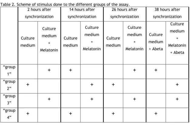
2.2 Treatment of the Z310 cell line with Abeta and Melatonin
In order to study if melatonin treatment has any modulatory effect in Bmal1 circadian expression in the presence of Abeta, the epithelial cell line was treated with these two
compounds. We used a low non-apoptotic concentration of Abeta (AnaSpec®;3ug/mL) already
tested by our group 172. For that, 1.5x10⁴ cells were seeded per well and cultured in 24 well
culture plates. After 24 hours, the cells were synchronized according to the protocol described above. Four different groups were established, and each group received a different melatonin treatment (“group 1” (+/-/+/-), “group 2” (-/+/-/+), “group 3” (+/+/+/+) and “group 4” -/-/-/-)) (Table 2). The culture medium was removed and to the “group 1” and “group 3” complete
15
culture medium supplemented with melatonin (Calbiochem®) at 10nM was added, while culture
medium with 0,002% ethanol (vehicle) was added to “group 2” and “group 4”. After 12 hours the culture medium was removed and to the “group 2” and “group 3” was added complete culture medium supplemented with melatonin at 10nM and to “group 1” and “group 4” was added culture medium with the vehicle. After 12 hours the culture medium was removed and complete culture medium supplemented with melatonin at 10nM was added to the “group 1” and “group 3” and culture medium with the vehicle was added to “group 2” and “group 4”. After 12 hours the culture medium was removed and complete culture medium supplemented with melatonin at 10nM was added to the “group 2” and “group 3” and culture medium with the vehicle was added to “group 1” and “group 4”. At this point Abeta was added to the cells at a final concentration of 3µg/mL. Scheme of these stimuli done during the three 12 hours cycles of melatonin treatment are represented in Table 2. Cells were then collected at different time points (6 hours, 12 hours, 18 hours, 24 hours, 30 hours, 36 hours, 42 hours, 48 hours, 54 hours, 60 hours, 66 hours and 72 hours after treatment with Abeta). For that, the cell culture medium was removed and the wells washed with room temperature PBS. TRI-reagent was added to each well and the cells transferred to a microtube. The microtubes were maintained at -80º C until RNA extraction.
Table 2. Scheme of stimulus done to the different groups of the assay. 2 hours after synchronization 14 hours after synchronization 26 hours after synchronization 38 hours after synchronization Culture medium Culture medium + Melatonin Culture medium Culture medium + Melatonin Culture medium Culture medium + Melatonin Culture medium + Abeta Culture medium + Melatonin + Abeta “group 1”
+
+
+
+
“group 2”+
+
+
+
“group 3”+
+
+
+
“group 4”+
+
+
+
3. tRNA collection
3.1 In vivo Assays: tRNA extraction
Samples were thawed and 300µL of TRIzol™ added to each microtube. With the aid of a pestle, the CP samples were homogenized, followed by 5 minutes incubation at room temperature to enable for complete dissociation of nucleoprotein complexes. 60µL of chloroform (200µL
16
chloroform for each 1mL of TRIzol™) were then added to the samples followed by vortex homogenization. Samples were left to incubate at room temperature for 15 minutes before undergoing centrifugation (4ºC, 12000g, 15 minutes). The supernatant (transparent phase) was carefully collected for a new microtube and 150µL of isopropyl (500µL of isopropyl for each 1mL of TRIzol™) were added, and homogenization by inversion performed enabling the ribonucleic acid (RNA) to precipitate. The microtubes were left at room temperature for 10 minutes before undergoing another centrifugation (4ºC, 12000g, 10 minutes). After centrifugation the supernatant was discarded and 200µL of 75% ethanol in diethylpyrocarbonate (DEPC) water was added to the pellet followed by another centrifugation (4ºC, 7500g, 5 minutes). The supernatant was discarded and, after total removal of the excess ethanol, the tRNA pellet was rehydrated in DEPC water. This was followed by tRNA quantification using a nanospectrophotometer (NanoPhotometer™, Implen). The equipment displays tRNA concentration (ng/µL) and also the 260nm/280nm ratio. tRNA is considered to be pure with a 260nm/280nm ratio between 1.8 and 2.1. If the ratio is <1.8 the tRNA is probably contaminated with protein or phenol and if the ratio is >2.1 tRNA is probably contaminated with genomic deoxyribonucleic acid (DNA).
3.2 In vitro Assays: tRNA extraction
Samples were thawed and 200µL chloroform for each 1mL of TRIzol™/ TripleXtractor (GRiSP®)
were added. The microtubes were then subjected to vortex homogenization. They were left to incubate at room temperature for 15 minutes before undergoing centrifugation (4ºC, 12000g, 15 minutes). The supernatant (transparent phase) was carefully collected for a new microtube and 150µL of isopropyl (500µL of isopropyl for each 1mL of TRIzol™) were added, and homogenization by inversion performed. The microtubes were left at room temperature for 10 minutes before undergoing another centrifugation (4ºC, 12000g, 10 minutes). After centrifugation the supernatant was discarded and 200µL of 75% ethanol in DEPC water was added to the pellet followed by another centrifugation (4ºC, 7500g, 5 minutes). The supernatant was discarded and, after total removal of the excess ethanol, the tRNA pellet was rehydrated in DEPC water. This was followed by tRNA quantification using a nanospectrophotometer (NanoPhotometer™, Implen) as reported above.
4. cDNA Synthesis
Complementary DNA (cDNA) synthesis is done using a reverse transcriptase enzyme, using RNA as a template. This enzyme has the capacity of producing an exact copy of the expressed genes
but without the intron segments. NZY M-MuLV Reverse Transcriptase (NZYTech®) was chosen
and used according to fabricant’s recommendations. A mix denominated MIX1 (n+2 reactions)
composed by 2µL of Random Primers (NZYTech®) and 1µL of DNTPs (NZYTech®) for each sample,
was prepared in a microtube. Polymerase chain reaction (PCR) tubes were prepared with approximately 500ng of tRNA extracted of each sample plus sterile water performing a final volume of 14µL. Next, 3µL of MIX1 were added to each PCR tube and the tubes were placed in
17
the MultiGene™ OptiMax Thermal Cycler (Labnet®) at 65ºC for 5 minutes, and immediately after
deposited in ice. Meanwhile, in a new microtube, MIX2 (n+2 reactions) was prepared with 2µL of Reverse Transcriptase Buffer (5x buffer) and 1µL of MMLV for each sample. 3µL of MIX2 was added to each PCR tube, followed by a 10 minutes incubation at 25ºC and 50 minutes at 37ºC. To stop the reaction, samples underwent 15 minutes at 70ºC. cDNA was then stored at -20ºC until use.
5. Conventional PCR
Using conventional PCR, the expression of Bmal1, Cry2 and Per2 was confirmed in the in vivo
samples and in the cell line Z310. NZYTaq II 2x Green Master Mix (NZYTech®) was selected for
DNA fragments amplification, following the fabricants recommendations. For each sample it was prepared a mix containing 10µL of NZYTaq II 2x Green Master Mix, 0.6µL of both Forward and Reverse Primers at 0,25µmol each, 6.8µL of sterile water and finally 2µL of cDNA, with the exception of the negative control to which was added 2µL of sterile water. The oligonucleotides initiators (Primers) were chosen using Primer-Blast-NCBI-NIH program (Table 3). The
amplification was performed in the MultiGene™ OptiMax Thermal Cycler (Labnet®). The process
consisted of 5 minutes at 95ºc followed by 40 cycles of amplification. These amplification cycles consisted in 95ºC for 30 seconds, 45 seconds at the optimal annealing temperature, and 30 seconds at 72ºC. After the completion of the 40 cycles of amplification, a final 5 minutes at 72ºC were programed. PCR products were run on a 1.5% agarose gel in the presence of
GreenSafe (GRiSP®) to stain DNA. 10µL of each PCR product were deposited in individual wells
18
Table 3. Primers and respective annealing temperatures used on the Conventional PCR and for Real-Time RT-PCR. Origin of the cDNA Gene accession no. Fragment Size (bp) Annealing Temperature (ºC) Primer Forward (5’-3’) Primer Reverse (5’-3’) Animal Samples GAPDH XM_017321385.1 169 58 TCACCACCAT GGAGAAGGC GCTAAGCAGT TGGTGGTGCA mBmal1 NM_007489.4 201 58 GCAGTGCCACT GACTACCAAGA TCCTGGACAT TGCATTGCAT mCry2 NM_009963.4 151 58 AGGGCTGCCA AGTGCATCAT AGGAAGGGACA GATGCCAATAG mPer2 NM_011066.3 75 58 CAACACAGAC GACAGCATCA TCCTGGTCC TTCAACAC Z310 Cell Line GAPDH XM_017593963.1 169 60 TCACCACCAT GGAGAAGGC GCTAAGCAGT TGGTGGTGCA Bmal1 NM_024362.2 100 60 ACACTGCACC TCGGGAGCGA CGCCGAGCTC CAGAGCACAA Cry2 NM_133405.2 189 60 GCCCAGGAGC CACCAAGCAA GCATGCACAC GCAAACGGCA Per2 NM_031678.1 177 60 CGCACACGCA ACGGGGAGTA AACGCTGGGG TGCGGAGTCT
6. Real-Time RT-PCR
Real-Time RT-PCR allowed for relative quantification of the different circadian genes of interest in the samples. The assay was optimized for each individual gene and also for each different cDNA origin (animal sample or cell line). Primer efficiency was tested using different dilutions of cDNA (1:1 stock; 1:2; 1:4 and 1:8). After each cycle, SYBR™ Green’s I fluorescence
was detected and quantified by the CFX Connect™ Real- Time System (Bio-Rad®) software. The
melting curves that were generated at the end of each assay, allowed for a strict control of possible contaminations and/or dimer primer formation. To normalize the level of expression of the genes of interest, the GAPDH gene was used as a housekeeping gene. Each mix and conditions used on the different assays are described on Table 4. The primers used are referred in Table 3.
19 Table 4. Each assay’s specific Real-Time RT-PCR specifications and reaction mixes.
Assay Mix Components
Set-up qPCR cycling Nº cycles Temperature (ºC) Time Animal Samples NZYSpeedy qPCR Green Master Mix (2x) (NZYTech®) 5 µL 1x 95 5 min Forward Primer 0.25 µmol (0.4 µL) 40x 95 15 sec Reverse Primer 0.25 µmol (0.4 µL) 58 45 sec
Sterile Water 3.2 µL 72 10 sec
cDNA 1 µL Dissociation/Melt Analysis According to manufacturer’s guidelines Cells Synchronization NZYSpeedy qPCR Green Master Mix (2x) (NZYTech®) 5 µL 1x 95 5 min Forward Primer 0.25 µmol (0.4 µL) 40x 95 15 sec Reverse Primer 0.25 µmol (0.4 µL) 60 45 sec
Sterile Water 3.2 µL 72 10 sec
cDNA 1 µL Dissociation/Melt Analysis According to manufacturer’s guidelines Treatment of Z310 cell line with Abeta and
Melatonin Xpert Fast SYBR (Uni) 2x Mastermix (GRiSP®) 5 µL 1x 95 3 min Forward Primer 0.25 µmol (0.4 µL) 40x 95 5 sec Reverse Primer 0.25 µmol (0.4 µL) 60 30 sec
Sterile Water 3.2 µL 72 10 sec
cDNA 1 µL Dissociation/Melt
Analysis
According to manufacturer’s guidelines
20
7. Statistical Analysis
To analyze the resulting data of the different assays four different statistical analysis were done. Animal samples Real-Time RT-PCR data was analyzed using CircWave to check for rhythmicity in clock genes expression. In vitro cells synchronization Real-Time RT-PCR data was evaluated using one-way ANOVA to determine significant differences in the clock genes expression between the different ZTs. CircWave analysis was done to verify if the clock genes expression presented circadian rhythmicity after cells synchronization protocol. Finally, in the treatment of Z310 cell line with Abeta and melatonin, the Bmal1 circadian expression was analyzed using Harmonic Regression Analysis with R software allowing for rhythmicity, amplitude, period and phase to be assessed. Data were considered statistically significant at p<0.05.
21
IV. Results
1. Changes in CP Clock Genes Expression in AD
WT 6-month-old male mice have rhythmical expression of Bmal1 (p<0.0001; Figure 5), and the same happens in WT 12-month-old male mice (p<0.05; Figure 5). The peak of expression of this gene in these samples happens approximately between ZT13 and ZT15. In WT 6-month-old females, Bmal1 is rhythmically expressed (p<0.05; Figure 5) just like in WT 12-month-old females (p<0.0001; Figure 5). In females Bmal1 peak expression occurs approximately between ZT13 and ZT16. In APP/PS1 male mice, Bmal1 expression is rhythmic at 6-months of age (p<0.001; Figure 6), with peak expression between ZT13 and ZT15, but at 12-month-old Bmal1 rhythmicity is lost (Figure 6). APP/PS1 female mice present Bmal1 rhythmicity at 6-months of age (p<0.001; Figure 6), with peak expression between ZT13 and ZT14, but at 12-months of age they lose its rhythmic expression (Figure 6), also. Per2 is rhythmically expressed in WT 6-month-old female mice (p<0.001; Figure 7), and in WT 12-month-6-month-old female mice (p<0.01; Figure 7), and the peak of expression is visible between ZT1 and ZT3 for 6-month-old and ZT0 and ZT2 for 12-month-old animals. In APP/PS1 female mice, Per2 is expressed with a circadian rhythmicity at 6-months of age (p<0.05; Figure 7) and at 12-months of age (p<0.01; Figure 7), and the peak of expression occurs between ZT0 and ZT2. Cry2 never presented rhythmic expression in any of the conditions analyzed.
22
Figure 5. CircWave curves of Bmal1 expression in the CP of WT animals. ZT1 and ZT25 are double plotted.
Figure 6. CircWave curves of Bmal1 expression in the CP of APP/PS1 animals. Absence of the CircWave curve shows absence of significant rhythmicity in Bmal1 expression. Both male and female APP/PS1 12-month-old mice don’t present rhythmic expression of Bmal1. ZT1 and ZT25 are double plotted.
WT
Male
WT
Female
6-month-old
12-month-old
Bmal1
N o rm al iz ed E xpr ess io n No rm al iz ed E xpr ess io n No rm al iz ed E xpr ess io n N o rm al iz ed E xpr ess io nAPP/PS1
Male
APP/PS1
Female
6-month-old
12-month-old
Bmal1
N o rm al iz ed E xpr ess io n N o rm al iz ed E xpr ess io n N o rm al iz ed E xpr ess io n N o rm al iz ed E xpr ess io n23 Figure 7. CircWave curves of Per2 expression in the CP of females. Male mice, both APP/PS1 and WT at 6 and 12 months of age don’t show rhythmicity and the lack of Circwave graphics is representative of that. Female mice Per2 expression presents a circadian rhythmicity in all the conditions. ZT1 and ZT25 are double plotted.
2. In vitro Assays
2.1 Dexamethasone is a Synchronizer Agent for the Z310 cell line
In order to study clock genes expression in vitro, cells’ synchronization was performed using a brief treatment with dexamethasone. The cells treated evidenced several significant differences between the time points tested (Figure 8). One-way ANOVA data analyses are showed in Table 5.For the control group, without the dexamethasone treatment, no significant differences in clock genes expression between time points were observed (Figure 8).
These data was also analyzed for clock genes expression circadian rhythmicity, using the CircWave software. Analysis of Bmal1 revealed, in cells treated with dexamethasone, a rhythmic expression (p<0.01; Figure 8 A) with peak occurring approximately around 12h. Per2 expression was also rhythmic (p<0.001; Figure 8 B) as well as Cry2 expression (p<0.05; Figure 8 C), with peak occurring approximately at 23h and 24h, respectively. No rhythmicity was detected in the control cells.
WT
Female
6-month-old
12-month-old
Per2
APP/PS1
Female
N o rm al iz ed E xpr ess io n N o rm al iz ed E xpr ess io n N o rm al iz ed E xpr ess io n N o rm al iz ed E xpr ess io n24
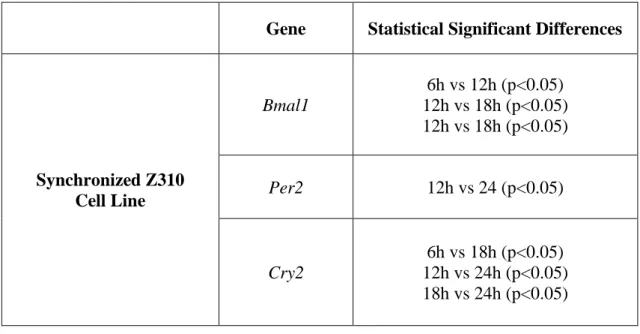
Table 5. Statistical differences between ZTs for each gene and condition tested in synchronized Z310 cell line.
Gene
Statistical Significant Differences
Synchronized Z310
Cell Line
Bmal1
6h vs 12h (p<0.05)
12h vs 18h (p<0.05)
12h vs 18h (p<0.05)
Per2
12h vs 24 (p<0.05)
Cry2
6h vs 18h (p<0.05)
12h vs 24h (p<0.05)
18h vs 24h (p<0.05)
Figure 8. One-way ANOVA analysis of the clock genes treated with dexamethasone and vehicle, and CircWave curves of Bmal1, Per2 and Cry2 expression in the Z310 cell line treated with dexamethasone. In one-way ANOVA each data set shows the mean of the target gene expression relative to control gene and error bars are ± standard error mean and 6h and 30h are double plotted. Presence of the CircWave curve shows significant rhythmicity in the expression of all the clock genes tested. A) One-way ANOVA reveals significant differences between: 6h vs 12h (p<0.05), 12h vs 18h (p<0.05) and 12h vs 24h (p<0.05); CircWave reveals Bmal1 rhythmic expression (p<0.01). B) One-way ANOVA reveals
6 12 18 24 30 0.8 1.2 1.6 2.0 Time (h) R e la tiv e B m a l1 m R N A e x p re s s io n * * * A) Dexamethasone Vehicle Bmal1 Per2 Cry2 ZT (h) ZT (h) ZT (h)
One-way ANOVA CircWave
6 12 18 24 30 0.0 0.5 1.0 1.5 2.0 2.5 Time (h) R e la ti v e P e r2 m R N A e x p re s s io n * B) Dexamethasone Vehicle N o rm al iz ed E xpr ess io n N o rm al iz ed E xpr ess io n N o rm al iz ed E xpr ess io n 6 12 18 24 30 0.0 0.5 1.0 1.5 2.0 2.5 3.0 Time (h) R e la ti v e C ry 2 m R N A e x p re s s io n * * C) * Dexamethasone Vehicle
25 significant differences between: 12h vs 24h (p<0.05); CircWave reveals Per2 rhythmic expression (p<0.001). C) One-way ANOVA reveals significant differences between: 6h vs 18h (p<0.05), 12h vs 24h (p<0.05) and 18h vs 24h (p<0.05); CircWave reveals Cry2 rhythmic expression (p<0.05).
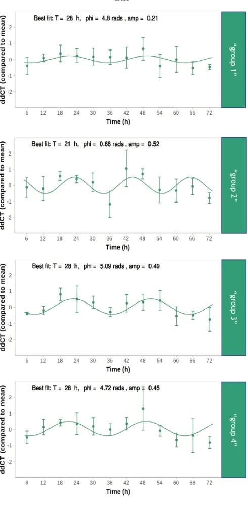
2.2 Melatonin Treatment Modulates Bmal1 Expression in the Presence of
Abeta
Real-Time RT-PCR results from cells treated with Abeta and melatonin were analyzed using Harmonic Regression Analysis. Like with the CircWave software, gene presents rhythmic expression if p<0.05 and this rhythmic expression is visually expressed by a sinusoidal curve. We choose this software since it enables the input of three continuous days of data while CircWave does not allow it. We only analyzed Bmal1 expression since it was the only clock gene that lost its rhythmicity in both sexes in APP/PS1 animals. Each group represents a different previous melatonin treatment (“group 1” (+/-/+/-), “group 2” (-/+/-/+), “group 3” (+/+/+/+) and “group 4” -/-/-/-) (Table 2). In “group 1” Bmal1 presented no rhythmicity (p=0.1404; Figure 9). In “group 2”, on the other hand, Bmal1 expression was rhythmic (p<0.01; Figure 9), with a period of 21 hours, a phase at 0.68 radians and with an amplitude of 0.52. In “group 3” Bmal1 also displayed rhythmic expression (p<0.0001; Figure 9), with a period of 28 hours, a phase at 5.09 radians and amplitude of 0.49. Finally, in “group 4”, Bmal1 also presented rhythmicity (p<0.01; Figure 9), displaying a period of 28 hours, a phase at 4.72 radians and an amplitude of 0.45. The strongest rhythmicity (represented by the lowest p-value) was presented by “group 3” and the widest amplitude in Bmal1 expression was presented by “group 2”. The Abeta stimulus, at least in the concentration used, was not capable of deregulating the circadian rhythm of Bmal1 expression.
26
Figure 9. Harmonic Regression Analysis of Bmal1 expression in cells treated with Abeta and melatonin.
“g ro up 1 ” “g ro up 2” “g ro up 3 ” “g ro up 4 ”
27 For each condition and at each time point, the dots represent the average data of gene expression value over all replicates and shows error bars ± standard deviation. The sinusoidal curve was plotted with the best fitting parameters of each condition. T: period in hours, phi: phase in radians, amp: amplitude.