BJRS
RADIATION SCIENCES
07-03B (2019) 01-21ISSN: 2319-0612 Accepted: 2019-07-17
Radiometric Survey in Mammography: Review of
regulations, recommendations, field and laboratories
practices and first tests
Macedo
a,bE.M., Navarro
aM.V.T., Jesus
aD.A.C., Garcia
aI.F.M.,
Leite
cH.J.D., Peixoto
bJ.G.P.
a Laboratório de Produtos para a Saúde do Instituto Federal de Educação, Ciência e Tecnologia da Bahia
(Labprosaud/IFBA), 41.745-715, Salvador, Bahia, Brasil
b Instituto de Radioproteção e Dosimetria, 22.783-127, Rio de Janeiro, Rio de Janeiro, Brasil
c Polo de Inovação Salvador, Instituto Federal de Educação, Ciência e Tecnologia da Bahia (PIS/IFBA), 41.745-715,
Salvador, Bahia, Brasil [email protected]
ABSTRACT
A review about Radiometric Survey in Mammography (RSM) in terms of regulatory documents, recommendations, radiation meters, besides field and laboratories practices shows that there is not many specific information about how these radiation area monitoring tests shall be performed. A search on the website of the most known manufacturers of radiation meters was made, but only one fully comply the requirements for RSM application. Tests with four laboratorial reference chambers, but none specific to RSM, were made submitting them to ISO N 20, 25 and 30 calibration setup (Based on document ISO 4037-1:1996) and their behavior in terms of HVL’s and air kerma rate measurements was compared to endorse some evaluations. The result of this work suggests that some RSM performed in Brazil, with nonspecific chambers and calibration can have underestimation of until 60% in those measurements, once a lack of specific information and specific calibration services in this country contributes for this scenario.
INTRODUCTION
Historically, radiation protection, especially occupational exposures, have been having increasingly addressed in scientific committees and communities. Even because a suitable program of radiation protection is an essential requirement for a safe and proper use of radiation. [1]
When the matter is radiometric survey in mammography (RSM) services, the discussion become more sensible, even because 5.799 mammography equipments are registered in Brazilian National Registry of Health Establishments (Cadastro Nacional de Estabelecimentos de Saúde, CNES/DATASUS). [2]
1.1. Regulation and Recommendations
About this theme, two very relevant points are going to be taken account. First is the publication of mammography room shielding by National Council on Radiation Protection and Measurements (NCRP), on report n° 147, Structural Shielding Design for Medical X-Ray Imaging Facilities, 2004 [3], recommends that permanent mammography facilities generally do not require additional protection besides common grass walls around the mammograph, plus acrylic or glass plumbing plates are for protection from operators inside the room. The fact of don’t need additional shield is not the same of don’t provide additional care.
The second point is the requirement to have a quality radiometric survey report by national Brazilian regulation Portaria SVS/MS 453/98 (ordinance), that becomes compulsory this evaluation to require Health Licensing. [4] The issue is lack of specific requirements, as calibration or instrumentation, in this regulation to guide the radiometric survey tests in mammographic rooms.
According Navarro et al [5], 20 years have passed since the publication of Portaria 453 and protocols of radiometric survey have not been established for the different areas of diagnostic and interventional radiology, standardizing the operational, instrumentation and calibration procedures.
Most recent regulation published in Brazil about diagnostic radiology was Normative Resolution N° 002/DIVS/SES [6], in Santa Catarina – Brazil, but nothing specifically about practices and instrumentation used in mammography rooms survey tests, both in terms of energy
application and in terms of calibration for the radiation meters. Same occurs with Portaria CVS – 18, São Paulo 2009. [7]
According to IAEA SRS 16: [1]
“Radiation protection monitoring instruments should be calibrated in terms of dose equivalent quantities. Area dosimeters or dose rate meters should be calibrated in terms of the ambient dose equivalent, H*(10), or the directional dose equivalent, H′(0.07), without any phantom present, i.e. free in air. “
The quantity and radiation qualities that are intended to use is based on ISO 4037-1:1996, depending on the application. For photons of energy in the mammography applications, is recommended to calibrate in terms of ambient dose equivalent, with 10 mm of deep penetration, which is nearest of photon interaction with matter. Radiation quality often used to diagnostic radiology application is narrow spectrum quality (ISO N), by its lower resolution, is better used to evaluate the variation of the response of radiation meter with photon energy. And it standard air-kerma rates, between 10–3 to 10–2 Gy.h-1 is suitable for radiometric survey applications. [8]
The AAPM Report no. 25, published in 1988 [9], presents general recommendations to Safety Surveys of Diagnostic Radiological Equipment, and does not treat specifically the applications, including mammography rooms. Same occurs with ARCAL XLIX Protocol. [10]
1.2. Radiation Meters
According to Yang et al [11], two challenges about instrumentation are facing currently. First one, the most common ionization chambers have not suitable sensitivity to scattered radiation measurements, and second one, survey meters with large volumes have enough sensitivity, but hard to place with good accuracy the right point of measurement.
In scientific publications, some works used Radcal 10X5-6M (mammography dedicated ion chamber), as Künzel et al (2006), Künzel et al (2008) and Judge et al (2013) [12–14]. Figueiredo (2012) [15] used Radcal Reference Chamber RC-6M to set ISO 4037 low-energies radiation qualities (10 – 30 kV of tube voltage), which are recommended to calibrate meters that will be used in mammo radiometric survey tests. Yang et al (2016) [11] used “a linear-array x-ray detector (X-Scan 0.8f3-512, Detection Technology, Inc., Finland)”, to evaluate scattered radiation generated from digital breast tomosynthesis exams. They used 30 and 35 kV tube voltage to the tests. None of these meters are commonly used or recommended to RSM tests in field.
In regulatory field, a recently search made in Brazilian calibration laboratories showed that has no service of calibration of radiation meters in terms of low-energies ambient dose equivalent, H*(10), offered to public. And that is the regulatory requirement. According to Portaria SVS/MS 453/98 [4], item 3.44, the operational quantity that must be used to verify the compliance with the dose restriction levels in area monitoring is ambient dose equivalent. Portaria CVS – 18, São Paulo 2009 [7] states that radiation meters used to this end must have sensibility of 0,01 nGy and nothing about energy range. Normative Resolution n. 002/DIVS/SES [6], requires that radiation meters to radiometric survey must have minimum sensibility of 10 nGy/s (on dose rate mode) and 10 nGy (on accumulated dose mode), time response lower than 1 s, energy dependence lower than 20% and nothing specifically about mammography energy range.
According ARCAL XLIX Protocol [10], meters should have a sensible volume of 1,800 cm³, range of 1 fC to 999 nC, precision ± 5%, reproducibility ± 1%. Nothing about mammography energy range, or its calibration.
According AAPM Report no. 25 [9], the survey meter shall be carefully selected, with characteristics more specifically described on AAPM Report no. 35 [16]. Generally, it shall be “an ionization chamber with a sensitive volume of one liter coupled to an electrometer with integrating capabilities”. Some factors must be evaluated too, as indicated below:
“(a) Meter's energy response (b) Meter's directional response (c) Meter's intensity response (d) Ion chamber cross sectional area corrections (e) Meter's calibration (f) Meter's response time in the rate mode on its most sensitive scales.”
This kind of approach could be incorporated on Brazilian regulation, since it contemplates all characteristics necessary to select the suitable survey meters to be applied in RSM.
Given the above, is not too hard to think that regulatory gaps and shortage of specific equipment strongly contribute to the lack of provision of calibration services in laboratories in Brazil.
According to Navarro et al [5], only three Brazilian laboratories perform calibration applied to radiometric survey conditions, and with follow features: Air kerma and ambient dose equivalent quantities, continuous beam, dose rate measure mode and to qualities between ISO N 60 (kV) to 150 (kV). These features diverge from field applications, based in instruments that do exposures shorter than 1 s, and, in mammography cases, energies lower than 40 kV.
1.3. Practices
According to Navarro et al [5], majority of practices in radiometric survey tests in mammographic rooms are performed using dosimeters on dose rate measurement mode, calibrated on ISO N 60 (kV) to 100 (kV) radiation qualities, exposure times lower than 1 s and the air kerma to H*(10) conversion factor not used appropriately. On the same study, they performed laboratorial tests that indicates possible underestimates measurements of up to 10 times when uses it on dose rate mode. This deviation was found comparing this with measures on integrated dose mode, in ISO N 25 beam quality. Leyton et al [17] tested the same equipment in the same laboratory and found results with the same behavior. Differences between measures with dose rate and integrated dose mode, but this time using exposures with ISO N60, N80 and N100, which corresponds to 60, 80 and 100 kV of Tube voltage.
1.4. Physical details in mammo scattering beams
According to Künzel et al [12], the acknowledgement about the spectra of scattering radiation of a mammograph is essential to evaluate quantities as air kerma, ambient dose equivalent [H*(10)] and effective dose [Ep]. An example is that Mo/Mo anode/filter combination has an air kerma value greater than Mo/Rh. Nevertheless, Mo/Rh showed H*(10) and Ep values greater than Mo/Mo. Such fact can be making a little harder to take confident evaluation of measures performed in the system. On the other hand, all we know that is impracticable make previous spectra catch of scattered beam and then measures of radiometric survey tests, mainly because the unavailability of time and resources in a commercial routine of image diagnosis services.
According to Judge et al [14], the scatter radiation coming from phantoms irradiated in a mammography system has a non-isotropic behavior, and this has to be account in tests. According to Yang et al [11], “due to the highly conservative initial assumptions, the current recommended scatter air kerma level from NCRP Report 147 is still suitable for wall barriers”, when the matter is digital breast tomosynthesis systems.
The aim of this work is evaluating the panorama of RSM in our country and discusses about possible solutions about the gaps found. It considers a search about the offer of specific radiation
meters with specific features to this application. Besides, an evaluation of the behavior of reference instruments under ISO N 20, 25 and 30 radiation conditions.
MATERIALS AND METHODS
2.1. Search of meters applied to RSM
This is a documental research and account with an overview of the regulatory situation, international recommendations and specific equipment to apply in radiometric survey tests in mammographic rooms. First step of this research was search on websites of the main manufactures of these radiation meters which one is applicable to that end (table 1).
Table 1. Manufacturers and websites of search.
Manufacturer Website RTI [18] http://rtigroup.com/ PTW [19] http://ptw.de/ Radcal [20] http://radcal.com/ Nuclear Enterprises [21] http://nuclearenterprisesllc.com/ Unfors/Raysafe [22] http://www.raysafe.com/ Fluke Biomedical [23] http://www.flukebiomedical.com/ Atomtex [24] http://atomtex.com/en Sun Nuclear [25] https://www.sunnuclear.com/ Ludlums [26] http://ludlums.com/ IBA [27] https://www.iba-dosimetry.com/ MRA [28] http://www.mra.com.br/
In terms of calibration conditions, taken and adapted from ISO 4037-1:1996 [8] on table 2, is the information about mean energy of each radiation quality that indicates the requirement of energy range of the dosimeters under study. This information will be considered for the assessment of energy range requirements. The sensitivity shall be considered suitable if it has less than 1 mSv/h, as also required as a calibration requirement in ISO 4037-1:1996. The specific intended to use will be considered too.
In terms of field usability, will be considered the energy range of at maximum 20.2 keV, according the evaluation of scattered spectra made by Künzel et al [12]. About meter sensitivity, many protocols and regulations indicate different minimum values to fulfill [6,7,9,10,16]. We will assume 1 nGy (or 1 nSv) for this parameter.
2.2. ISO N 20, 25 e 30 qualities implantation
To establish the radiation qualities ISO N 20, 25 e 30 on laboratory, it was used an X-ray tube with a tungsten anode, and inherent filtration of 1 mm Be, and requirements based on table 2.
The test setup was based on ISO 4037-2:1999 [29], as showed on figure 1. The used setup is in the picture below, on figure 2.
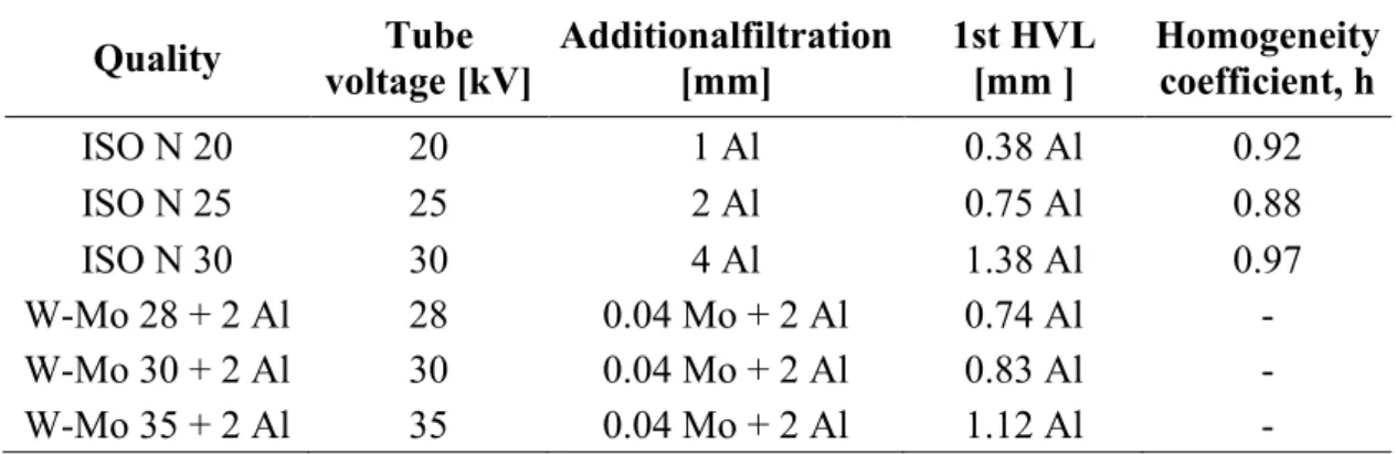
Table 2. Table of radiation qualities ISO N from ISO 4037-1:1996. [8]
Mean
Energy, 𝑬𝑬� tion. RResolu-E
Tube
Voltage Additional Filtration 1st HVL 2nd HVL Homogeneity coefficient, h keV % kV mm Al mm mm 16 34 20 1.0 0.32 Al 0.37 Al 0,75 – 1,0 20 33 25 2.0 0.66 Al 0.73 Al 24 32 30 4.0 1.15 Al 1.30 Al
The Peak Practical Voltage (PPV), based on IEC 61267:2005 [30], will be determined to ensure good values of tube voltage to be used on this work. The meter used was the PTW Nomex, a non-invasive kV meter (PTB Traceability 2018).
Figure 1. Typical setup recommended by ISO 4037-2:1999.
Source: ISO 4037-2:1999 [29]
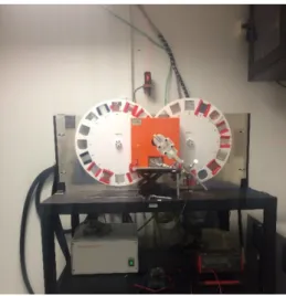
Figure 2. Setup used with PTW 10 liters ion chamber, on Labprosaud/IFBA.
Figure 3. Setup used on PPV determination tests using PTW NOMEX, on
Labprosaud/IFBA.
Source: Labprosaud/IFBA The instrumentation used on tests is described on table 3.
Table 3. Reference instrumentation, on Labprosaud/IFBA.
Instrument Model Application
X ray system -
Constant potential ISOVOLT TITAN E 160 M2 X-ray generation 10-160 kV
Monitor Chamber PTW - TM786 Beam monitoring
Electrometer UNIDOS webline PTW [PTB traceability] Charge or current Ion Chamber 6 cm³ MAMMO (“PTW 6cc”) PTW TM34069-2,5 Air Kerma
Establishment of radiation qualities [PTB traceability to RQA-M 2] Ion Chamber 10 liters
(“PTW 10 L”) TM32003 PTW [PTB traceability to ISO N 60] Air Kerma Ion Chamber 6 cm³
MAMMO Radcal RC6M [RBC traceability to RQA-M 2] Air Kerma Ion Chamber 1800 cm³ Radcal RC1800 [RBC traceability to ISO N 60] Air Kerma Noninvasive multimeter
ISOVOLT TITAN E [31,32] is an X-ray generator of constant potential, with ripple lower than 1%. The 160 M2 X-ray tube has a voltage range from 1 to 160 kV, tungsten anode and an inherent filtration of 1 mm of Beryllium. The fixed apparatus used on tests (X-ray tube window and monitor chamber has an inherent filtration measured, as ISO 4037-1:1996 [8] recommendation, of 0.1 mm of Aluminum.
These tests were performed to compare the results found on 1st and 2nd HVL tests, and measurements of air kerma rate in a point of test of 2.5 m far from X-ray source, based on ISO N calibration setup.
RESULTS AND DISCUSSION
3.1. Search of meters applied to RSM
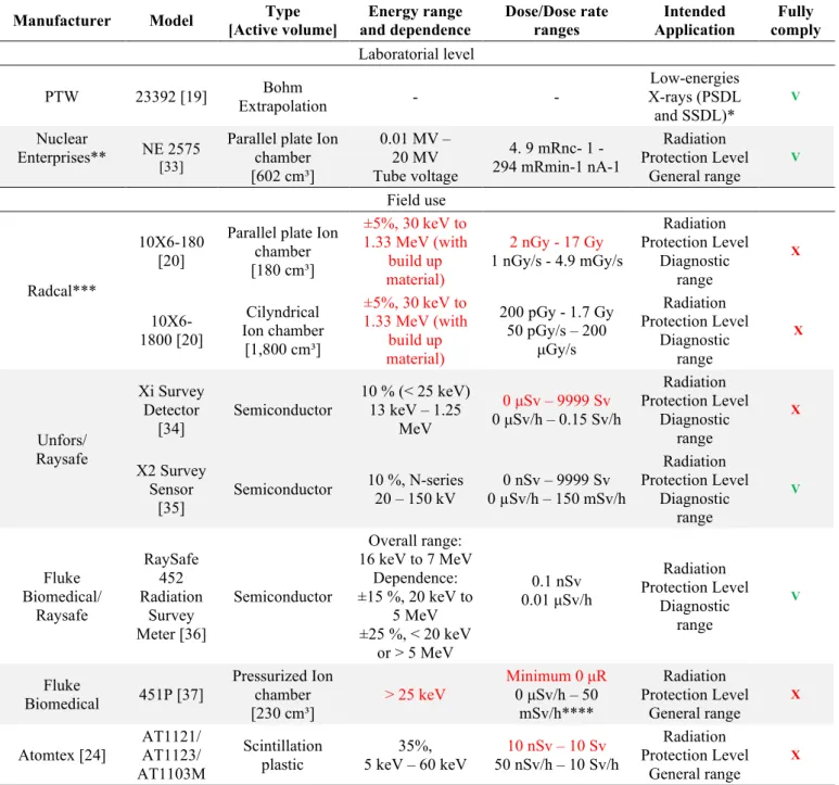
The results of the specific search are described on table 4. The columns are indicating the information about these radiation meters, when treating of calibration and field measurements.
PTW Bohm Extrapolation chamber was considered in this work because it was used in other publication about low-energies x-ray with ISO 4037-1 radiation qualities, but we won’t deepen on it. NE 2575 fulfills the requirement. But both is recommended to laboratorial use.
As showed in table 4, there are only two dosimeters that fulfills the metrological requirements to radiometric survey tests in mammographic rooms, the X2 Survey Sensor and the most recent RaySafe 452 Radiation Survey Meter. Xi Survey Detector and Atomtex (3 models) do not comply with minimum sensitivity of 1 nGy or 1 nSv. First showed μSv scale and Atomtex showed 10 nGy of minimum value of dose measurement. But other features were well complied with. It’s valid to emphasize that Atomtex dosimeters got the energy range feature, but with high energy dependence (35%), that can take to the tests a high uncertainty.
Others three meters listed, according to Navarro et al [5], are the widely used radiation meters in RSM tests. Additionally, he cited electrometers Radcal models: 2025, 2026, 9010, 9015, 9095, 9096 and AccuGold, with 180 cm³ and 1800 cm³ ion chambers, to respective electrometers models; Fluke/Victoreen portable monitors models 450, 450P, 451P and 451B, besides electrometer model 660 with ion chamber 660-5.
Table 4. List and features of the detectors found with range on low energies (10 keV – 30
keV) and/or used in field tests.
Manufacturer Model [Active volume] Type and dependence Energy range Dose/Dose rate ranges Application Intended comply Fully Laboratorial level
PTW 23392 [19] Extrapolation Bohm - - X-rays (PSDL Low-energies
and SSDL)* V Nuclear
Enterprises** NE 2575 [33] Parallel plate Ion chamber [602 cm³] 0.01 MV – 20 MV Tube voltage 4. 9 mRnc- 1 - 294 mRmin-1 nA-1 Radiation Protection Level General range V Field use Radcal*** 10X6-180 [20]
Parallel plate Ion chamber [180 cm³] ±5%, 30 keV to 1.33 MeV (with build up material) 2 nGy - 17 Gy 1 nGy/s - 4.9 mGy/s Radiation Protection Level Diagnostic range X 10X6-1800 [20] Cilyndrical Ion chamber [1,800 cm³] ±5%, 30 keV to 1.33 MeV (with build up material) 200 pGy - 1.7 Gy 50 pGy/s – 200 μGy/s Radiation Protection Level Diagnostic range X Unfors/ Raysafe Xi Survey Detector [34] Semiconductor 10 % (< 25 keV) 13 keV – 1.25 MeV 0 μSv – 9999 Sv 0 μSv/h – 0.15 Sv/h Radiation Protection Level Diagnostic range X X2 Survey Sensor [35] Semiconductor 10 %, N-series 20 – 150 kV 0 µSv/h – 150 mSv/h 0 nSv – 9999 Sv Radiation Protection Level Diagnostic range V Fluke Biomedical/ Raysafe RaySafe 452 Radiation Survey Meter [36] Semiconductor Overall range: 16 keV to 7 MeV Dependence: ±15 %, 20 keV to 5 MeV ±25 %, < 20 keV or > 5 MeV 0.1 nSv 0.01 μSv/h Radiation Protection Level Diagnostic range V Fluke Biomedical 451P [37] Pressurized Ion chamber [230 cm³] > 25 keV Minimum 0 μR 0 μSv/h – 50 mSv/h**** Radiation Protection Level General range X Atomtex [24] AT1121/ AT1123/
AT1103M
Scintillation
plastic 5 keV – 60 keV 35%, 50 nSv/h – 10 Sv/h 10 nSv – 10 Sv
Radiation Protection Level
General range X Highlighted in red the requirements which the meter didn’t comply with.
* PSDL and SSDL are Primary (and Secondary) Standard Dosimetry Laboratories. ** Not found meters on proper website, but evaluated in laboratory application.
*** Despite exists others models of Radcal sensor with same volume and structure, only differing the connector (10X5, 10X6, 10X9, 20X5, 20X6), we are considering these 10X6, that is stated as available on website. And these sensors come with their respective electrometers.
Radcal 180 cm³ ion chamber and Fluke 451P do not comply with minimum sensitivity of 1 nGy or 1 nSv and with energy range. Radcal 1800 cm³ ion chamber would be the most appropriate meter to use, since that complies with the most requirements to make RSM tests, including large volume (good feature to low intensity beams) recommended by two international protocols, AAPM and ARCAL. [9,10] Nevertheless, the it energy range is intended to beams with energies greater than 30 keV, while mammography scattered beams have mean energy around 20 keV. [12]
The question of energy range needs to be more evaluated, because the fact of the manufacturer doesn’t state the information about lower energies than 30 keV, let doubts about reliability of the measurements. All of field meters have dose rate sensitivity able to perform a good calibration, according ISO 4037-1 requirement. [8] However, this feature alone is not enough to perform a good RSM. As shown in section 2.1, dose sensitivity of 1 nGy or 1 nSv and their intended use should be also complied.
When we combine this with the gap in the provision of specific calibration services, in terms of equivalent dose ambient in ISO 4037 narrow spectrum radiation qualities, in the range of 20 to 30 kV, the lack of reliability in those measurements became even worst. Another question is the distance between the field use condition and calibration conditions that, sometimes, can mask it real behavior, given that most of meters has very good performance when measuring continuous x-ray beam from a constant potential generator, in dose rate mode and controlled ambient conditions. But the behavior of meters under short times exposures, with poor ambient control and unknown generator conditions can perhaps lead to dubious results.
On their work, Navarro et al recommend that calibration be carried out with conditions as close as possible to field use, provided they do not deviate from the regulatory requirements. [5] This action can improve the reliability of tests results in mammography radiometric survey practices.
3.2. PPV determination
The point of 20 kV is not traceable, but to determinate PPV value, an extrapolation line was performed with the R² = 0,9572 using PTB traceable points of 25, 28, 30 and 35 kV. The reference quality of PTB calibration was WAVa (Tungsten anode and 0.7 mm of aluminum as additional filtration), and uncertainty of 2.0 % (k = 2).
The results found are showed on table 5.
Table 5. PPV determination.
Radiation Quality Nominal Voltage PPV Measured Uncertainty (k = 1)
ISO N 20 20 kV 20,0 R² = 0,9572* ISO N 25 25 kV 25,0 1,1% ISO N 30 30 kV 30,0 1,1% W-Al 28 + 2 Al 28 kV 28,0 1,1% W-Al 30 + 2 Al 30 kV 30,0 1,1% W-Al 35 + 2 Al 35 kV 35,1 1,1%
* Adjust quality value stated since NOMEX is not able to measure PPV in this level of tube voltage.
3.3. Establishment of radiation qualities
Once PPV evaluated, the next step is implant ISO N radiation qualities, determining the first and second HVL values for each quality. The chamber used on the tests is PTW 6CC, because mainly of its PTB traceability in terms of air kerma in RQA-M qualities. A comparison among the laboratorial reference chambers and nominal values is an easy way to evaluate the behavior of them under ISO N low energies conditions. First one comparing HVL’s obtained in ISO N qualities and second is comparing air kerma rate obtained in ISO N and W-Mo + 2 Al qualities, at 2,5 m from the source, and air kerma rate of 5,0 mGy/h.
Table 6. Data of ISO N and W-Mo + 2 Al radiation qualities.
Quality voltage [kV] Tube Additionalfiltration [mm] 1st HVL [mm ] Homogeneity coefficient, h
ISO N 20 20 1 Al 0.38 Al 0.92 ISO N 25 25 2 Al 0.75 Al 0.88 ISO N 30 30 4 Al 1.38 Al 0.97 W-Mo 28 + 2 Al 28 0.04 Mo + 2 Al 0.74 Al - W-Mo 30 + 2 Al 30 0.04 Mo + 2 Al 0.83 Al - W-Mo 35 + 2 Al 35 0.04 Mo + 2 Al 1.12 Al -
3.4. HVL determination with different chambers
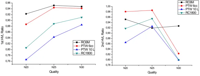
Once PPV evaluated, the next step on establishment of the ISO N radiation qualities is the determination of first and second HVL values for each quality. The chambers used on the tests are described on table 3. A comparison among the laboratorial reference chambers and nominal values is an easy way to evaluate the behavior of them under ISO N low energies conditions. The results found are showed on table 7. The figure 4 shows a graphical view of chambers response.
Table 7. Results of HVL tests.
Ratio between nominal and measured values
1stHVL 2ndHVL Radcal RC6M 0.923 0.952 0.952 0.919 0.948 0.927 PTW 6cc 0.887 0.981 0.944 0.986 0.941 0.913 Radcal RC1800 0.806 0.918 0.889 0.955 0.911 0.800 PTW 10 L 0.766 0.865 0.843 0.926 0.885 0.799 Color graduation: 1.05-0.95; 1.10-1.05 or 0.95-0.90;1.20-1.10 or 0.90-0.80;1.30-1.20 or 0.80-0.70.
Clearly can be seen the effect of energy dependence on results of the 1st HVL and 2nd HVL, evaluation. Radcal RC6M [38] had the best performance as expected, since it’s energy dependence is ±5%, 10 keV to 40 keV, as stated on its specification sheet. The ratio between nominal value of 1st HVL on qualities ISO N 20, 25 and 30 were 0.923, 0.952 and 0.948, respectively. In terms of 2nd HVL, 0.952, 0.919 and 0.927.
The PTW 6cc [39] (±2%, 25 keV to 35 keV), Radcal RC1800 [40] (±5%, 33 keV to 1.33 MeV) and PTW 10 L [41] (≤ ± 3% in the range 40 keV to Co-60) had, in this order, the best to worst result found on this work. The results were expected, since the energy response ranges stated on their instruction manuals are out of mean energy range of ISO N 20, 25 and 30 radiation qualities, that are 16, 20 and 24 keV respectively.
Figure 4. Comparison between 4 reference chambers in calculation of 1st and 2nd HVL, and
the nominal value.
N20 N25 N30 0,76 0,78 0,80 0,82 0,84 0,86 0,88 0,90 0,92 0,94 0,96 1s t H VL R at io Quality RC6M PTW 6cc PTW 10 L RC1800 N20 N25 N30 0,78 0,80 0,82 0,84 0,86 0,88 0,90 0,92 0,94 0,96 0,98 1,00 2nd H VL R at io Quality RC6M PTW 6cc PTW 10 L RC1800
A possible cause for the different response of chambers can be the thick and/or material of chamber walls, that contributes to a less accurate measurement. An example can be the grater absorption of low energies photons by those chamber walls, that makes the beam harder and consequently a higher HVL is obtained. [42–44]
3.5. Kerma rate measurements
By way of comparison, a kerma rate measurements test was made, with “MAMMO chambers” Radcal RC6M and PTW 6cc were calibrated in terms of air kerma in RQA-M 2 quality, and “Radiation Protection chambers” Radcal RC1800 and PTW 10 L were calibrated in terms of H*(10) in ISO N 60 quality. This is a representative calibration to field chambers [34,35,37,45,46], since a single calibration factor is imputed on electrometers and all charge or current measured with sensor and multiplied by the calibration factor.
The measurements, showed on table 8 and illustrated graphically on figure 5, converge with HVL’s results. Radcal RC6M and PTW 6CC had good performance, with maximum deviation of 3,2%. On the other hand, Radcal RC1800 and PTW 10 L had not a good performance, with measurements underestimation of until 60%.
Table 8. Measurements of air kerma rate [mGy/h] @ 2.5 m. ISO N 20 ISO N 25 ISO N 30 PTW 6cc 5.06 ± 0.45% 4.96 ± 0.22% 5.11 ± 0.28%
RC6M 4.87 ± 0.57% 4.84 ± 0.54% 4.90 ± 0.15%
PTW 10 L 2.04 ± 0.79% 3.05 ± 0.43% 3.89 ± 0.10%
RC1800 3.28 ± 0.17% 3.86 ± 0.62% 4.35 ± 0.42%
W-Mo 28 + 2 Al W-Mo 30 + 2 Al W-Mo 35 + 2 Al PTW 6cc 5.09 ± 0.33% 5.08 ± 0.22% 5.17 ± 0.21%
RC6M 5.04 ± 0.40% 5.00 ± 0.91% 4.91 ± 0.47%
PTW 10 L 3.15 ± 0.14% 3.30 ± 0.10% 3.63 ± 0.15%
RC1800 3.99 ± 0.01% 4.03 ± 0.24% 4.19 ± 0.18%
Figure 5. Comparison between 4 reference chambers in Air kerma rate measurements
[mGy/h] @ 2,5 m (ISO N 20, 25 and 30 qualities on orange square, W-Mo 28, 30 and 35 + 2 Al on blue square).
CONCLUSION
The overview about the national panorama and international recommendations on radiometric survey in mammography services showed us certain gaps involving regulatory issues, current practices and radiation meters used in this application.
Those results do not indicate that RC1800 and PTW 10 L do not function properly, since their performance in the higher energy qualities, such as ISO N 60, 80 and 100, is extremely satisfactory. What the results indicate is that they are not suitable for radiation qualities applied in radiometric surveys in mammography. Just as the PTW 6cc and RC6M that don’t have their application stated for RSM, but to Diagnostic Radiology – Mammography. Their use in this work was only for comparison between instruments that are dedicated to low energies and those that are not.
Additionally, the result of this work suggests that some RSM tests performed in Brazil, with nonspecific chambers and nonspecific calibration can have underestimation of until 60% in those measurements. Since these two radiation protection reference instruments evaluated can represent most equipment used in field, instead of the technical specifications of them state that mammo energy range application is not indicated. Specific calibrations with field radiation meters listed on table 4, and widely used in radiometric survey tests should be performed to confirm this evaluation, including mammography and conventional radiology energy range (ISO N 20 to ISO N 100).
It’s hard to find field instruments applied to RSM. Then, an accurate study of behavior of most used radiation meters is important to improve the practices, since the measurement is impaired by the features of most radiation meters used. Specific calibration and right evaluation of features as right energy range and sensibility of dose (or rate) are crucial features to evaluate to perform traceable and more accurate RSM tests.
ACKNOWLEDGMENT
The authors acknowledge Coordenação de Aperfeiçoamento de Pessoal de Nível Superior (CAPES), Instituto de Radioproteção e Dosimetria (IRD), Comissão Nacional de Energia Nuclear (CNEN), Ministério da Ciência, Tecnologia, Inovações e Comunicações (MCTIC) and Laboratório
de produtos para a Saúde (LABPROSAUD) do Instituto Federal de Educação, Ciência e Tecnologia da Bahia (IFBA), for the financial and structural support of this work.
REFERENCES
[1] International Atomic Energy Agency. Safety Reports Series 16 - Calibration of Radiation
Protection Monitoring Instruments. Vienna; 2000.
[2] DATASUS. Ministério da Saúde/SAS - Cadastro Nacional de Estabelecimentos de Saúde (CNES). 2018.
[3] National Council on Radiation Protection and Measurements. Structural Shielding Design for Medical X-Ray Imaging Facilities. NCRP no 147. 2004.
[4] Secretaria de Vigilância Sanitária, Ministério da Saúde. Diretrizes de Proteção Radiológica em Radiologia Médica e Odontológica. 1998;Portaria/MS/SVS no 453, de 01 de junho de 1998.
[5] Navarro MVT, Navarro VCC, Garcia IFM, Macedo EM. Levantamento radiométrico em mamografia : problemas e desafios. In 2015. p. 1–4. Available from:
http://media.metrologia2015.org.br/media/uploads/trabalhos/Levantamento_Radiométrico_e m_Mamografia.pdf
[6] Estado de Santa Catarina. Resolução Normativa n. 002/DIVS/SES. 2015;
[7] Estado de São Paulo. Portaria CVS - 18, de 7-10-2009 [Internet]. 2009. Available from:
http://www.cvs.saude.sp.gov.br/zip/Portaria CVS no 18, de 07out09.pdf
[8] International Organization for Standardization. X and γ Reference Radiations for Calibrating Dosemeters and Dose Rate Meters and for Determining their Response as a Function of Photon Energy - Part 1: Radiation characteristics and production methods. ISO 4037-1.
1996;(ISO 4037-1).
[9] American Association of Physicists in Medicine. Protocols for the Radiation Safety Surveys of Diagnostic Radiological Equipment Protocols for the Radiation Safety Surveys of
Diagnostic Radiological Equipment X-Ray Imaging Committee. AAPM REPORT NO. 25.
1988.
Tecnología Nuclear en Latinoamérica y el Caribe. Protocolos de Control de Calidad en Radiodiagnóstico - ARCAL XLIX. 2001.
[11] Yang K, Li X, Liu B. Scatter radiation intensities around a clinical digital breast tomosynthesis unit and the impact on radiation shielding considerations. Med Phys.
2016;43(3):1096–110.
[12] Künzel R, Herdade SB, Costa PR, Terini RA, Levenhagen RS. Ambient dose equivalent and effective dose from scattered x-ray spectra in mammography for Mo/Mo, Mo/Rh and W/Rh anode/filter combinations. Phys Med Biol. 2006;51(8):2077–91.
[13] Künzel R, Levenhagen RS, Herdade SB, Terini RA, Costa PR. X-ray spectroscopy applied to radiation shielding calculation in mammography. Med Phys. 2008;35(8):3539–45.
[14] Judge MA, Keavey E, Phelan N. Scatter radiation intensities around full-field digital mammography units. Br J Radiol. 2013;86(1021):1–8.
[15] Figueiredo MTT de. Análise dos Procedimentos e Critérios de Implantação de Feixes de Raios X de Referência ISO 4037 em Baixas Energias [Internet]. 2012. Available from:
http://www.iaea.org/inis/collection/NCLCollectionStore/_Public/44/073/44073110.pdf [16] American Association of Physicists in Medicine. Recommendations on performance
characteristics of diagnostic exposure meters. AAPM REPORT NO. 35. 1992.
[17] Leyton F, Macedo EM, Ferreira MJ, Navarro VCC, Garcia IFM, Pereira LCS, et al.
Temporal Dependence of the Radiation Monitoring Instruments for Area Monitoring Used At Radiodiagnostic and Intervention Facilities. Radiat Prot Dosim [Internet].
2015;1–11. Available from:
http://rpd.oxfordjournals.org/content/early/2015/11/14/rpd.ncv467.abstract [18] RTI Group AB. RTI [Internet]. 2016 [cited 2018 Mar 24]. Available from:
http://rtigroup.com/
[19] PTW Freiburg GmbH. PTW Freiburg [Internet]. 2012 [cited 2018 Mar 24]. Available from:
http://ptw.de/
[20] Radcal Corporation. Radcal [Internet]. 2016 [cited 2018 Mar 24]. Available from:
http://radcal.com/
[21] Nuclear Enterprises. Nuclear Enterprises [Internet]. 2005 [cited 2018 Mar 24]. Available
[22] Raysafe. Unfors/Raysafe [Internet]. 2018 [cited 2019 Mai 24]. Available from:
http://www.raysafe.com/
[23] Fluke Corporation. Fluke Biomedical [Internet]. 2019 [cited 2019 Jul 2]. Available from:
http://www.flukebiomedical.com/
[24] Atomtex. Atomtex [Internet]. 2018 [cited 2018 Mar 24]. Available from:
http://atomtex.com/en
[25] Sun Nuclear Corporation. Sun Nuclear [Internet]. 2017 [cited 2018 Mar 24]. Available
from: https://www.sunnuclear.com/
[26] Ludlum Measurements. Ludlum [Internet]. 2018 [cited 2018 Mar 24]. Available from:
http://ludlums.com/
[27] IBA. IBA Dosimetry [Internet]. 2017 [cited 2018 Mar 24]. Available from:
https://www.iba-dosimetry.com/
[28] MRA Indústria. MRA [Internet]. 2017 [cited 2018 Mar 24]. Available from:
http://www.mra.com.br/
[29] International Organization for Standardization. X and γ Reference Radiations for Calibrating Dosemeters and Dose Rate Meters and for Determining their Response as a Function of Photon Energy - Part 2: Dosimetry for radiation protection over the energy ranges 8 keV to 1,3 MeV and 4 MeV to 9 MeV. ISO 4037-2. 1997;(ISO 4037-2).
[30] International Electrotechnical Commission. Medical diagnostic X-ray equipment – Radiation conditions for use in the determination of characteristics. IEC 61267. 2005.
[31] General Electric Company. The ISOVOLT Titan E X-ray Generator. 2009.
[32] General Electric Company. SEIFERT X-ray Tubehousing ISOVOLT 160 M2 / 0.4-1.5.
2004.
[33] NE Technology Limited. INSTRUCTION MANUAL for 2575 600cc THIN WINDOW IONIZATION CHAMBER and 2576 ST ABILITY CHECK SOURCE. 1995. p. 1–35.
[34] Unfors/Raysafe. RaySafe Xi, USER MANUAL [Internet]. 2014. Available from:
http://www.raysafe.com/Products/Equipment/RaySafe Xi#Downloads
[35] Unfors/Raysafe. RaySafe X2, Technical Specifications [Internet]. 2017. Available from:
http://mediabank.raysafe.com/A/RaySafe+Media+Bank/1828
Available from: https://www.flukebiomedical.com/sites/default/files/6011930a-en-452-ds-w_0.pdf
[37] Fluke Biomedical. Victoreen 451P & 451P-DE-SI Ion Chamber Survey Meter - Operators Manual [Internet]. 2005. Available from:
http://assets.fluke.com/manuals/451P____omeng0000.pdf
[38]. Radcal Corporation. Specification Sheet - RC6M LOW ENERGY X-RAY MEASUREMENTS. 2014. p. 4500045.
[39] PTW. User Manual SFD Chambers Type 34060 and Type 34069. 2008;1–14.
[40] Radcal Corporation. Specification Sheet - RC1800 Low Dose Rate Radiation Applications. 2014.
[41] PTW. User manual 1 l Spherical chamber Type 32002 10 l Spherical chamber Type 32003 Spherical chamber TK30 Type 32005. 2009. p. 1–15.
[42] Knoll GF. Radiation Detection and Measurement. 4th ed. 2010.
[43] Attix FH. Introduction to Radiological Physics and Radiation Dosimetry. Wiley-VCH;
2004. 607 p.
[44] International Commission on Radiation Units and Measurements. Radiation Protection Instrumentation and Its Application. ICRU Report 20. J Int Comm Radiat Units Meas
[Internet]. 2016 Apr 27; Available from: https://doi.org/10.1093/jicru/os11.1.Report20 [45] Fluke Biomedical. Victoreen 660 Digital Radiation Survey Meter - Operators Manual
[Internet]. Cleveland, Ohio; 2005. Available from:
http://assets.fluke.com/manuals/660_____omeng0000.pdf

![Table 2. Table of radiation qualities ISO N from ISO 4037-1:1996. [8]](https://thumb-eu.123doks.com/thumbv2/123dok_br/18278141.881333/7.892.127.763.718.897/table-table-radiation-qualities-iso-n-iso.webp)


![Figure 5. Comparison between 4 reference chambers in Air kerma rate measurements [mGy/h] @ 2,5 m (ISO N 20, 25 and 30 qualities on orange square, W-Mo 28, 30 and 35 +](https://thumb-eu.123doks.com/thumbv2/123dok_br/18278141.881333/16.892.277.673.762.1055/figure-comparison-reference-chambers-measurements-qualities-orange-square.webp)
