Universidade de Lisboa
Faculdade de Motricidade Humana
Effects Of Neural Tension In The Sciatic Nerve Stiffness, In
Healthy People And People With Low Back Related Leg Pain
Tiago Gonçalves Neto
Orientador: Professor Doutor Raul Alexandre Nunes da Silva Oliveira Co-orientador: Professor Doutor Sandro Remo Martins Neves Ramos Freitas
Dissertação elaborada com vista à obtenção do Grau de Doutor no ramo Motricidade Humana, na especialidade de Comportamento Motor
Tese por compilação de artigos, realizada ao abrigo da alínea a) do nº2 do artº 31º do Decreto-Lei º 230/2009
Júri:
Presidente:
Doutor Francisco José Bessone Ferreira Alves
Professor Catedrático e Presidente do Conselho Científico Faculdade de Motricidade Humana da Universidade de Lisboa
Vogais:
Doutor Jaime da Cunha Branco Professor Catedrático
Faculdade de Ciências Médicas da Universidade Nova de Lisboa Doutora Colette Ridehalgh
Senior Lecturer
School of Health Sciences da Universidade de Brighton (Reino Unido) Doutor Raul Alexandre Nunes da Silva Oliveira
Professor Auxiliar
Faculdade de Motricidade Humana da Universidade de Lisboa Doutora Filipa Oliveira da Silva João
Professora Auxiliar
Faculdade de Motricidade Humana da Universidade de Lisboa Doutora Sandra Cristina Fernandes Amado
Professora Adjunta
Escola Superior de Saúde do Instituto Politécnico de Leiria Doutor João Pedro Casaca de Rocha Vaz
Professor Adjunto Universidade Europeia
Declaração de Reprodução da Tese
Nome: Tiago Gonçalves Neto
Endereço eletrónico: [email protected] Telefone: +351 96 825 07 84
Número do Cartão de Cidadão: 12163035
Título da Tese: Effects of neural tension in the sciatic nerve stiffness, in healthy
people and people with low back related leg pain
Orientador: Professor Doutor Raul Alexandre Nunes da Silva Oliveira
Co-orientador: Professor Doutor Sandro Remo Martins Neves Ramos Freitas
Ano de conclusão: 2016
Ramo de conhecimento do Doutoramento: Motricidade Humana, especialidade de
Comportamento Motor
É AUTORIZADA A REPRODUÇÃO INTEGRAL DESTA DISSERTAÇÃO APENAS PARA EFEITOS DE INVESTIGAÇÃO, MEDIANTE DECLARAÇÃO ESCRITA DO INTERESSADO, QUE A TAL SE COMPROMETE.
Faculdade de Motricidade Humana – Universidade de Lisboa Cruz Quebrada, 2016
Assinatura:____________________________
(Tiago Gonçalves Neto)
i
DEDICATÓRIA
ii
iii
AGRADECIMENTOS / ACKNOWLEDGEMENTS
O doutoramento é um processo composto por diferentes capítulos, assemelhando-se a uma história. E para esta história importam não apenas os métodos e técnicas que se desenvolvem, os equipamentos que se aprende a dominar, ou os laboratórios que servem como segunda casa, mas importam sobretudo as pessoas que se conhece e que dão significado a toda esta história. Mais que um acto de obrigação ou dever, estas linhas expressam um profundo reconhecimento da importância que essas pessoas tiveram durante este doutoramento.
Em primeiro lugar quero agradecer à minha família, em particular aos meus pais e a minha irmã, por serem os pilares da minha vida.
Não posso também deixar de destacar a importância que os amigos têm neste longo, e muitas vezes desgastante, processo. Obrigado a todos vós, em particular aqueles que diariamente partilharam esta história, como o Amândio, o Luis, o Nery, a Joana, o Fred, a Vanda, e mais recentemente a Monica, e o Ricardo D.
Gostaria igualmente de agradecer a todos os professores do laboratório de Comportamento Motor, com destaque para os professores Filipe Melo e Pedro Pezarat, que enquanto elementos das CATs em muito contribuiram para esta tese através dos seus valiosos feedbacks. Uma palavra de apreço também para as professoras Ana Isabel Carita, e Paula Bruno pela preciosa ajuda na componente de estatística; e também à Marta Marques e ao Luis Gomes pela valiosa contribuição num dos capítulos desta hitória.
A todo o staff do Benfica LAB, uma palavra de apreço. Em particular quero agradecer ao Bruno Mendes e ao Telmo Firmino por toda a ajuda. Também o João Gomes foi uma pessoa muito importante no processo de recolha de dados, obrigado João!
I also want to thank all investigators from the Laboratory MIP, of the University of Nantes, especially professor Antoine Nordez for the hospitality and for all the advices which were of great value during this process. Also, a big thank you for Guillaume Le Sant for his help during data collection.
Quero também agradecer aos “mestres” João Vaz e Ricardo Andrade. Eles são dois dos mais brilhantes investigadores que tive a oportunidade e previlégio de conhecer durante estes últimos 4 anos, e tenho a sorte de os poder considerar meus amigos. O seu papel neste doutoramento foi fundamental, e agradeço profundamente a ambos pela sua ajuda.
Last but certainly not least, reconhecer que sem a ajuda dos meus orientadores jamais
iv
Freitas! Dificilmente alguém poderá ter uma dupla de orientadores tão distintos, e ao mesmo tempo tão complementares. Ao professor Raul, o yin, agradeço imenso os seus conselhos, a calma, ponderação, e experiência, que se revelaram fundamentais em inúmeras situações. Consegue sempre vislumbrar a melhor solução mesmo perante os cenários mais adversos. Ao professor Sandro, o yang, agradeço a sua energia, motivação, e ambição. Dizia ele, “para quê jogares na liga Europa, quando
podes jogar na Champions?!”. Ensinou-me a ser autónomo e independente, e a não
v
TITULO: Efeitos da tensão neural na rigidez do nervo ciático, em pessoas saudáveis e
em pessoas com dor lombar dor irradiada para o membro inferior
RESUMO
As manobras de neurodinâmica são utilizadas no âmbito clínico como forma de avaliação dos nervos periféricos, bem como de intervenção em patologias que afectam o quadrante inferior (e.g. dor lombar irradiada para o membro inferior - DLIMI), e ainda como treino de flexibilidade em populações saudáveis. Contudo, não existe evidência suficiente sobre os efeitos clínicos e mecânicos das manobras de neurodinâmica, nomeadamente das de tensão neural, dirigidas ao quadrante inferior. Assim, o objectivo principal desta tese foi determinar os efeitos agudos de uma técnica de tensão neural na rigidez do nervo ciático, estimada por elastografia de shear wave, em pessoas saudáveis e em pessoas com DLIMI. Para tal, três estudos foram realizados: 1) Uma revisão sistemática, com meta-análise, que demonstrou elevada evidência das manobras de neurodinâmica no alívio da dor e melhoria da incapacidade em pessoas com lombalgia, bem como evidência moderada no aumento da flexibilidade em pessoas saudáveis; 2) Um estudo em sujeitos saudáveis e sem história de dor lombar que revelou ausência de efeitos imediatos significativos da aplicação de tensão neural na posição de slump na redução da rigidez do nervo ciático; 3) Um estudo onde se verificou que pessoas com DLIMI apresentaram uma rigidez do nervo ciático mais elevada no membro afectado comparativamente ao não afectado, e a controlos saudáveis; e que uma técnica de tensão neural permitiu restabelecer a simetria de rigidez do nervo ciático entre membros. Estes resultados evidenciam os efeitos clínicos e mecânicos das manobras de neurodinâmica, nomeadamente de tensão neural. Os efeitos da tensão neural na redução da rigidez do nervo ciático em pessoas com DLIMI parecem estar relacionados com alterações nas propriedades mecânicas do nervo. No entanto, investigações futuras deverão confirmar esta hipótese, bem como analisar os efeitos a médio e longo prazo das manobras de neurodinâmica sobre as propriedades mecânicas do nervo ciático.
PALAVRAS-CHAVE: Nervo ciático; Neurodinâmica; Slump; Lombalgia; Velocidade de
vi
vii
TITTLE: Effects of neural tension in the sciatic nerve stiffness, in healthy people and
people with low back related leg pain
ABSTRACT
Neurodynamics techniques, such as neural tension maneuvers, are often used by health professionals to assess the peripheral nerves properties. They are also used in the rehabilitation of several lower body quadrant disorders (i.e. as in low back related leg pain – LBRLP), or as a training method in healthy individuals. Nevertheless, there is insufficient evidence of the neurodynamics effects, mainly neural tension, when applied to the lower body quadrant. This thesis aimed to determine the immediate effects of neural tension in the sciatic nerve stiffness, estimated by shear wave elastography, in both healthy people and people with LBRLP. Three studies were conducted to meet with this purpose: 1) a systematic review, with meta-analysis, which revealed evidence favoring the use of neurodynamics techniques for pain relief and disability improvement in people with low back pain, and for flexibility improvements in healthy people; 2) a study that showed no significant effects of neural tension in a slump position in reducing the sciatic nerve stiffness of healthy people; and 3) a study which determined that people with LBRLP present greater sciatic nerve stiffness in the affected limb compared to the unaffected limb, and to healthy controls; and that neural tension immediatly reduced the sciatic nerve stiffness of the affected limb. This thesis provides evidence of the clinical and mechanical effects of neurodynamics techniques, mainly neural stiffness. The effect of neural tension in reducing the sciatic stiffness in people with LBRLP seems to be related with changes in the nerve mechanical properties, however future research should confirm this finding while also determining the long-term effects of neurodynamics techniques.
KEYWORDS: Sciatic nerve; Neurodynamics; Slump; Low back pain; Shear wave
viii
ix
TABLE OF CONTENTS
CHAPTER I - Introduction ... 1
Objectives and Hypothesis ... 3
Structure of the thesis ... 5
CHAPTER II - Review of the Literature ... 7
1. Peripheral nerve anatomy ... 7
1.1. Sciatic nerve anatomy ... 7
2. Peripheral nerves biomechanics ... 8
2.1. Biomechanics of the sciatic nerve in people with low back related leg pain ... 9
3. Neurodynamics: Definition and types of techniques ... 10
4. Effects of Neurodynamics ... 11
4.1. Effects on blood flow ... 12
4.2. Effects on pain/mechanosensitivity ... 13
4.3. Mechanical effects ... 14
5. Ultrasonography in the assessment of peripheral nerves characteristics ... 16
6. Elastography and the advances in measuring the stiffness of soft tissues ... 17
6.1. Shear wave elastography ... 17
6.2. Shear wave elastography in the assessment of peripheral nerves ... 18
CHAPTER III - Effects of Lower Body Quadrant Neural Mobilization in Healthy and Low Back Pain Populations: A Systematic Review and Meta-Analysis ... 21
ABSTRACT ... 22 INTRODUCTION ... 23 METHODS ... 24 RESULTS ... 27 DISCUSSION ... 36 CONCLUSIONS ... 39 REFERENCES ... 40
CHAPTER IV - Immediate effects of neural tension in a slump position in the sciatic nerve stiffness ... 45
ABSTRACT ... 46
INTRODUCTION ... 47
MATERIALS AND METHODS ... 48
x
DISCUSSION ... 54
CONCLUSIONS ... 57
REFERENCES ... 58
CHAPTER V - Sciatic nerve stiffness is increased in people with low back related leg pain and can be acutely reduced using a neural tension technique ... 61
ABSTRACT ... 62 INTRODUCTION ... 63 METHODS ... 64 RESULTS ... 69 DISCUSSION ... 72 CONCLUSIONS ... 75 REFERENCES ... 76 APPENDIX... 79
CHAPTER VI - General Discussion ... 83
1. Summary of main findings ... 83
2. Limitations and recommendations for future research ... 86
3. Implications for clinical practice ... 87
3.1. Evidence-based supporting the use of neurodynamics techniques ... 87
3.2. Clinical potential of SWE ... 87
CHAPTER VII - Conclusions ... 89
xi
LIST OF FIGURES
Figure 1. Four examples of neurodynamics techniques used in a slump position: A –
slider technique, alternating cervical movement with knee movement; B – single joint slider technique (knee); C – single joint slider technique (cervical); D – tensioner
technique. Image retrieved from Ellis et al. (2012). ... 15
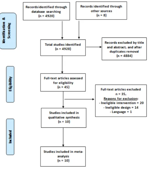
Figure 2. Flow of studies through the review ... 28
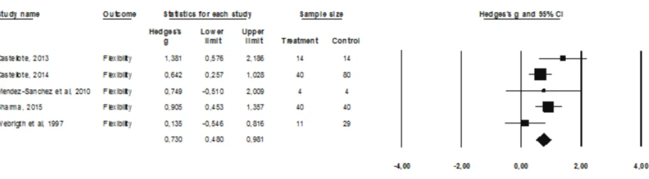
Figure 3 Forest plot of NM effects on lower limb flexibility ... 34
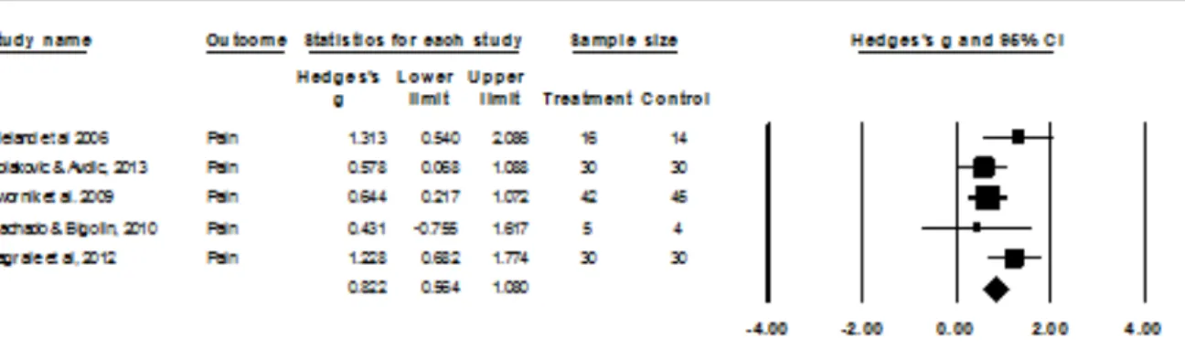
Figure 4. Forest plot of NM effects on Pain ... 35
Figure 5. Forest plot of NM effects on Disability ... 36
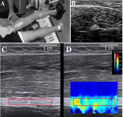
Figure 6. (A) Testing setup. B-mode sonograms of sciatic nerve in (B) cross-sectional and (C) longitudinal views. The (D) elastogram window was defined over the nerve section, and the largest area within the epineurium was considered as region of interest ... 49
Figure 7. Slump test position ... 51
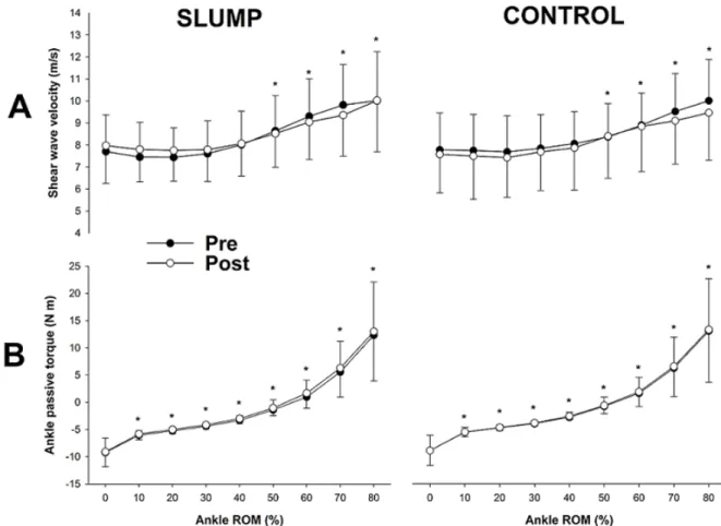
Figure 8. (A) Shear wave velocity of the sciatic nerve and (B) ankle passive torque during the ankle passive rotation from 40° of plantar flexion (0%) to 80% of maximal dorsiflexion range of motion (ROM) before (pre) and after (post) the slump and control interventions ... 54
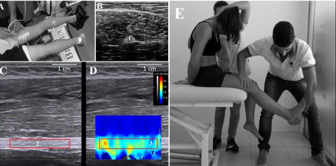
Figure 9. A – Experimental setup; B – Cross-sectional view of the sciatic nerve (1) in B-mode; C – Longitudinal view of the sciatic nerve in B-mode; D – Elastogram window was defined above the nerve section, and the largest area within the epineurium was considered as region of interest (2) and (3); E – Example of the intervention in a slump position. ... 67
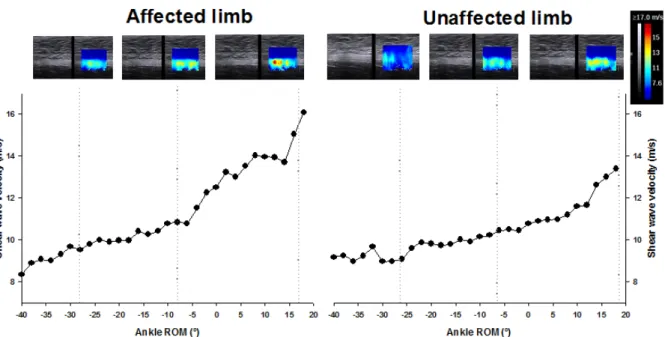
Figure 10. Sciatic shear wave velocity response during an ankle dorsiflexion, in both the affected (left image) and unaffected (right image) limbs of one participant (#7) with low back related leg pain. For each graphic examples of the elastogram are provided for 3 different amplitudes, 30° and 10° of plantarflexion, and 15° of dorsiflexion. ... 70
Figure 11. Between-groups and within-subjects comparisons of the sciatic shear wave velocity (mean ± SD) throughout the ankle motion percentiles for the pre-intervention condition. Significant differences were found between the affected and unaffected limbs of the LBRLP group. ... 71
Figure 12. Sciatic shear wave velocity (mean ± SD) before (PRE) and after (POS) the
xii
were found between the PRE and POS shear wave velocity measurements, in the LBRLP group. ... 72
xiii
LIST OF TABLES
Table 1. PEDro scores of the included studies ... 30
Table 2. Characteristics of the included RCTs on healthy populations ... 31
Table 3. Characteristics of the included RCTs on people with LBP ... 32
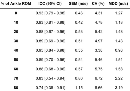
Table 4. Intra-rater reliability values of the sciatic nerve shear wave velocity, for every 10% interval of the total ankle range of motion. ... 53
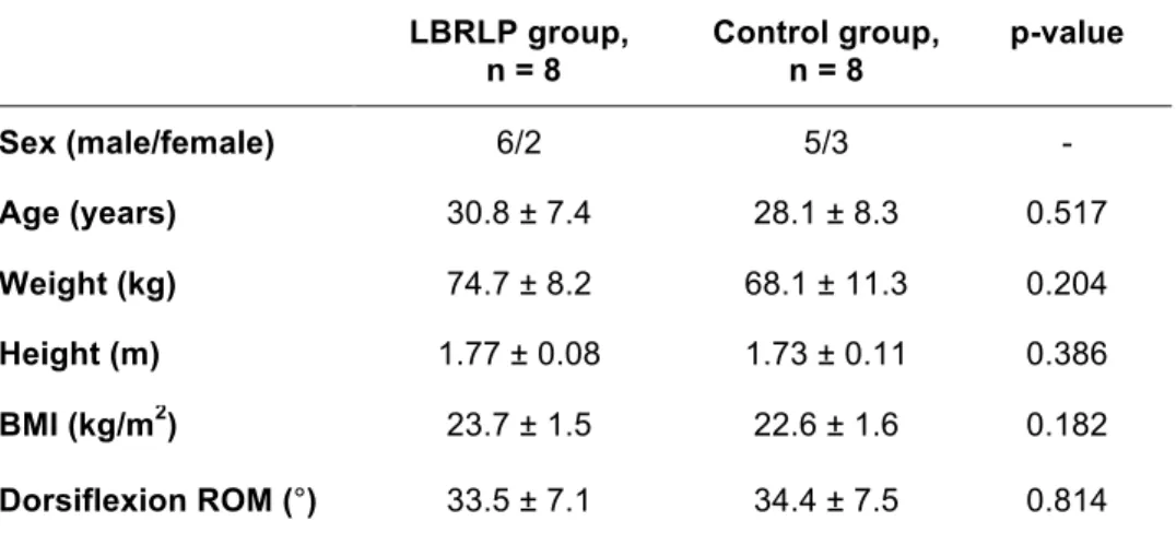
Table 5. Demographic variables from LBRLP and healthy participants ... 69
Table 6. Clinical variables of the participants with LBRLP (n=8) ... 69
Table 7. Reproducibility of the SWV measurements in LBRLP people ... 79
Table 8. Within-group, and between-groups, difference of sciatic SWV before the intervention, throughout the ankle ROM percentiles ... 80
Table 9. Sciatic shear wave velocity, before (PRE) and after (POS) the intervention, in both groups, and associated effect size ... 81
xiv
xv
LIST OF ABBREVIATIONS
AKE – Active Knee Extension Test BMI – Body Mass Index
CI – Confidence intervals CSA – Cross Sectional Area CTS – Carpal Tunnel Syndrome CV – Coefficient of Variation EMG – Electromyography
ICC – Intraclass Correlation Coefficient LBP – Low Back Pain
LBRLP – Low Back Related Leg Pain MDD – Minimal Detectable Difference
MIVC – Maximal Isometric Voluntary Contraction NM – Neural Mobilization
ODI – Oswestry Disability Index RCT – Randomized Controlled Trials ROM – Range of Motion
RMDQ – Roland Morris Disability Questionnaire SD – Standard Deviation
SEM – Standard Error of Measurement SLR – Straight Leg Raise
SWE – Shear Wave Elastography SWV – Shear Wave Velocity SSI – Supersonic Shear Imaging ULTT – Upper Limb Tension Test
US – Ultrasonography/Ultrasonographic/Ultrasound VAS – Visual Analogic Scale
xvi
1
CHAPTER I - Introduction
Neurodynamics techniques are frequently used to evaluate the peripheral nerves (Shacklock, 1995), both in the upper and the lower body quadrant. Neurodynamics uses a combination of movements that gradually load the peripheral nerve until the subject reaches his maximal range of motion (ROM) (Butler, 2000) in order to assess the nerve mechanosensitivity (Boyd, Wanek, Gray, & Topp, 2009).
Besides being used as an assessment tool, neurodynamics techniques, which can be grouped as neural tension, or neural gliding techniques (Shacklock, 1995), are also used with a rehabilitation purpose (e.g. in people with peripheral neuropathy) (Oskay et al., 2010), and as a training method in healthy people (e.g. for flexibility improvements) (Castellote-Caballero, Valenza, Puentedura, Fernández-de-Las-Peñas, & Alburquerque-Sendín, 2014). In the past years, a considerable number of studies have been published reporting the effects of neurodynamics interventions (Kitteringham & Christine, 1996; Scrimshaw & Maher, 2001; Tal-Akabi & Rushton, 2000).
While the majority of the studies using neurodynamics techniques aimed at the upper body quadrant report positive effects (Akalin et al., 2002; Coppieters, Stappaerts, Wouters, & Janssens, 2003; Nee, Vicenzino, Jull, Cleland, & Coppieters, 2012), the same has not been reported for the lower body quadrant. The reasons for this lack of evidence are related to the few number of studies, and also to conflicting results. Some studies report effects of neurodynamics techniques superior to the control, or minimal interventions, in both healthy (Castellote-Caballero et al., 2014) and clinical populations (i.e. mainly in people with low back pain - LBP) (Cleland, Childs, Palmer, & Eberhart, 2006; Nagrale, Patil, Gandhi, & Learman, 2012). On the other hand, some studies concluded that neurodynamics interventions were not superior to the control, or minimal interventions, in both populations (Scrimshaw & Maher, 2001; Webright, Randolph, & Perrin, 1997).
Thus, it is important to determine the evidence associated with the effects of neurodynamics techniques applied to the lower body quadrant, in either healthy individuals or clinical populations, particularly people with LBP, which is one of the most common problems affecting the lower body quadrant (Hoy et al., 2012).
In addition there is also a lack of knowledge surrounding the mechanical and physiological effects that neurodynamics, mainly neural tension, immediately produces following its application. Several theories can be found in the literature related, for instance, to pain mechanisms (Dilley, Lynn, & Pang, 2005) or to intraneural fluid
2
dispersion (Gilbert et al., 2015). However, these are mere hypothesis driven by investigations in human cadavers or in animal models. There are few studies that analyze, in vivo, the acute effects of neurodynamics maneuvers. In the recent years the mechanical properties of the peripheral nerves (e.g. neural strain, or excursion) have been fairly examined, mostly in animal models (Mason & Phillips, 2011; Phillips, Smit, De Zoysa, Afoke, & Brown, 2004) and in human cadavers (Boyd, Topp, & Coppieters, 2013; Coppieters et al., 2006), but also using ultrasonography (US) brightness-mode imaging (Carroll, Matthew, Janet, Keith, & Wayne, 2012; Hough, Moore, & Jones, 2000). In fact, US allows to measure peripheral nerves’ morphology [e.g. cross-sectional area (CSA)] (Bathala, Kumar, Kumar, Shaik, & Visser, 2014), and to quantify neural excursion in different planes (Ellis, Hing, Dilley, & McNair, 2008), but these studies fail to provide direct evidence about the nature and magnitude of the forces that act upon the human peripheral nerves. This has recently been possible, since shear wave elastography (SWE) was developed. This technique measures shear wave velocity (SWV) within soft tissues, which in turn corresponds to the tissues’ shear modulus (Drakonaki, Allen, & Wilson, 2012). Briefly, SWE transmits ultrasound waves to interact with the tissues. In response, shear waves will be produced, and their velocity can be measured and used to estimate the stiffness of tissues (Bercoff, Tanter, & Fink, 2004). Recently, SWE was used with the objective of estimate the stiffness of peripheral nerves, such as the median (Kantarci et al., 2014), tibial (Greening & Dilley, 2016), and sciatic (Andrade et al., 2016) nerves. Moderate (Greening & Dilley, 2016) to excellent (Andrade et al., 2016) reliability values have been reported for SWV measurements, which support the use of this method to assess the mechanical properties of peripheral nerves. Consequently, it would be relevant to use SWE to determine the acute effects of neural tension in the stiffness of the sciatic nerve, both in healthy and clinical populations.
Additionally, SWE has also been used to estimate the stiffness of nerves in people with several neuropathies, such as carpal tunnel syndrome (Kantarci et al., 2014), or diabetic neuropathy (Dikici et al., 2016). Yet, there is no information about the stiffness of the sciatic nerve in people with low back related leg pain (LBRLP), often termed
sciatica, a very common neuromuscular disorder that reaches lifetime prevalence rates
up to 43% (Konstantinou & Dunn, 2008). Hence, it would be important to determine if there are changes in the sciatic nerve stiffness in people with LBRLP.
3
Objectives and Hypothesis
The main purpose of this thesis was to investigate the immediate changes to sciatic nerve stiffness after a slump mobilisation technique in healthy participants and in those with LBRLP. In order to reach this primary objective, three studies were carried out, each one with its specific goals, but all following a logic sequence, and dependent from each other.
The study 1 was conducted with the objectives of:
1) Systematically evaluate the current body of evidence into the effectiveness of neurodynamic mobilizations applied to the lower body quadrant, in healthy people and in people with LBP;
2) Determine the most used parameters for neurodynamics protocols.
In order to meet with these purposes we performed a systematic review, with meta-analysis, of the effects of neurodynamics in healthy individuals and in people with LBP. Considering the lack of evidence regarding the effects of neurodyanimcs, in particular targeting the lower body quadrant, we searched the literature for randomized controlled trials (RCT) which used any form of neurodynamics aimed to the lower body quadrant. We also used the systematic review to determine the most used parameters in neurodynamics interventions, related for instance with the number and frequency of sessions, duration of the intervention, and type of technique used. This information was later used in the second study to establish the experimental protocol.
The hypothesis formulated for this first study was that the meta-analysis would yield significant effect sizes favoring the use of neurodynamics techniques in both healthy individuals and in people with LBP.
The study 2 was performed to meet with the following objectives:
1) To determine the immediate effects of a slump mobilization technique in the sciatic nerve stiffness of healthy individuals;
2) To ascertain the reproducibility of the sciatic stiffness assessments;
3) To establish the reliability of the experimental setup in order to replicate it in clinical populations
This second study had a quasi-experimental design, and it was conducted in healthy participants. Considering the lack of knowledge regarding the mechanical effects of
4
neurodynamics techniques in human nerves, we measured the sciatic nerve stiffness (i.e. estimated through the measurement of SWV) before, and immediately after, a neural tension intervention. The parameters of the neural tension intervention were selected accordingly to the findings of the study 1. Consequently, a sustained slump position was used to induce neural tension in the sciatic nerve. In addition, reproducibility of the measurements was determined by performing two SWV assessments (1 min between measurements) in each limb.
The following hypotheses were tested in this study:
a) A sustained slump position would lead to an acute decrease in the sciatic nerve stiffness, in healthy participants;
b) The measurements of SWE would yield substantial intra-rater reproducibility
, Finally, the study 3 aimed to address the following objectives:
1) To measure the sciatic nerve stiffness in people with unilateral LBRLP, and compare it between limbs;
2) To compare the sciatic nerve stiffness between healthy individuals and people with LBRLP;
3) To determine the acute effects of neural tension in the sciatic nerve stiffness of people with LBRLP
Similarly to the second study, this one was also a quasi-experimental study. Given that the literature shows changes to the median and tibial nerves stiffness as a consequence of several neuropathies, this study intended to analyze if the sciatic nerve stiffness was also altered in people with unilateral LBRLP. The experimental protocol used and validated in the second study for the sciatic SWV assessment was replicated in this one. Moreover, the acute effects of neural tension were also determined in this population, being the affected limb subjected to the intervention, while the unaffected limb served as control.
Three hypotheses were formulated for this study, as follows:
a) The affected limb of people with unilateral LBRLP would present higher stiffness than the unaffected limb;
b) The affected limb of people with unilateral LBRLP would present higher stiffness when compared to either limb of healthy controls;
c) A neural tension intervention would lead to an acute decrease in the sciatic nerve stiffness, in people with LBRLP.
5
Structure of the thesis
This thesis follows a study compilation organization, and is presented as follows:
● Chapter I, shows a Introduction, where the problem of the investigation is identified, and the objectives are presented;
● Chapter II, shows a Review of the Literature, where the main concepts are defined and explained. The topics related to the neurodynamics, peripheral nervous system, and shear wave elastography are explored and detailed to the reader;
● Chapters III to V, show, respectively, the three studies mentioned above. These chapters share the same organization: Introduction, Methods, Results, Discussion, Conclusions, and References;
● Chapter VI and VII, shows the General Discussion and Conclusions, respectively, where we establish a link between the 3 studies presented, and the Introduction. A summary of the main findings observed in the studies is performed, followed by a rationale of how these results can be useful for clinicians in their practice. The limitations of the thesis are also discussed in this chapter, as well as the recommendations for future research. The final considerations regarding this thesis are presented in the Conclusions section; ● And, Chapter VIII, which represents the list of References used in the chapters
I, II, and VI. This chapter does not include the references used in the studies, given that each one of them has its own reference list.
6
7
CHAPTER II - Review of the Literature
1. Peripheral nerve anatomy
Peripheral nerves have a fascicular organization, meaning that axons are bundled together along the nerve’s length (Topp & Boyd, 2012). Protecting the nerve there are 3 different layers of connective tissue: the endoneurium, the perineurium, and the epineurium (Topp & Boyd, 2006). The endoneurium is the innermost connective tissue, and is found between individual nerve fibers; the perineurium bundles nerve fibers into a fascicle, and provides mechanical strength to the nerve, being reinforced where the nerve crosses joints (Lowry, Wilcox, Masson, & Williams, 1997); the epineurium is the outermost layer, and holds fascicles together to form a nerve trunk. When the nerve has more than one fascicle the epineurium is divided into epifascicular and interfascicular (Stolinski, 1995). The first forms an interface between the nerve and their surrounding tissues, whereas the interfascicular epineurium allows gliding between nerve fascicles (Stolinski, 1995).
The blood supply to the peripheral nerves is assured by arterioles and venules that run across the length of the nerve, within the epineurial space (Olsson, 1990). From here, the vessels pass obliquely into the perineurial compartment, transporting perineural cells (Olsson, 1990). The amount of blood vessels is not similar throughout the nerve. The density of capillaries is higher in the regions with larger metabolic needs, such as the dorsal root ganglia, and smaller in the endoneurial space of the peripheral nerve (Bell & Weddell, 1984).
The interface formed between the blood vessels and the nerve, known as the blood-nerve barrier, is of major importance. Intact endoneurial capillaries help to regulate an adequate intraneural pressure, counteracting the oncotic pressure from surrounding tissues (Sunderland, 1978). If this equilibrium is compromised, changes to intraneural blood flow will occur, causing for instance ischemia, or an accumulation of intraneural fluid, which will be difficult to drain since there are no lymphatic capillaries in the endoneurial compartment (Sunderland, 1978).
1.1. Sciatic nerve anatomy
The sciatic nerve has its origin in the sacral plexus. The sacral plexus is formed by the lumbo-sacral cord, and the anterior divisions of the 3 upper sacral nerves. The upper nerves of the plexus (i.e. lumbo-sacral cord, the 2 first, and part of the third, sacral
8
nerves) will prolong into the sciatic nerve. This nerve will leave the pelvis through the great sacro-sciatic foramen, traveling along the posterior thigh covered by the long head of the biceps femoris. When the sciatic reaches the lower third of the thigh it divides into the external popliteal nerve and the internal popliteal nerve, which will distally continue as the tibial nerve (Gray, 1977). The sciatic has numerous branches responsible for innervating most of the articular and muscular structures of the lower limb, and also for the cutaneous innervation of the leg and foot (VanPutte et al., 2013). The path and territory innervated by the sciatic is directly related to the symptomatology of the people with LBRLP, which will be later described.
2. Peripheral nerves biomechanics
Peripheral nerves exhibit some biomechanical properties which enables them to withstand various mechanical stresses, imposed during joint movement (Topp & Boyd, 2006). Nerves have the ability to glide (or slide) relatively to the surrounding tissues. That ability is termed excursion, and its direction varies accordingly to the axis of rotation in the moving joint (Dilley, Lynn, Greening, & DeLeon, 2003). The nerve glides towards the moving joint (i.e. convergence) when there is elongation of the nerve bed; on the other end, when tension is released from the nerve bed the nerve will glide away from the moving joint (i.e. divergence) (Wright, Glowczewskie, Cowin, & Wheeler, 2001). Neural excursion occurs first in the segments adjacent to the moving joint, and progress distally to the axis of rotation (Wright et al., 2001). Similarly, excursion levels are greater in the adjacent segments when compared to the segments distal to the moving joint (Boyd, Puttlitz, Gan, & Topp, 2005).
Joint movement not only produces neural excursion, but also causes changes in neural strain, i.e. nerve deformation caused by longitudinal stress (Driscoll, Glasby, & Lawson, 2002). Nerves subjected to tensile stresses show a strain increase, which can be described in a stress-strain curve. The first phase of the curve is characterized by a toe region, representing the straightening of the wavy neural connective tissues following minimal stress. With gradual stress increase, neural strain will grow at a steady rate until ultimate strain is reached, causing permanent nerve deformation (Kwan, Wall, Massie, & Garfin, 1992). The slope of the stress-strain curve indicates the resistance of the nerve to deformation, known as stiffness (Topp & Boyd, 2006). Increasing neural strain will cause a phenomenon known as transverse contraction, which is a reduction in the nerve’s cross-sectional area. This results in an increase of the intraneural pressure (Millesi, Zöch, & Reihsner, 1995).
9
Neural excursion, strain, and stiffness are not homogeneous throughout the nerve’s length. An animal study analyzed the local variations in mechanical properties of the median and sciatic nerve. The authors found that both nerves show greater strain in the joint regions, while stiffness values were significantly smaller, compared to the non-joint regions (Phillips et al., 2004). In addition, it is known that nerve stiffness is dependent from the elongation velocity (Driscoll et al., 2002), which is consistent with a viscoelastic and time-dependent behavior. As seen in the muscle-tendon unit (Kubo, Kanehisa, & Fukunaga, 2002; Magnusson, Simonsen, Aagaard, & Kjaer, 1996; Mizuno, Matsumoto, & Umemura, 2013), nerves also present stress relaxation and creep (Driscoll et al., 2002). This means that when nerves are stretched and maintained in an elongated position, their stiffness will decrease, and their length will increase. Interestingly, it was found, in animals, that stress relaxation is greater when smaller elongation rates are applied, and when performed at slower velocities. However, we must take into consideration that these findings were mostly observed in animal studies, and in cadaveric investigations. The living tissues surrounding peripheral nerves may have a strong influence on the nerve biomechanics, and more information from studies in vivo is necessary.
2.1. Biomechanics of the sciatic nerve in people with low back related leg pain
Low back pain is one of the most common musculoskeletal disorders, with lifetime prevalence over 70% (Burton et al., 2006). It is defined as the presence of pain and discomfort below the costal margin and above the gluteal fold (Koes, 2006). Frequently, LBP is accompanied by irradiating symptoms, such as pain or numbness, along the regions innervated by the sciatic nerve and their nerve roots, which is characteristic of LBRLP (Bogduk, 2009). Several conditions are linked with the development of radicular symptoms, mainly herniated disks, but also spinal stenosis, spondylolisthesis (Schoenfeld, Laughlin, Bader, & Bono, 2012), or nerve root inflammation (Bogduk, 2009). Low back related leg pain is usually unilateral, and involves irradiating pain that travels dorsolaterally in the thigh when there is compression of the L5 nerve root, and posteriorly when the S1 nerve root is compressed. Pain is anterolateral in the thigh following L4 nerve root compression (Ropper & Zafonte, 2015). Additionally, muscle weakness and reflex changes may also be present in lumbar radiculopathies, mainly in L4/L5 nerve roots compression (Konstantinou & Dunn, 2008).
10
Information is scarce concerning the mechanical behaviour of the peripheral nerves in people with LBRLP. Recently, studies analysing people with unilateral radiculopathy have observed an increased cross-sectional area of the affected sciatic nerve in comparison to the unaffected sciatic (Frost & Brown, 2016; Kara et al., 2012). Another study showed that the transverse displacement direction of the sciatic nerve was altered in people with LBRLP (Ridehalgh, Moore, & Hough, 2015). Despite these changes found, it is unknown to this date, if the sciatic nerve in people with LBRLP has any change to its mechanical properties, mainly stiffness.
Next, we will characterize the neurodynamics techniques, which are amongst the most used interventions to evaluate and treat people with LBRLP. Its various effects on the peripheral nerves will also be presented.
3. Neurodynamics: Definition and types of techniques
The nervous system has mechanical and physiological functions which interact closely, being dynamically interdependent. The concept of Neurodynamics joins the mechanical and the physiological components of the nervous system (Shacklock, 1995). A change in one or in both functions will cause impairment for proper neurodynamics (Shacklock, 2014). For instance, inadequate and constant mechanical stresses (e.g. excessive strain, or compressive forces) applied to the peripheral nerves will result in intraneural blood flow changes, inflammation, and mechanosensitivity (Shacklock, 2014). On the other hand, physiological changes as the one seen in diabetic patients that develop distal symmetric polyneuropathy, causes an increase in the nerve’s CSA, which will compromise its mechanical properties (i.e. longitudinal excursion) (Boyd & Andrew, 2014).
Neurodynamics interventions include several techniques used for assessment and treatment of peripheral nervous system related problems (Butler, 2000). These techniques use a combination of joint movements in order to promote neural gliding and neural tension. Exercises aimed for neural gliding induce excursion of the nerve relative to its surrounding structures, by elongating the nerve bed at one end and reducing it at the other end; neural tension exercises promote nerve elongation at both ends, which results in an increase of neural strain (Coppieters & Butler, 2008).
The most common neurodynamics techniques for the upper body quadrant include the Upper Limb Tension tests, which target the peripheral nerves from the brachial plexus (e.g. median, ulnar, or radial nerves) (Kleinrensink et al., 2000). Regarding the lower body quadrant, the Straight Leg Raise (SLR) test, and the Slump test, are the most
11
common techniques applied. The SLR test consists in a passive hip flexion movement, while the knee remains in full extension, performed in supine (Breig & Troup, 1979). This test is traditionally applied to evaluate lumbosacral nerve roots disorders, with some variations being used as sensitizing, or differentiation, maneuvers (e.g. medial hip rotation, or ankle dorsiflexion) to distinguish the symptoms originated in neural and non-neural structures (Boyd et al., 2009).
The slump test is used to determine the relationship between the patient’s symptoms and the restriction of movement of the pain-sensitive structures (Maitland, 1979). During this test, patients are seated with the thoracolumbar spine in a slump position, and the cervical spine flexed. Afterwards, knee extension and ankle dorsiflexion are performed, adding additional mechanical stress to the nervous system. Finally, cervical extension is performed as a differentiation maneuver (Maitland, 1985). Some variations of the slump test are described, by changing the test order (Miller, 1999): ankle dorsiflexion may be applied prior to knee extension, and a “long-sitting” position may be considered to initiate the slump test sequence.
The slump test position is also used to apply neurodynamics techniques. Some studies use a sustained slump test position to induce neural tension (Cleland et al., 2006; Nagrale et al., 2012). In this case, cervical flexion is maintained during the period of intervention, while the ankle is maximally dorsiflexed by the examiner (Cleland et al., 2006). Neural gliding exercises are also performed in a slump position (Castellote-Caballero et al., 2013). While maintaining the thoracolumbar spine slumped, patients are instructed to alternate cervical with knee movements (Sharma, Balthillaya, Rao, & Mani, 2016). The combination of cervical extension with knee extension will cause a distal excursion of the sciatic, whereas proximal neural excursion occurs during cervical flexion/knee flexion (Ellis, Hing, & McNair, 2012).
4. Effects of Neurodynamics
The effects of neurodynamics are reported in the literature both in clinical (Colakovic & Avdic, 2013; Scrimshaw & Maher, 2001) and healthy populations (Méndez-Sánchez et al., 2010; Sharma et al., 2016). This means that these techniques are used either to rehabilitate neuromuscular disorders (Allison, Nagy, & Hall, 2002; Cleland et al., 2006), or to enhance physical performance, like joint flexibility (Castellote-Caballero et al., 2013). A systematic review determined that 8 out of 11 studies reported positive effects from the use of neurodynamics, mostly in upper body quadrant disorders (Ellis & Hing, 2008). More recently, a meta-analysis determined moderate and large effect sizes
12
favoring the use of neurodynamics techniques in pain relief and disability improvements, respectively (Su & Lim, 2015). Again, these findings were mostly retrieved from studies in the upper body quadrant.
As with clinical populations, the effects of neurodynamics in healthy individuals are positive, resulting in acute lower limb flexibility improvements (Sharma et al., 2016), even when compared to performing static stretching, a method with well-known results in increasing flexibility (Castellote-Caballero et al., 2014; Méndez-Sánchez et al., 2010).
The reasons for the clinical effects of neurodynamics in pain and disability, and in flexibility, are not entirely known. Several physiological and mechanical explanations are proposed, that will be detailed.
4.1. Effects on blood flow
As discussed earlier, the peripheral nerves have a network of blood vessels present in every layer of its fascicular organization. When a longitudinal force is applied to the nerve, it will begin to elongate and to strain. Consequently, the blood vessels will also elongate (Shacklock, 1995). This happens within a physiological range (i.e. 6% to 8%), during joint motion (Topp & Boyd, 2006). Animal studies showed that with 8% strain venous return starts to decline, and by 15% there is a complete occlusion in blood flow (Lundborg & Rydevik, 1973; Ogata & Naito, 1986). In addition, data from a cadaveric study revealed that a position similar to the upper limb tension test for the median nerve (i.e. shoulder abduction and external rotation + forearm supination + elbow, wrist, and fingers extension) resulted in 7.6 ± 8.2 % strain in the median nerve (Byl, Puttlitz, Byl, Lotz, & Topp, 2002). The same position, but using elbow flexion and forearm pronation, resulted in strain levels of 9.9 ± 10.9 % for the ulnar nerve (Byl et al., 2002). Similar strain values were found for the sciatic nerve, during the SLR maneuver in human cadavers (Coppieters et al., 2006).
Apparently, these strain values may be superior to the ones indicated for total blood flow occlusion. Following a constant compressive or tensile force applied to the nerve, the resulting blood flow restriction may cause endoneurial edema and fibrosis (Rydevik & Lundborg, 1977).
Accordingly to Wang et al. (2015), neurodynamics favors peripheral nerve regeneration. These techniques facilitate intraneural blood flow, which will improve the availability of oxygen and nutrients to the neural tissue (Wang et al., 2015). Previously to this investigation, a cadaveric study described the effects of neural mobilization as a
13
“pumping action” (Brown et al., 2011). In this study, Brown et al (2011) used repetitive
passive ankle mobilization, which caused an intermittent change in the tibial nerve internal pressure (Brown et al., 2011). This pumping effect may have a direct influence on intraneural blood flow, favoring fluid dispersion, thereby preventing the deposition of mechanosensitivity elements (Brown et al., 2011).
4.2. Effects on pain/mechanosensitivity
Mechanosensitivity is defined as the generation of pain impulses as a consequence of mechanical stimulus (e.g. compressive or tensile stresses) applied to neural structures (Shacklock, 1995). It can affect both myelinated and unmyelinated fibers (Dilley et al., 2005), and is caused by the accumulation of mechanically sensitive elements (e.g. colchicine). This leads to a disruption in axoplasmic transport (Dilley & Bove, 2008), and consequently to the development of local mechanosensitivity.
Neurodynamics maneuvers aim to assess the mechanosensitivity associated with range of motion restriction (Butler, 2000), that was recently found in patients with CTS (Jaberzadeh & Zoghi, 2013) and diabetic polyneuropathy (Boyd, Wanek, Gray, & Topp, 2010). Consequently, neurodynamics interventions have the purpose of restoring normal nerve mechanosensitivity, enabling pain free ROM. Several studies report positive effects of neurodynamics techniques on pain relief, either in upper body (Colakovic & Avdic, 2013; Nee et al., 2012; Pinar, Enhos, Ada, & Güngör, 2005), or lower body (Nagrale et al., 2012) quadrant disorders. The physiological reasons for this benefit of neurodynamics are probably related with hypoalgesic effects, already shown in healthy subjects (Beltran-Alacreu, Jiménez-Sanz, Fernández Carnero, & La Touche, 2015). Beltran et al. (2015) compared the immediate mechanical hypoalgesic effects of neurodynamics, with a placebo intervention, in healthy individuals. They found an immediate widespread hypoalgesic effect, following 7 min of neural gliding or neural tensioning maneuvers (Beltran-Alacreu et al., 2015). A recent study, in animals, also showed the effect of neurodynamics techniques in reducing hyperalgesia (Santos et al., 2012). This effect on pain sensation is probably a consequence of an inhibition of temporal summation, as a result of central sensitization in the dorsal horn of the spinal cord (Staud, Price, Robinson, Mauderli, & Vierck, 2004; Woolf, 2011)
14
4.3. Mechanical effects
As described earlier, peripheral nerves exhibit viscoelastic properties which enable them to glide and elongate, in order to adapt to human movement (Topp & Boyd, 2006). These characteristics have been extensively studied in animal (Kwan et al., 1992; Mason & Phillips, 2011; Wall, Kwan, Rydevik, Woo, & Garfin, 1991) and cadaveric (Boyd et al., 2013; Byl et al., 2002) research, but only recently studies in vivo analyzed neural excursion and strain of the peripheral nerves (Carroll et al., 2012; Coppieters, Hough, & Dilley, 2009; Shum, Attenborough, Marsden, & Hough, 2013). A recent systematic review (Silva et al., 2014) analyzed studies in vivo that quantified neural excursion and strain. It was concluded that the sciatic nerve has, in average, 3.5 mm of longitudinal excursion, when alternate movements are used in the cervical spine and knee joint, in a slump position. These results are quite different from the ones determined in cadaveric studies, where values up to 28 mm (Coppieters et al., 2006) are reported. This illustrates the influence that the tissues surrounding the nerves have on their mechanical properties.
One of the main objectives in using neurodynamics is to restore proper nerve biomechanics (Butler, 2000), despite the lack of evidence of such effect. Literature shows that peripheral nerves mechanical properties are altered in some pathological conditions. It was determined that patients with carpal tunnel syndrome have a reduction of the longitudinal excursion in the median nerve, when compared to healthy controls (Hough, Moore, & Jones, 2007). In addition, it was also found that the median nerve CSA (Lopes, Lawson, Scott, & Keir, 2011) was increased, and its transverse movement was reduced (van Doesburg, Henderson, Mink van der Molen, An, & Amadio, 2012), in people with CTS.
Regarding the lower body quadrant, the sciatic nerve showed less transverse movement, but not longitudinal, in people with spinally referred leg pain (Ridehalgh et al., 2015), while the tibial nerve presented an increased CSA and smaller longitudinal excursion, in people with diabetic polyneuropathy (Boyd & Andrew, 2014).
To date, we know of no studies that assessed the long-term effects of a neurodynamics intervention on the restoration of altered nerve biomechanics. Nevertheless, the immediate impact of neurodynamics on the excursion and strain is known, as well as the influence of the movement sequence during the technique (Boyd et al., 2013; Nee, Yang, Liang, Tseng, & Coppieters, 2010). Using human cadavers, Boyd et al (2013) studied 2 sequences of SLR: a) performing the SLR maneuver with hip flexion followed by ankle dorsiflexion; and b) invert the sequence, performing ankle dorsiflexion before
15
hip flexion. It was observed that, at the knee region, there were no differences between the 2 sequences in maximal strain or excursion of the sciatic nerve. Additionally, it was found that strain and excursion increased earlier, and maintained increased for longer periods, in the regions closest to the moving joint (Boyd et al., 2013). This follows the convergence phenomenon referred earlier, where a nerve under tensile stress tends to glide towards the moving joint (Wright et al., 2001).
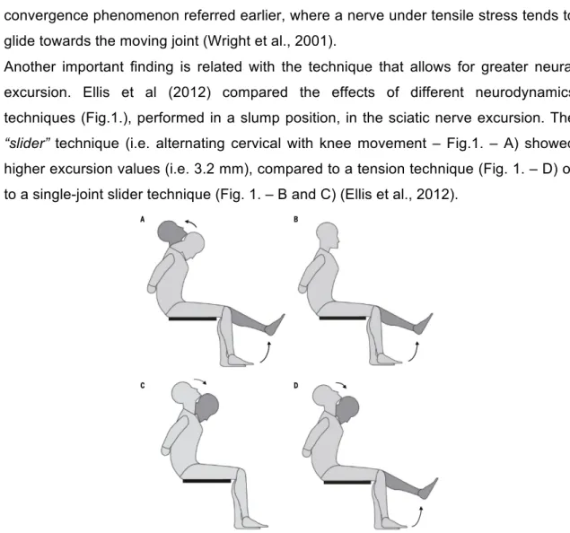
Another important finding is related with the technique that allows for greater neural excursion. Ellis et al (2012) compared the effects of different neurodynamics techniques (Fig.1.), performed in a slump position, in the sciatic nerve excursion. The
“slider” technique (i.e. alternating cervical with knee movement – Fig.1. – A) showed
higher excursion values (i.e. 3.2 mm), compared to a tension technique (Fig. 1. – D) or to a single-joint slider technique (Fig. 1. – B and C) (Ellis et al., 2012).
Figure 1. Four examples of neurodynamics techniques used in a slump position: A – slider
technique, alternating cervical movement with knee movement; B – single joint slider technique (knee); C – single joint slider technique (cervical); D – tensioner technique. Image retrieved from Ellis et al. (2012).
These findings are relevant to a clinical context given that provide valuable information regarding the neurodynamics parameters which have more impact on the nerve biomechanics. However, information is still scarce concerning the magnitude and effect of the forces (e.g. stiffness) transmitted to the neural tissue during and after neurodynamics techniques.
16
5. Ultrasonography in the assessment of peripheral nerves characteristics
Ultrasonographic imaging made it possible to measure in vivo the motion of soft tissues, in alternative to more expensive technologies such as magnetic resonance imaging (Hough et al., 2000). Using the brightness mode (i.e. B-mode) it is possible to have a continuous real-time image, displayed in a grey-scale image (Dilley, Greening, Lynn, Leary, & Morris, 2001). By comparing two consecutive images, distances and displacements between structures can be determined (Hough et al., 2000). This method was used by Fukunaga et al (1996) to measure the tendinous movement of the tibialis anterior muscle during voluntary contractions (Fukunaga et al., 1996). With a similar method the transverse motion of the median nerve was also measured (Nakamichi & Tachibana, 1992). However, the longitudinal motion of peripheral nerves only became possible to be determined with the advances in speckle tracking. This method recognizes and tracks two different points in the B-mode picture (Meunier, 1998), enabling to quantify their displacement in two consecutive images (Anderson & McDicken, 1999). Another important development to assess nerve excursion was the frame-by-frame cross correlation method (Dilley et al., 2001). Dilley et al (2001) developed an algorithm which quantifies the motion between the selected regions of interest, in two consecutive frames of the image sequence (Dilley et al., 2001).
The combination of high-frequency US imaging with the cross correlation analysis, allowed the estimation in vivo of the longitudinal nerve excursion. Neurodynamics studies benefited from these technological advances, as it became possible to analyse neural biomechanics during neurodynamics techniques, such as the ULTT (Coppieters & Butler, 2008), SLR (Ridehalgh, Moore, & Hough, 2012), or Slump tests (Ellis et al., 2012). It also allowed to characterize, from a biomechanical point of view, the peripheral nerves affected by some of the most common neuropathies (Boyd & Andrew, 2014; Ridehalgh et al., 2015).
Although studies using US to measure nerve dynamics represented important information for clinicians and researchers, there are limitations associated with this method, specifically when it comes to infer about nerve stiffness. Considering a nerve being elongated in its slack length, the excursion will be considerable, while little change occurs in stiffness (Andrade et al., 2016). Thus, it is important to understand in
vivo the effects of joint motion in neural stiffness, which only recently has been possible
17
6. Elastography and the advances in measuring the stiffness of soft tissues
Elastography is a technique developed over the past 20 years, that allows to estimate the Young’s modulus which is the physical parameter equivalent to stiffness (Gennisson, Deffieux, Fink, & Tanter, 2013). This technology has been in constant improvement with the purpose of providing faster analysis at higher resolutions. Quasi-static elastography methods (e.g. strain elastography) were initially used to assess soft tissues stiffness, by applying a constant compressive force to superficial tissues (Drakonaki et al., 2012). This method has some disadvantages such as being unable to quantify the stiffness because the applied stress distribution is unknown (Brandenburg et al., 2014). In addition, strain elastography is limited to the superficial tissues given that the stress applied is operator dependent (Gennisson et al., 2013).
Instead of using an external compressive force, acoustic radiation force impulses is another elastography method that estimates the stiffness of tissues by using one focalised US beam that will cause tissue displacement (Drakonaki et al., 2012). The transducer will detect and follow the resultant displacement (i.e. by speckle tracking), allowing the reconstruction qualitative maps of tissue stiffness (Gennisson et al., 2013). Although this method is more reliable than strain elastography, and allows for the assessment of deeper tissues, it still does not provide quantitative data on tissue stiffness (Brandenburg et al., 2014).
6.1. Shear wave elastography
Shear wave elastography is a method that enables to quantitatively measure tissue stiffness (Bercoff et al., 2004). Shear waves are the product of tissue displacement following US push beams (Brandenburg et al., 2014; Drakonaki et al., 2012), and their velocity is correlated with the stiffness of tissues (Brandenburg et al., 2014).
Supersonic shear imaging (SSI) is perhaps the state of the art of SWE techniques. Developed by the Institute Langevin SSI uses ultrafast imaging (i.e. up to 30.000 images per second) to obtain a full acquisition all at once, allowing for higher image quality at real time, in only few milliseconds (Gennisson et al., 2013). Supersonic shear imaging has been used in the past years to assess the stiffness of several organs, with good reproducibility (Cosgrove et al., 2012) and specificity (Berg et al., 2012).
Recently, SWE has been used in musculoskeletal applications, especially following the determination of excellent correlation between shear modulus and muscle passive tension (Eby et al., 2013; Koo, Guo, Cohen, & Parker, 2013). The validation study
18
conducted by Eby et al (2013) determined that the transducer orientation is crucial for reliable measures of shear modulus. Only a parallel orientation of the transducer in relation to the muscle fibers showed good correlation with the muscle passive tension. When the transducer was perpendicular, or at 45º, with the muscle fibers, there was poor correlation between the shear modulus and passive tension (Eby et al., 2013). As a consequence of these validity and reliability studies, SWE, and in particularly SSI, started to be the chosen method of several investigations regarding muscle-tendon stiffness assessment, mainly: effects of stretching in muscle shear modulus (Freitas, Andrade, Antoine, Bruno, & Pedro, 2016; Freitas, Andrade, Larcoupaille, Mil-homens, & Nordez, 2015; Koo, Guo, Cohen, & Parker, 2014; Le Sant, Ates, Brasseur, & Nordez, 2015; Miyamoto, Hirata, & Kanehisa, 2015; Nakamura et al., 2014; Umegaki et al., 2015); determination of muscle shear modulus during muscle contraction (Ateş et al., 2015; Muraki et al., 2015; Raiteri, Hug, Cresswell, & Lichtwark, 2016; Yoshitake, Takai, Kanehisa, & Shinohara, 2014) assessment of tendon shear modulus (Cortes, Suydam, Silbernagel, Buchanan, & Elliott, 2015; Fu, Cui, He, & Sun, 2016; Rosskopf et al., 2016; Slane, Martin, DeWall, Thelen, & Lee, 2016); and even the stiffness of the shoulder joint capsule (Takenaga et al., 2015). Shear wave elastography has also been used to study a number of clinical conditions (Dirrichs et al., 2016; Lee et al., 2014; Leong, Hug, & Fu, 2016) with the purpose of characterizing the affected structure regarding its stiffness, and to establish cut-off values for distinguish between physiological or pathological conditions. This may be important not only for injury prevention, but also as a guide for the rehabilitation process.
6.2. Shear wave elastography in the assessment of peripheral nerves
There is little doubt that tendons, and especially muscles, are the focus of the majority of published studies using SWE. To date, there are still few studies that use SWE to assess peripheral nerves. Not due to its lack of relevance or interest, but because it is more difficult to measure nerves. A recent study used US to measure several nerves CSA and observed that the median nerve has an average CSA of 11 mm2 measured in the carpal tunnel; the tibial nerve, at the popliteal fossa, has 33 mm2 ; and the sciatic nerve has 59.5 mm2, measured below the gluteal fold (Jang, Cho, Yang, Seok, & Kim, 2014). To better understand how nerves are thin, the tendon of the tibialis anterior muscle, close to its distal insertion, has 26.4 mm2 (Morales-Orcajo, Becerro de Bengoa Vallejo, Losa Iglesias, & Bayod, 2016), which is similar to the tibial nerve; the patellar tendon has 90 mm2 (Wiesinger, Rieder, Kösters, Müller, & Seynnes, 2016), which
19
represents almost twice the sciatic nerve, and the tibialis anterior muscle has a CSA of 777 mm2 (Maddocks et al., 2014), 13 times the sciatic CSA. Therefore, even the larger nerve is a relatively thin structure, which together with its depth (i.e. approximately 4-5 cm) makes it difficult to measure, especially in a longitudinal view.
Nevertheless, some studies have recently been published using SWE to analyze the peripheral nerves stiffness, mainly the median (Greening & Dilley, 2016), tibial (Dikici et al., 2016), and sciatic (Andrade et al., 2016) nerves. Kantarci et al (2014) used SSI to measure the stiffness of the median nerve in people with CTS, in a resting condition (Kantarci et al., 2014). They found that people with CTS had higher median nerve stiffness when compared to healthy controls (Kantarci et al., 2014). The stiffness of the median nerve was also measured by Greening & Dilley, (2016), in different postures of the ULTT, in healthy participants. These assessments were also performed in a resting condition, and showed a significant increase in the nerve stiffness as the limb was moved into positions that elongate the nerve bed (Greening & Dilley, 2016). These authors reached the same conclusion for the tibial nerve, during a SLR maneuver (Greening & Dilley, 2016). Recently, Dikici et al (2016) studied the tibial nerve stiffness of people with diabetic neuropathy, also in a resting condition (i.e. supine, with the foot relaxed in slight plantar flexion), and concluded that it was significantly increased comparing to healthy controls (Dikici et al., 2016).
The first study, to our knowledge, that used SWE to measure a peripheral nerve during a dynamic action was performed by Andrade et al. (2016). In this study, participants laid prone, while a dynamometer passively rotated the ankle into dorsiflexion. This motion caused a distal excursion of the sciatic nerve, and a corresponding increase in its stiffness (Andrade et al., 2016). This investigation proved that it is reliable to non-invasively assess the sciatic nerve stiffness, in healthy participants, during a dynamic action (Andrade et al., 2016).
Following this up to date, and brief review, where the basic concepts and terminology were detailed, we will present the studies of this thesis. Each study will be presented in separate, but all will share the same organization: an Introduction stating the problem, relevance and objectives of the study; a Methods section, where the materials, experimental procedures, and statistical analysis will be detailed; a section where the main Results will be presented; and a section with the Discussion of the results found, as well as the limitations of the investigation, and recommendations for future research.
20
21
CHAPTER III -
Effects of Lower Body Quadrant Neural Mobilization in Healthy and Low Back Pain Populations: A Systematic Review and Meta-AnalysisTiago Neto, Sandro Freitas, Marta Marques, Luis Gomes, Ricardo Andrade, Raúl Oliveira
Status: Published in the Manual Therapy journal (relaunched as Musculoskeletal Science & Practice journal from January, 2017) with the DOI:
22
ABSTRACT
Background: Neural mobilization (NM) is widely used to assess and treat several neuromuscular disorders. However, information regarding the NM effects targeting the lower body quadrant is scarce. Objectives: To determine the effects of NM techniques targeting the lower body quadrant in healthy and low back pain (LBP) populations. Design: Systematic review with meta-analysis. Method: Randomized controlled trials were included if any form of NM was applied to the lower body quadrant. Pain, disability, and lower limb flexibility were the main outcomes. PEDro scale was used to assess methodological quality. Results: Forty-five studies were selected for full-text analysis, and ten were included in the meta-analysis, involving 502 participants. Overall, studies presented fair to good quality, with a mean PEDro score of 6.3 (from 4 to 8). Five studies used healthy participants, and five targeted people with LBP. A moderate effect size (g = 0.73, 95% CI: 0.48 - 0.98) was determined, favoring the use of NM to increase flexibility in healthy adults. Larger effect sizes were found for the effect of NM in pain reduction (g = 0.82, 95% CI 0.56-1.08) and disability improvement (g = 1.59, 95% CI: 1.14 - 2.03), in people with LBP. Conclusion: Evidence suggests that there are positive effects from the application of NM to the lower body quadrant. Specifically, NM shows moderate effects on flexibility in healthy participants, and large effects on pain and disability in people with LBP. Nevertheless, more studies with high methodological quality are necessary to support these conclusions.