Received on 02/09/2009. Approved on 01/11/2010. The author declares no conlict of interest.
1. Full Professor of Rheumatology of the Universidade Federal do Triângulo Mineiro (UFTM). Ad hoc medical consultant of the National Council for Scientiic and Technological Development (CNPq, from the Portuguese) (1995-2000)
Correspondence to:Hamid Alexandre Cecin. Rua Nacib Cury, 419. São Sebastião, Uberaba, MG – CEP: 38060-380. E-mail: [email protected]
Cecin’s Sign (“X” Sign): improving the diagnosis of
radicular compression by herniated lumbar disks
Hamid Alexandre Cecin1
iNTRoDUCTioN
Due to the high prevalence, negative repercussions on working capacity, and negative effects on the quality of life, disorders of the lumbar spine represent a challenge in many aspects.
Four out of ive people will have lumbar pain sometime in
their lifetime. Complete history and physical exam are still the diagnostic pillars of those disorders.1 Imaging diagnostic methods and other tests are just complementary. Approximately 120 diseases affect the lumbar spine. Herniated lumbar disk is one of them. Lumbar pain, lumbosciatalgia, cruralgia, and sciatic pain represent the main symptoms.2
Lasègue’s sign, also called “straight leg raising” in English
speaking countries, is the maneuver performed more often to
detect mechanical compression and inlammation of lumbar
nerve roots by protruding and extruding herniated disks. However, how it should be determined, its mechanism, and the true meaning of its clinical application are still surrounded
ABSTRACT
While reafirming that the clinical exam still is the best medical practice, the author has proposed a new maneuver (Cecin‘s sign or “X” sign) to help the diagnosis of herniated lumbar disk, describing its biomechanical bases. However, the diagnostic performance of this maneuver has not been formally tested. Patients and Methods: The maneuver, which consists on the lexion of the lumbar spine while simultaneously performing the Valsalva maneuver, was tested in 45 patients with typical sciatic pain and herniated lumbar disk conirmed by magnetic resonance imaging (MRI), and in 21 patients with simple mechanical back pain with normal MRI. Lasègue’s sign was investigated concomitantly and the concordance with the “X” sign was assessed. Results: Concordance between the two tests was very low (Kappa = 0.17, Kappa < 0.4) and discordance was statically signiicant (P < 0.001). The “X” sign had a sensitivity of 73.3%, speciicity of 95.2%, positive predictive level of 97.1%, and negative predictive level of 62.5% in the diagnosis of herniated lumbar disk by MRI, while Lasègue’s sign showed sensitivity of 22.2%, speciicity of 95.2% (P < 0.001), positive predictive value of 90.9%, and negative predictive value of 36.4% (P = 0.153). Conclusion: Cecin’s sign had higher sensitivity,
positive predictive value, and negative predictive value than Lasègue’s sign in the diagnosis of symptomatic herniated lumbar disk.
Keywords: low back pain, sciatic pain, herniated lumbar disk, diagnostic performance.
by divergence. Until the 1990’s, agreement on the angle of
the leg in relation to the horizontal plane that was considered positive, 70°, 60°, 45°, or less, did not exist. Nowadays, many authors admit that the maneuver can only be considered as indicative of effective root compression at a 45° angle.3,4,5 False-positive results represent other confounding factor of
Lasègue’s sign: referred pain in the path of the sciatic nerve
would result from increased tension of the thigh muscles and not radicular compression. Although it is still very important in the diagnosis of herniated lumbar disks, due to the current tendency to use the 45° angle as criterion for radicular
compression, the sensitivity and speciicity of Lasègue’s
other semiotic maneuver could minimize those divergences, increase reliability, and validate the superiority of the clinical exam in the diagnosis of symptomatic herniated lumbar disks.
In 1996, at the XX Brazilian Rheumatology Congress in Curitiba,7 the author stated, based on clinical observations,
Pascal’s principle, and torque, that, when patients with
herniated lumbar disk perform the Valsalva maneuver while
maintaining the lumbar spine lexed, this could cause greater
compression of the cauda equina and nerve roots emerging from it to form the sciatic nerve. This would result from the summation of torque and shearing forces on the disk and the pressure exerted on the cauda equina secondary to the action of those two physical principles: the moments of force, which
increase during lumbar lexion, and elevation of the pressure of
the CSF, secondary to the Valsalva maneuver.7,8 This maneuver was initially called “X” sign, since the results of a long-term clinical study were unknown. The objective of the present study was to evaluate the concordance, discordance, sensitivity,
speciicity, and positive and negative predictive values between the “X” sign and Lasègue’s sign in the diagnosis of herniated
lumbar disks, by recruiting patients from private practice. In an editorial of the Brazilian Rheumatology Journal, Dr. Fernando Neubarth suggested that the “X” sign should be
called Cecin’s sign.9
PATiENTS AND METHoDS
Test Group
Inclusion criteria
1. Patients of both genders, ages between 20 and 80 years, with lumbar pain irradiating to the buttocks, thighs, and extending below the knees, with or without motor and sensorial
deicit of the affected limb.
2. Pain should be sudden, of medium to high intensity –
from 6 to 9 in the visual analogue scale- supericial and well-deined, similar to an “electrical shock”. Irradiation should
affect the dermatomes corresponding to the sciatic nerve roots
and/or its ramiications.3,4,8,10,11
3. Patients who fulilled the above mentioned criteria
(which characterize the clinical diagnosis of acute and/or subacute herniated lumbar disk) had an MRI to determine
whether the indings conirmed the clinical diagnosis, if the
anatomical level of the herniated disk was compatible with the dermatome distribution corresponding to the pain, and
sensorial, motor and deep tendon relexes changes of the
affected nerve root. The MRIs were evaluated by radiologists
with speciic experience on this subject and blinded for the diagnosis. All reports were conirmed by a second observer.
Exclusion criteria
Patients with “alert signs” (infection, tumor, fracture) Imaging diagnosis of the herniated disk followed the criteria recommended in the literature.12,13
Control group
Inclusion criteria
Patients of both genders, ages between 20 and 80 years with chronic lumbar pain.
Exclusion criteria
1. Patients with lumbar pain that increased with extension of the lumbar spine for more than 30 seconds.
2. Patients with sciatalgia and/or sciatic pain with intermittent neurogenic claudication.
3. Sciatalgia secondary to narrowing of the lateral recesses due to arthrosis.
4. Patients with any of the above mentioned criteria and “alert signs” (infection, tumors, fracture).
Patients who fulilled those criteria had an MRI, and those
who presented herniated lumbar disk were excluded.
Procedures
Patients who fulilled the inclusion criteria were examined by the author, who performed Cecin’s or “X” sign and Lasègue sign.
Lasègue’s sign
Lasègue’s sign was performed with the patient in the supine
position, relaxed, and with the neck in neutral position. The iliac bone was immobilized with one of the hands while the other hand held the ankle; the leg was raised with the knee straight. It was considered positive if the patient experienced increased pain – with the characteristics described in the inclusion criteria – at an angle between 5° and 45° between the affected limb and the horizontal plane.
Cecin’s sign (or “X” sign)
The investigation of Moments of Force with simultaneous
Valsalva maneuver (biomechanical fundament of Cecin’s Sign)
or in the territory of the sciatic nerve, as shown in Figure 1A. As soon as the patient started to experience pain irradiation, he was asked to cough. If coughing did not change pain severity, sneezing was induced by asking the patient to sniff pepper or snuff. The maneuver was considered positive if the patient experienced the onset and/or worsening of pain in the buttock, in the dermatome of the ipsilateral crural and/or sciatic nerve,
i.e., pain of greater severity than that caused by simple lexion. If
the patient did not experience pain at a 75° angle, he was asked to bend his spine further, decreasing the angle between 75° to 30° or less (Figure 1B). He was then asked to cough or sneezing was stimulated. Similarly, the development of pain or worsening
of preexisting pain indicated positive Cecin’s sign. This second
phase is necessary since, depending on the size and location of the herniated disk, it might not be positive at 75°.
Statistical analysis
The McNemar test was used to determine the discordance
between Cecin’s and Lasègue’s signs on MRI. The level of
concordance between both techniques was determined by
the Kappa index. To calculate the sensitivity and speciicity coeficients, and positive and negative predictive values of the “X” sign and Lasègue’s sign, the Chi-square test with Yates correction and Fisher’s exact test were used, respectively. Differences were considered signiicant when P < 0.05.
This study was approved by the Ethics Committee of the Universidade Federal do Triângulo Mineiro, and registered at CNPq under the number 106670/93-0. Patients in both groups signed an informed consent.
RESULTS
Demographic parameters
Between 1999 and 2007, 66 consecutive patients with acute,
subacute and/or increasingly worse pain who fulilled the
inclusion and exclusion criteria; the test group had 45 patients with symptomatic herniated lumbar disk and the control group was composed of 21 patients with chronic mechanic-degenerative diseases of the same segment, all seen at the
private ofice.
The test group was composed by 31 males (68.9%) and 14 females (31.1%), with mean age of 51.4 ± 17.7 years (ranging from 21 to 83 years). The control group was composed by 7 males (33.3%) and 14 females (66.7%), with mean age of 59.0 ± 17.7 years (ranging from 23 to 83 years). Statistically
signiicant differences in the mean age of both groups were
not observed (P > 0.07).
Concordance and discordance between Cecin’s and Lasègue’s signs
Table 1 shows that 24 out of 34 patients with positive Cecin’s
Sign had negative Lasègue sign. On the other hand, among
10 patients with positive Lasègue’s sign, none had positive “X” Sign. McNemar test showed a statistically signiicance difference between both maneuvers (χ2 = 22.042; P < 0.001).
Kappa coeficient demonstrated the low concordance between Lasègue’s Sign and Cecin’s Sign (Kappa = 0.17; Kappa < 0.4).
Diagnostic performance
The sensitivity and speciicity, as well as positive and negative predictive values, of Cecin’s Sign (Table 2) and Lasègue’s sign
(Table 3) in 66 cases of lumbar pain (test and control groups),
in which the presence of herniated lumbar disk was conirmed
by MRI in 45 cases and ruled out in the remaining 21 cases, were evaluated.
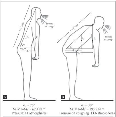
Figure 1
Exacerbation of pain along the path of the crural and sciatic nerves secon-dary to an increase in Moments of Force (M) when bending the lumbar spine at 75° angle, in A, and 30°, in B, with an increase in intra-spinal pressure secondary to coughing or sneezing in the Valsalva maneuver. L = length of the spine; M1 = mass of the trunk; M2 = mass of head and arms; F1 = M1 x 9.8 sec; F2 = M2 x 9.8 sec; dL = distance perpendicular to F1; d2 = distance perpendicular to F2; alpha 1 and alpha 2 = angle between the length of the spine and the horizontal plane. Pressure with cough and sneeze17 (modiied).
A B
Pressure with cough: 13.6 atmospheres Pressure on sneezing: 14.8 atmospheres
α
1 = 75°
M: M1+M2 = 62.4 N.m Pressure: 11 atmospheres
α
2 = 30°
M: M1+M2 = 193.9 N.m Pressure on coughing: 13.6 atmospheres ℓ = 70 cm
d1
d1
α1
α2 d2
d2 Sneeze
or cough
F1 F2 F1 F2 Sneeze
or cough
In 45 cases of herniated lumbar disk by MRI, 33 had
positive Cecin’s Sign, indicating a sensitivity of 73.3%. On the other hand, Cecin’s Sing was negative in 20 out of 21
patients without herniated lumbar disk on MRI, indicating
95.2% speciicity. This sign had a positive predictive value of
97.1% and negative predictive value of 62.5%.
In comparison, among 45 cases of herniated lumbar disk
on MRI, 10 presented positive Lasègue’s sign, indicating a
sensitivity of 22.2%. On the other hand, 20 out of 21 cases
of negative MRI had negative Lasègue’s sign, indicating a speciicity of 95.24%. This maneuver had a positive predictive
value of 90.9% and negative predictive value of 36.4%. Distribution of the cases in the contingency table above,
evaluated by Fisher’s exact test, did not show statistically signiicant differences (P = 0.153).
Therefore, comparing the sensitivity and positive and negative predictive values of both clinical maneuvers,
despite similar speciicity, superior diagnostic capacity can be attributed to Cecin’s sign (sensitivity = 73.3%; positive
predictive value = 97.1%; and negative predictive value =
62.5%) and not to Lasègue’s sign (sensitivity 22.2%, positive
predictive value of 90.9%, and negative predictive value of 36.4%) for the detection of herniated lumbar disk in patients with lumbar pain, sciatalgia, and/or sciatic pain.
DiSCUSSioN
From the results presented in Table 1, one can observe a
low concordance and patient discordance between Lasègue’s sign and Cecin’s sign, concluding that one of them had better
diagnostic performance than the other. This performance of
Cecin’s sign is shown in Table 2, in which the distribution of the
cases in the contingency table was shown to be not a result of a
casual event, since a signiicant difference was observed by the Chi-square test with Yates correction (χ2 = 24.279; P < 0.001).
Those facts did not occur by chance; they most certainly were the result of biomechanical factors that differentiate the physiopathogeny of both maneuvers, which, as it will be discussed later, affect the natural history of the disease.
Similarly, on table 3, differences in sensitivity (73.3
versus 22.2%), positive predictive value (97.1 versus 90.9%),
and negative predicted value (62.5 versus 36.3%) between
Cecin’s sign and Lasègue’s sign, respectively, indicate that
those differences cannot be attributed to chance either. As
for the difference between the sensitivity of Lasègue’s sign
observed in the present study, of only 22%, to that reported in the literature, from 30 to 80%, the variation in performance depended on the criteria used to characterize it.
When straight leg raising is considered positive with
pain at a 70° angle, it has a higher sensitivity, but speciicity
is much lower, between 20 and 40%,14,15,16 i.e., at this angle the prevalence of false-positive results is very high. When one considers an angle of 45° (adopted nowadays in several studies), the sensitivity is much lower, explaining the low
sensitivity of Lasègue’s sign (22%) in the present study. Here, Lasègue’s was considered positive at an angle of 45° and only
in the presence of the characteristic irradiation to the affected
dermatome, increasing speciicity considerably, but with a
reduction in sensitivity.
The differences between both maneuvers can be explained:
Lasègue’s sign is caused by mechanical deformation and
stretching of the sciatic nerve by the herniated disk while
Cecin’s sign is secondary to two phenomena – torque, resulting
Table 1
Concordance and discordance between Cecin’s sign and Lasègue’s sign in the test grou
LasèguePositive Negative Lasègue Total
Positive Cecin’s Sign 10 24 34
Negative Cecin’s Sign 0 11 11
Total 10 35 45
Table 2
Results of Cecin’s sign in 66 patients with
lumbar pain evaluated by MRI
Test group (herniated disk on MRI)
Control group (MRI negative for herniated
disk) Total
Positive Cecin’s Sign 33 1 34
Negative Cecin’s Sign 12 20 32
Total 45 21 66
Table 3
Results of Lasègue’s sign in 66 cases of
lumbar pain evaluated by MRI
disk on MRIHerniated Negative MRI Total
Positive Lasègue’s 10 1 11
Negative Lasègue’s 35 20 55
If the patient sneezes with the lumbar spine at a 30° angle (Figure 1B), pressure increases by 0.38 MPa and intraspinal pressure reaches 1.48 MPa or 14.8 atmospheres.17,21 Sneezing increases pressure on the cauda equina from 0.12 to 0.38 MPa (3.8 atmospheres).17
It occurs in the Valsalva maneuver because the cauda equina
is a closed cylinder to which Pascal’s principle is applied: “increases in pressure exerted on a point of a balanced luid are transmitted integrally to all points of the luid and to the
walls of the recipient.”
To understand what this pressure on the structures of the lumbar spine means and, consequently, on the herniated tissue, one atmosphere is approximately equivalent to the pressure of a 10-meter water column on a person on the sea surface. Add the Moments of Force on the lumbar spine
during investigation of Cecin’s sign to this pressure of
14.8 atmospheres, the intensity of preexisting pain (lower lumbar region, buttocks, and thigh) and/or typical sciatalgia/ sciatic pain (dermatomeric) will increase. If the pain does not irradiate, such as in central herniated disks and/or in other location, it will appear.22 It is as if the disk received an additional torque of 62.4 (Figure 1A), 193.9 (Figure 1B), and 214.0 N.m (at a 15° angle, not shown). Besides, the cauda equina will also receive and additional pressure of 14.8
atmospheres. This will relect not only on the intervertebral
disks and nerve roots, but also on all intra- and extra-dural structures, aggravating the radicular compression by the herniated disk, not only in the intervertebral foramen, but also in central hernias, central posterior-lateral, posterior-lateral, and paramedian posterior-lateral herniated disks, aggravating preexisting symptoms or making them appear by working as facilitators of the approximation between the nerve tissue and the dislodged disk material.5,23
The sensitivity of Cecin’s sign can also be higher in patients with lower anatomofunctional reserve during lexion of the
lumbar spine, in which the sagittal diameter and area of the vertebral canal decreases.24,25
CoNCLUSioNS
Cecin’s sign is easy to perform, has a high sensitivity, and
elevated negative predictive value, and better diagnostic
performance than Lasègue’s sign in the diagnosis of
symptomatic herniated lumbar disks. Validation of the procedure in clinicalpractice and its role in other disorders should be better investigated in future prospective studies.
from the lexion of the lumbar spine, and increased pressure
on the herniated lumbar disk during the Valsalva maneuver. Torque increases the shearing forces on the intervertebral disk, causing its deformity and dislocation in all planes, while maintaining a constant volume.
As for Lasègue’s sign, it is done with the patient in the
supine position, in which only a minimal load is applied on the disk,3,17 while Cecin’s sign is done with the patient in the standing position. In this initial position, erect, the Moment of Force applied on the lumbar spine is close to zero. When bending the spine – considering the length of the spine equal to 70 cm (l), mass of the trunk (m1) of 35 kg, and the mass of the head and of both upper limbs of 15 kg (m2) – with a 75-degree angle between the spine and the horizontal plane (Figure 1A) – the Moment of Force [(m1 + m2) x 9.8 m/s2 x cosine of alpha] results in 62.4 N.m. If the
lexion of the lumbar spine increases, decreasing the angle to
30° (Figure 1B), the Moment of Force goes to 193,9 N.m. If
lexion is increased even further, to an angle of 15°, Moment
(or torque) will be 214.0 N.m.18-20 One can see that greater
lexion is associated with higher “Moments of Force” on
the intervertebral disk. Thus, it can be inferred that small
hernias, which would not have positive Cecin’s sign with greater lexion angles, could become positive with smaller lexion angles.
Therefore, while in the ”X” Sign, progressively higher torques are applied on the intervertebral disks at angles of 75°, 30°, and 15°, respectively, torque does not exist in
Lasègue’s sign.
Those Moments of Force, that occur when the lumbar spine is bent, are due to the concept of torque: “Torque or Moment (M) of Force (F) is the force associated with the possibility of rotation around an axis (pole), caused by applying force to a body. The module (or value) is calculated by the product of intensity of the force applied by the lever arm. It is the perpendicular distance between the line of action of the force and the rotation axis [(d1,d2 [Figures 1A and 1B)]. This distance is called lever force or lever arm, i.e., M = F.X.18,19,20
The second moment of Cecin’s Sign, with the patient
ACKNoWLEDGEMENTS
The author is thankful to the suggestions of Professors Ricardo Machado Xavier (UFRGS), Ricardo Fuller (FMUSP), and Mittermayer Barreto Santiago (EBMSP).
REFERÊNCIAS REFERENCES
1. Kosteljanetz M, Bang F, Schmidt-Olsen S. The clinical signiicance
of straight-leg raising: (Lasègue’s sign) in the diagnosis of prolapsed
lumbar disc. Spine 1988; 13(4):393-5.
2. Cecin HA. Diretriz I: fundamentos do diagnóstico das doenças da coluna vertebral lombar. Rev Bras Reumatol 2008; 48(supl 1):2-7. 3. Wiesel SW, Weinstein JN, Herkowitz HN, Dvorák J, Bell GR. The lumbar Spine. 2 ed. Philadelphia: W.B. Saunders 1996; 1:235-51. 4. Rebain R, Baxter GDD, McDonough S. A Systematic Review of the
Passive Straight Leg Raising Test as a Diagnostic Aid for Low Back Pain (1989 to 2000). Spine 2002; 27(17):E388-E95.
5. Summers B, Malhan K, Cassar-Pullicino V. Low Back Pain on Passive Straight Leg Raising. Spine 2005; 30(3):342-5.
6. Vroomen PCAJ, Krom MCTEM, Knottnerus JA. Diagnostic value of history and physical examination in patients suspected of sciatica due to disc herniation: a systematic review. J Neurol 1999; 246:899-906. 7. Cecin HA. O que interessa da anamnese e do exame físico no
diagnóstico das doenças degenerativas e inlamatórias da coluna
vertebral lombar. A coluna vertebral em todas as idades. In: XXI Congresso Brasileiro de Reumatologia, Curso Bienal de Reumatologia, 1996, pp. 23-39.
8. Wiesel SW, Weinstein JN, Herkowitz HN, Dvorák J, Bell GR. The lumbar Spine. 2 ed. Philadelphia: W.B. Saunders 1996; 1:123-141; 235-58.
9. Neubarth F. A propósito da revisão das diretrizes sobre lombalgias e lombociatalgias e a propriedade de dar nome a achados relevantes. Rev Bras Reumatol 2008; 48(supl 1):1.
10. Scham SM, Taylor TKF. Tension signs in lumbar disc prolapse. Clin Orthop Relat Res 1971; 75:195-204.
11. Gunnar BJ, Deyo A, Deyo RA. History and physical examination in patients with herniated lumbar discs. Spine 1996; 21(24s):10s-18s.
12. Milette PC. The proper terminology for reporting lumbar intervertebral disk disorders. American Society of Neuroradiology. 1997; 18:1859-66.
13. Milette PC, Fontaine S, Lepanto L, Cardinal E, Breton G. Differentiating lumbar disc protusions, disc bulge, and discs with normal contour but abnormal signal intensity: magnetic resonance imaging with discographyc correlations. Spine 1999; 24(1):44-53. 14. Devillé WLJM, van der Windt DAWM, Dzaferigic A, Bezemer PD,
Bouter LM. The Test of Lasègue: systematic review of the accuracy in diagnosing herniated discs. Spine 2000; 25(9):1140-7.
15. Kosteljanetz M, Espersen JO, Halaburt H, Miletic T. Predictive value
of clinical and surgical indings in patients with lumbago-sciatica:
a prospective study (part I). Acta neurochir 1984; 73(1-2):67-76. 16. Jackson RP, Cain JE Jr, Jacobs RR, Cooper BR, McManus GE. The
neuroradiographic diagnosis of lumbar herniated nucleus pulposus: II. A comparison of computed tomography (CT), myelography, CT-myelography, and magnetic resonance imaging. Spine 1989; 14:1362-7.
17. Wilke HJ, Peter N, Caimi M, Hoogland T, Claes LE. New In Vivo Measurements of Pressures in the Intervertebral Disc in Daily Life. Spine 1999; 24(8):755-62.
18. Hocheberg M, Silman A, Smolen JS, Weinblatt ME, Weisman MH (eds.). Rheumatology. 3 ed. Edinburgh: Mosby, 2003, pp. 562-3. 19. Okuno E, Frantin L. Desvendando a física do corpo humano:
biomecânica. São Paulo: Manole, 2003, pp. 131-3; 156-60. 20. Tipler PA. Física para cientistas e engenheiros. 4 ed., v. 4. Rio de
Janeiro: LTC, 2000, pp. 240, 352.
21. Nachemson A. The load on lumbar disks in different positions of the body. Clin Orthop 1966; 45:107-22.
22. Takahashi K, Shima I, Porter RW. Nerve root pressure in lumbar disc herniation. Spine 1999; 24(19):2003-6.
23. Scham SM, Taylor TKF. Tension signs in lumbar disc prolapse. Clin Orthop Relat Res 1971; 75:195-204.
24. Cecin HA. Proposição de uma reserva anatomofuncional, no canal
raquidiano, como fator interferente na isiopatologia das lombalgias
e lombociatalgias mecânicodegenerativas. Rev Assoc Med Bras 1997; 43:295-310.