Avaliação da função diastólica no continuum cardiovascular- The evaluation of diastolic function in the cardiovascular continuum
194
0
0
Texto
(2)
(3) DISSERTAÇÃO DE CANDIDATURA AO GRAU DE DOUTOR APRESENTADA À FACULDADE MEDICINA DA UNIVERSIDADE DO PORTO PROGRAMA DOUTORAL EM CIÊNCIAS CARDIOVASCULARES. Avaliação da Função Diastólica no Continuum Cardiovascular The Evaluation of Diastolic Function in the Cardiovascular Continuum. Orientador: Prof. Doutor Adelino Leite-Moreira Co-orientadores: Prof. Doutora Ana Azevedo. Prof. Doutor Francisco Rocha Gonçalves. Ricardo Fontes-Carvalho Porto • 2015.
(4) Art.º 48º, 3º - “A Faculdade não responde pelas doutrinas expendidas na dissertação.” (Regulamento da Faculdade de Medicina da Universidade do Porto – Decreto-Lei n.º 19337 de 29 de Janeiro de 1931).
(5) Constituição do Júri da Prova de Doutoramento. Presidente DOUTORA MARIA AMÉLIA DUARTE FERREIRA Diretora da Faculdade de Medicina da Universidade do Porto (por delegação reitoral). Vogais DOUTOR FAUSTO JOSÉ DA CONCEIÇÃO ALEXANDRE PINTO Professor Catedrático, Faculdade de Medicina da Universidade de Lisboa DOUTOR JOAQUIM ADELINO CORREIA FERREIRA LEITE MOREIRA Professor Catedrático, Faculdade de Medicina da Universidade do Porto DOUTOR PAULO MIGUEL BETTENCOURT SARDINHA PONTES FERNANDO Professor Catedrático convidado, Faculdade de Medicina da Universidade do Porto DOUTOR LINO MANUEL MARTINS GONÇALVES Professor Associado, Faculdade de Medicina da Universidade de Coimbra DOUTOR LUÍS FILIPE VILELA PEREIRA DE MACEDO Professor Associado convidado, Faculdade de Medicina da Universidade do Porto DOUTOR ANDRÉ PEDRO LEITE MARTINS LOURENÇO Professor Auxiliar da Faculdade de Medicina da Universidade do Porto.
(6) Corpo Catedrático da Faculdade de Medicina da Universidade do Porto. Professores Catedráticos Efectivos DOUTOR MANUEL ALBERTO COIMBRA SOBRINHO SIMOES DOUTORA MARIA AMELIA DUARTE FERREIRA DOUTOR JOSÉ AGOSTINHO MARQUES LOPES DOUTOR PATRÍCIO MANUEL VIEIRA ARAÚJO SOARES SILVA DOUTOR DANIEL FILIPE LIMA MOURA DOUTOR ALBERTO MANUEL BARROS DA SILVA DOUTOR JOSE MANUEL LOPES TEIXEIRA AMARANTE DOUTOR JOSE HENRIQUE DIAS PINTO DE BARROS DOUTORA MARIA FÁTIMA MACHADO HENRIQUES CARNEIRO DOUTORA ISABEL MARIA AMORIM PEREIRA RAMOS DOUTORA DEOLINDA MARIA VALENTE ALVES LIMA TEIXEIRA DOUTORA MARIA DULCE CORDEIRO MADEIRA DOUTOR ALTAMIRO MANUEL RODRIGUES COSTA PEREIRA DOUTOR RUI MANUEL ALMEIDA MOTA CARDOSO DOUTOR ANTONIO CARLOS FREITAS RIBEIRO SARAIVA DOUTOR JOSE CARLOS NEVES DA CUNHA AREIAS DOUTOR MANUEL JESUS FALCAO PESTANA VASCONCELOS DOUTOR JOÃO FRANCISCO MONTENEGRO ANDRADE LIMA BERNARDES DOUTORA MARIA LEONOR MARTINS SOARES DAVID DOUTOR RUI MANUEL LOPES NUNES DOUTOR JOSÉ EDUARDO TORRES ECKENROTH GUIMARÃES DOUTOR FRANCISCO FERNANDO ROCHA GONÇALVES DOUTOR JOSE MANUEL PEREIRA DIAS DE CASTRO LOPES DOUTOR ANTÓNIO ALBINO COELHO MARQUES ABRANTES TEIXEIRA DOUTOR JOAQUIM ADELINO CORREIA FERREIRA LEITE MOREIRA DOUTOR RAQUEL ÂNGELA SILVA SOARES LINO.
(7) Professores Jubilados ou Aposentados DOUTOR ABEL VITORINO TRIGO CABRAL DOUTOR ALEXANDRE ALBERTO GUERRA SOUSA PINTO DOUTOR ÁLVARO JERONIMO LEAL MACHADO DE AGUIAR DOUTOR AMÂNDIO GOMES SAMPAIO TAVARES DOUTOR ANTONIO AUGUSTO LOPES VAZ DOUTOR ANTÓNIO CARVALHO ALMEIDA COIMBRA DOUTOR ANTÓNIO FERNANDES DA FONSECA DOUTOR ANTÓNIO FERNANDES OLIVEIRA BARBOSA RIBEIRO BRAGA DOUTOR ANTÓNIO JOSÉ PACHECO PALHA DOUTOR ANTÓNIO MANUEL SAMPAIO DE ARAÚJO TEIXEIRA DOUTOR BELMIRO DOS SANTOS PATRICIO DOUTOR CÂNDIDO ALVES HIPÓLITO REIS DOUTOR CARLOS RODRIGO MAGALHÃES RAMALHÃO DOUTOR CASSIANO PENA DE ABREU E LIMA DOUTOR DANIEL SANTOS PINTO SERRÃO DOUTOR EDUARDO JORGE CUNHA RODRIGUES PEREIRA DOUTOR FERNANDO TAVARELA VELOSO DOUTOR FRANCISCO DE SOUSA LÉ DOUTOR HENRIQUE JOSÉ FERREIRA GONÇALVES LECOUR DE MENEZES DOUTOR JORGE MANUEL MERGULHAO CASTRO TAVARES DOUTOR JOSÉ CARVALHO DE OLIVEIRA DOUTOR JOSÉ FERNANDO BARROS CASTRO CORREIA DOUTOR JOSÉ LUÍS MEDINA VIEIRA DOUTOR JOSÉ MANUEL COSTA MESQUITA GUIMARÃES DOUTOR LEVI EUGÉNIO RIBEIRO GUERRA DOUTOR LUÍS ALBERTO MARTINS GOMES DE ALMEIDA DOUTOR MANUEL ANTÓNIO CALDEIRA PAIS CLEMENTE DOUTOR MANUEL AUGUSTO CARDOSO DE OLIVEIRA DOUTOR MANUEL MACHADO RODRIGUES GOMES DOUTOR MANUEL MARIA PAULA BARBOSA DOUTORA MARIA DA CONCEIÇÃO FERNANDES MARQUES MAGALHÃES DOUTORA MARIA ISABEL AMORIM DE AZEVEDO DOUTOR MÁRIO JOSÉ CERQUEIRA GOMES BRAGA DOUTOR SERAFIM CORREIA PINTO GUIMARÃES DOUTOR VALDEMAR MIGUEL BOTELHO DOS SANTOS CARDOSO DOUTOR WALTER FRIEDRICH ALFRED OSSWALD.
(8)
(9) FAZEM PARTE INTEGRANTE DESTA DISSERTAÇÃO OS SEGUINTES TRABALHOS: 1. Ricardo Fontes-Carvalho, Alexandra Gonçalves, Milton Severo, Patrícia Lourenço, Francisco Rocha Gonçalves, Paulo Bettencourt, Adelino Leite-Moreira, Ana Azevedo. Direct, inflammation-mediated and blood-pressure-mediated effects of total and abdominal adiposity on diastolic function: EPIPorto study. Int J Cardiol 2015, in press.. Journal’s Impact Factor: 6.17 2. Ricardo Fontes-Carvalho, Joana Pimenta, Paulo Bettencourt, Adelino Leite-Moreira, Ana Azevedo. Plasma leptin and adiponectin levels and diastolic function in the general population: data from the EPIPorto study. Expert Opin Ther Targets. 2015 Mar 18:1-9.. Journal’s Impact Factor: 4.90. 3. Ricardo Fontes-Carvalho, Marta Fontes-Oliveira, Francisco Sampaio, Jennifer Mancio, Nuno Bettencourt, Madalena Teixeira, Francisco Rocha Gonçalves, Vasco Gama, Adelino Leite-Moreira. Influence of Epicardial and Visceral Fat on Left Ventricular Diastolic and Systolic Function in Patients After Myocardial Infarction. Am J Cardiol 2014, 114(11): 1663-9.. Journal’s Impact Factor: 3.43. 4. Ricardo Fontes-Carvalho, Ricardo Ladeiras-Lopes, Paulo Bettencourt, Adelino Leite-Moreira, Ana Azevedo. Diastolic Dysfunction in the Diabetic Continuum: Association with Insulin Resistance, Metabolic Syndrome and Diabetes. Cardiovasc Diabetol. 2015 Jan 13;14(1):4.. Journal’s Impact Factor: 3.71 5. Ricardo Fontes-Carvalho, Azevedo A, Leite-Moreira A. The new grade IA of diastolic dysfunction: is it relevant at the population level? JACC Cardiovasc Imaging. 2015 (2): 229-30.. Journal’s Impact Factor: 6.99 6. Ricardo Fontes-Carvalho, Francisco Sampaio, Madalena Teixeira, Francisco Rocha Gonçalves, Vasco Gama, Ana Azevedo, Adelino Leite-Moreira. Left Ventricular Diastolic Dysfunction and E/E’ ratio as the Strongest Echocardiographic Predictors of Reduced Exercise Capacity After Acute Myocardial Infarction. Clin Cardiol. 2015;38(4):222-9.. Journal’s Impact Factor: 2.22 7. Ricardo Fontes-Carvalho, Francisco Sampaio, Madalena Teixeira, Francisco Rocha Gonçalves, Vasco Gama, Ana Azevedo, Adelino Leite-Moreira. Left atrial deformation analysis by speckle tracking echocardiography to predict exercise capacity after myocardial infarction. [Under review, J Am Soc Echocardiog 2015]. 8. Ricardo Fontes-Carvalho, Francisco Sampaio, Madalena Teixeira, Vasco Gama, Adelino Leite-Moreira. The role of a structured exercise-training program on cardiac structure and function after acute myocardial infarction: study protocol for a randomized controlled trial. Trials. 2015 Mar 12;16(1):90.. Journal’s Impact Factor: 2.12 9. Ricardo Fontes-Carvalho, Francisco Sampaio, Madalena Teixeira, Francisco Rocha Gonçalves, Vasco Gama, Ana Azevedo, Adelino Leite-Moreira. The effect of exercise training on diastolic and systolic function after acute myocardial infarction: a randomized study. [Under Review, Medicine 2015]..
(10)
(11) À Luísa À Inês e ao Pedro. À minha Família. Aos meus “Mestres”, na Vida e na Medicina.
(12)
(13) Agradecimentos/ Acknowledgements A elaboração desta tese de doutoramento constituiu um esforço de superação pessoal. Mais do que um projeto individual, só foi possível com um amplo trabalho de equipa, a nível profissional e familiar. De entre tantos que contribuíram, directa e indirectamente, cumpre-me manifestar especial agradecimento: Ao Prof. Adelino Leite-Moreira pelo seu constante incentivo, disponibilidade e exemplo de competência científica e humanismo. Agradeço-lhe o apoio e autonomia que me deu ao longo destes anos e o voto de confiança quando, ainda enquanto aluno, me aceitou no Serviço de Fisiologia da Faculdade de Medicina do Porto. Recordo hoje esse momento como o primeiro pequeno passo desta longa viagem até à conclusão desta tese. Entre tantas outras coisas, com ele aprendi as mais-valias que a compreensão da investigação dita “básica” e de translação traz todos os dias à melhoria da minha prática clínica diária. À Prof. Ana Azevedo por toda a sua disponibilidade e ajuda, essenciais à realização dos trabalhos desta tese. Agradeço-lhe o grande enriquecimento pessoal trazido pelas estimulantes discussões científicas, pela análise rigorosa das questões e pela revisão crítica, e esclarecida, de todos os trabalhos. A sua exigência obrigou-me a um esforço contínuo de superação pessoal. Ao Prof. Rocha Gonçalves pelo contínuo apoio e estímulo na persecução dos trabalhos. Agradeço ainda as sempre estimulantes conversas e reflexões sobre a Vida e a Cardiologia. Ao Dr. Vasco Gama Ribeiro pela sua incansável força, dinamismo, visão e determinação. Em poucos anos construiu, à sua imagem, um serviço de Cardiologia que é uma referência a nível nacional e europeu. Nele aprendi, e cresci, enquanto Homem, médico e cardiologista! Ao Serviço de Cardiologia do Centro Hospitalar de Gaia. Dos colegas médicos, aos enfermeiros, aos técnicos e aos auxiliares. A todos eles devo muito do que sou, e consegui, até hoje! À Dra. Madalena Teixeira, pela ajuda na realização dos trabalhos clínicos. Ao Dr. Lino Simões pela sua amizade, pelas estimulantes discussões sobre a Cardiologia, a vida, a política e a cultura, e pelo seu exemplo pessoal de dedicação ao doente e à prática clinica. Ao Dr. Aníbal Albuquerque pelo exemplo de integridade, mas sobretudo pela sua amizade e pelo contínuo apoio e palavras estímulo à execução dos trabalhos. Ao Dr. Pedro Braga, meu orientador durante o internato de Cardiologia, tendo contribuído para fazer de mim um melhor cardiologista. Um agradecimento especial a todo o sector da Ecocardiografia. Ao Dr. José Ribeiro pela sua confiança, espírito de equipa e por me ter ensinado e incutido a paixão pelos desafios da ecocardiografia. À Dra. Conceição Fonseca pela permanente energia, disponibilidade e dedicação ao doente. Um agradecimento particular ao Dr. Francisco Sampaio – un compagnon de route – pelas discussões científicas, pela partilha de ideias e pela sua imprescindível participação nos estudos clínicos.
(14) A todos os colegas do Serviço de Fisiologia da Faculdade de Medicina do Porto pela partilha de conhecimentos e pela valiosa introdução ao raciocínio e método científico. Entre eles uma palavra particular ao Prof. Paulo Chaves que me introduziu ao trabalho laboratorial e me motivou o gosto pela ciência básica. Aos doentes, que são a razão final desta tese! Um agradecimento especial àqueles que aceitaram participar nos estudos clínicos. Agradeço a todos os meus “Mestres” (muitos deles já aqui citados) que na Vida, na Medicina, na Cardiologia e na Investigação – frequentemente de forma simultânea – contribuíram para o meu crescimento. Sempre os recordarei e para sempre lhes estarei grato!. •••. Deixo propositadamente para o fim, a mais importante das reflexões e o mais forte dos meus agradecimentos. Merecem-nos a minha Família! Agradeço-te a ti, Luísa, por tudo! Mas também por todo o teu carinho, amor, amizade e incansável compreensão. À Inês e ao Pedro, por terem dado um novo significado à Vida. E pela alegria que me trazem todos os dias. Aos meus pais. Por sempre me terem orientado para “o rumo certo” da vida. A eles devo tudo o que sou! Espero agora saber usar os vossos sábios conselhos, mas sobretudo o vosso exemplo, para guiar os meus filhos. Aos meus irmãos, Ni e João. Por toda a amizade, apoio incondicional e sentimento de partilha. Ao meu avô, Augusto. Uma fonte de inspiração. Pelo seu exemplo de vida, de inteligência e de determinação. Tenho a certeza que gostarias de estar aqui hoje!.
(15) Table of Contents. ABSTRACT | RESUMO. 17. CHAPTER I | INTRODUCTION . 27. 1. The Cardiac Cycle: Focus on Diastole. 29. 2. Diastolic Dysfunction Across the Cardiovascular Continuum . 30. 3. The Impact of Diastolic Dysfunction: Prognostic Implications 3.1. Diastolic Dysfunction and Heart Failure Risk. 31 31. 3.2. Diastolic Dysfunction in the Pathophysiology of Heart Failure with Preserved Ejection Fraction (HFpEF) 32. 3.3. The Impact of Diastolic Function on Exercise Capacity and Quality of Life . 32. 4. The Evaluation of Left Ventricular Diastolic Function. 34. 5. The Determinants of Diastolic Dysfunction: What is Known and What is Unknown 5.1. Established Determinants of Diastolic Dysfunction. 36 36. 36. 5.2. Obesity as a New Determinant of Diastolic Dysfunction: Understanding “Obesity Cardiomyopathy”. 5.3. Diastolic Function in the Diabetic Continuum: the Role of Insulin Resistance, Metabolic Syndrome and Diabetes on Diastolic Dysfunction 38 6. Review Article: “Heart Failure with Preserved Ejection Fraction: Fighting Misconceptions for a New Approach” . 39. 7. Review Article: “The Pathophysiology of Heart Failure Preserved Ejection Fraction and Its Therapeutic Implications”. 49. CHAPTER II | PURPOSE . 69. CHAPTER III | RESULTS/ PUBLICATIONS . 73. A) Determinants of Diastolic Dysfunction. 77. B) The Evaluation of Diastolic Dysfunction. 115. C) The Impact of Diastolic Function on Exercise Capacity. 127. D) Modulation of Diastolic Function by Exercise Training. 145. CHAPTER IV | DISCUSSION . 167. CHAPTER V | CONCLUSIONS . 175. CHAPTER VI | BIBLIOGRAPHY . 179. 13.
(16) 14. RICARDO FONTES-CARVALHO.
(17) Foreword. This thesis is organized in six main chapters.. Chapter 1 includes a general introduction to the knowns and unknowns of diastolic function across the several stages of the cardiovascular continuum including the determinants, methods of evaluation and prognostic implications. The role of diastolic dysfunction as an early step in the progression to heart failure is also discussed, particularly in the progression to heart failure with preserved ejection (HFpEF). Also, to provide a more detailed overview on the importance of diastolic dysfunction in the pathophysiology of HFpEF, two review articles were included at the end of this chapter.. Chapter 2 defines the main objectives of the thesis, which are schematized in the figure provided in this chapter.. Chapter 3 describes the results, which are shown in the form of several published or submitted articles. These manuscripts have been grouped into four sub-chapters: A) determinants of diastolic dysfunction; B) the evaluation of diastolic dysfunction; C) the impact of diastolic function on exercise capacity; D) the modulation of diastolic function by exercise training.. Chapter 4 provides a global overview and discussion of the major findings, how do they integrate with previously published studies, possible implications to clinical practice and future areas of research.. The conclusions are presented in chapter 5.. Finally, chapter 6 depicts the references that were used in the introduction and discussion sections of this thesis.. 15.
(18) 16. RICARDO FONTES-CARVALHO.
(19) Resumo Abstract.
(20)
(21) RESUMO | ABSTRACT. Introdução A disfunção diastólica (DD) do ventrículo esquerdo é frequente nas várias fases do continuum cardiovascular, atingindo cerca de 20% da população em geral. Apesar de ser muitas vezes assintomática, a DD é um importante preditor de prognóstico e do risco de progressão para insuficiência cardíaca. Por outro lado, nos doentes no outro extremo do continuum cardiovascular, a DD é o principal mecanismo envolvido na fisiopatologia da insuficiência cardíaca com fração de ejecção preservada (ICFEP), também conhecida como insuficiência cardíaca diastólica. Assim sendo, a identificação – e correção – dos principais determinantes de DD pode ser um importante passo para prevenir a progressão para insuficiência cardíaca. Esta abordagem preventiva poderá ser particularmente relevante na ICFEP uma vez que, até hoje, nenhuma terapêutica ou intervenção mostrou uma melhoria significativa do prognóstico destes doentes. O estudo da função diastólica nas várias fases do continuum cardiovascular é importante por múltiplas outras razões. Além do seu impacto prognóstico, desconhece-se o seu papel como determinante da capacidade funcional dos doentes, sobretudo após o enfarte do miocárdio. A própria avaliação ecocardiográfica da função diastólica é complexa, sobretudo a categorização em graus de DD. Ainda recentemente foi descrito um novo grau de DD, designado grau IA, cuja prevalência e caracterização clínica são desconhecidas na população em geral. Por outro lado, sabe-se que os volumes e função da aurícula esquerda são interdependentes da função diastólica do ventrículo esquerdo e, portanto, as novas técnicas ecocardiográficas de avaliação da função auricular esquerda por speckle tracking poderão ajudar na compreensão e na análise detalhada da função diastólica. Finalmente, estudos recentes realizados em doentes com ICFEP mostraram que o exercício físico poderá ser uma nova intervenção terapêutica capaz de melhorar a função diastólica, não estando ainda validada em outros grupos de doentes.. Objectivos Neste projeto pretendemos avaliar a importância da avaliação da função diastólica em várias fases do continuum cardiovascular, identificando e analisando os seus determinantes, o papel dos novos métodos de avaliação ecocardiográfica, o seu impacto na capacidade funcional e uma eventual modulação por um programa estruturado de treino de exercício. Foram objectivos específicos destes trabalhos: i) avaliar o papel da obesidade, e do padrão de distribuição de gordura, como determinante da função diastólica, assim como os principais mecanismos fisiopatológicos envolvidos nesta associação; ii) avaliar se o tecido adiposo epicárdico pode influenciar diretamente a função diastólica por um efeito local/parácrino; iii) determinar se a secreção de adipocinas (leptina e adiponectina) pode estar envolvida na associação entre a obesidade e a função diastólica; iv) avaliar o papel da insulinorresistência, da síndrome metabólica e da diabetes como eventuais novos determinantes de DD; v) avaliar, na população em geral, a prevalência e características clínicas dos doentes com o novo grau IA de DD; vi) avaliar o impacto da função diastólica como determinante da. 19.
(22) RESUMO | ABSTRACT. capacidade funcional em doentes após enfarte do miocárdio; vii) analisar a relação entre os volumes e a função da aurícula esquerda com a função diastólica, assim como o seu papel enquanto preditor da capacidade funcional; viii) avaliar se um programa estruturado de treino de exercício pode melhorar a função diastólica num grupo de doentes após enfarte do miocárdio.. População e Métodos Para avaliar os determinantes de disfunção diastólica foi analisado um grupo de 1063 indivíduos provenientes de uma coorte da população em geral (estudo EPIPorto), sem doença cardiovascular conhecida. Todos os doentes foram avaliados através da colheita de história clinica, exame físico, avaliação antropométrica (incluindo avaliação da massa gorda por bioimpedâcia), estudo analítico (incluindo doseamento da insulina, HOMA-IR score, leptina e adiponectina) e ecocardiografia transtorácica, com avaliação detalhada da função diastólica. Foi ainda analisado, de forma prospectiva, um outro grupo de 225 doentes um mês após enfarte do miocárdio. Todos os doentes foram submetidos, no mesmo dia, a avaliação clínica, antropométrica e ecocardiográfica (incluindo a análise dos volumes e função da aurícula esquerda por speckle tracking, num subgrupo destes doentes), assim como a prova cardiopulmonar (para determinação do consumo de oxigénio: VO2) e tomografia computorizada de 64 cortes (para determinação das áreas de gordura abdominal total, subcutânea e visceral e do volume de gordura epicárdica). Deste grupo de doentes após enfarte do miocárdio, 188 foram randomizados (1:1) para um programa supervisionado de 8 semanas de treino de exercício (combinando treino aeróbio e de resistência) versus tratamento convencional. No início e no fim do estudo, estes doentes foram submetidos a avaliação seriada da função diastólica por ecocardiografia e da capacidade de exercício por prova cardiopulmonar. Em todos os estudos a função diastólica foi avaliada de acordo com as últimas recomendações de consenso para a avaliação da função diastólica que incluem a determinação da velocidade E’ e da relação E/E’ do Doppler tecidular e a categorização em graus de disfunção diastólica.. Principais Resultados e Discussão O aumento da adiposidade, principalmente da gordura visceral, revelou-se um determinante. RICARDO FONTES-CARVALHO. independente da função diastólica. A associação entre a adiposidade e a DD parece ser mediada. 20. parcialmente por um efeito endócrino, envolvendo a secreção de adipocinas, já que que o aumento dos níveis de leptina – mas não de adiponectina – se associou de forma independente à presença de DD. Além disso, observamos que o aumento do volume de gordura epicárdica pode também modular a função diastólica, por um efeito local/parácrino. Estes dados permitem uma compreensão dos mecanismos envolvidos na “cardiopatia da obesidade” e reforçam a importância da associação entre a obesidade, a.
(23) RESUMO | ABSTRACT. disfunção diastólica e o aumento do risco de insuficiência cardíaca, observado nos indivíduos obesos. A função diastólica está também associada ao grau de insulinorresistência e à síndrome metabólica e não a um efeito “glicotóxico” induzido pela hiperglicemia. Observamos ainda que surgem alterações subclínicas da função diastólica logo em fases precoces do continuum diabético, ainda antes do aparecimento de diabetes. Contrariamente a anteriores descrições, verificamos que na população em geral a prevalência do novo grau IA de DD é baixa. Deste modo, em futuros estudos, será necessário reavaliar o papel de prognóstico deste novo grau de disfunção diastólica e a sua utilidade clínica. Os parâmetros de função diastólica, sobretudo a relação E/E’ septal (derivada da análise do Doppler tecidular), foram os principais determinantes ecocardiográficos de diminuição da capacidade funcional após enfarte do miocárdio. Além disso, observamos que os volumes da aurícula esquerda, a sua função de reservatório e o strain longitudinal (avaliados por speckle tracking) foram também determinantes da capacidade de exercício, devido à interdependência entre a função diastólica e a função auricular esquerda. Finalmente, num estudo prospectivo, randomizado, realizado em doentes um mês após enfarte do miocárdio, verificou-se que um programa de treino de exercício de 8 semanas, combinando treino aeróbio e de resistência, não melhorou significativamente a função diastólica.. Conclusões A disfunção diastólica tem uma prevalência crescente ao longo do continuum cardiovascular. Os seus principais determinantes são a idade, a hipertensão arterial e a doença coronária, mas também a obesidade e a insulinorresistência. Estas observações reforçam a visão da disfunção diastólica como uma doença com um componente cardiometabólico. Atendendo à crescente epidemia de obesidade, a compreensão dos mecanismos envolvidos na cardiopatia da obesidade poderá ser clinicamente útil para a prevenção do expectável aumento da incidência de insuficiência cardíaca nos obesos. No futuro será necessário avaliar se intervenções como a perda de peso (pela dieta, exercício e/ou cirurgia bariátrica), a toma de insulino-sensibilizadores (como a metformina) ou a administração de inibidores da leptina pode melhorar a função diastólica e prevenir a progressão para insuficiência cardíaca. Atualmente, estamos a realizar um ensaio clínico de fase II que avaliará se a administração precoce de metformina em doentes com síndrome metabólico pode melhorar a função diastólica. A análise da função diastólica fornece ao clínico informação adicional que tem impacto prognóstico e funcional. Deste modo, a avaliação da função diastólica, através do cálculo da relação E/E’ e da categorização em graus de função diastólica, deve fazer parte integrante do exame ecocardiográfico de rotina.. 21.
(24) RESUMO | ABSTRACT. Apesar de nos doentes após enfarte do miocárdio o treino de exercício não ter melhorado significativamente a função diastólica, é expectável que programas de exercício físico sustentado possam melhorar a estrutura e função do miocárdio, sobretudo se aplicados em fases mais precoces do continuum cardiovascular.. RICARDO FONTES-CARVALHO. •••. 22.
(25) RESUMO | ABSTRACT. Introduction Left ventricle diastolic dysfunction (LVDD) is common in the cardiovascular continuum. It affects about 20% of the adult population. Although frequently associated with a subclinical course, LVDD is an important predictor of heart failure and of long-term mortality. Moreover, in patients at the other end of the cardiovascular continuum, LVDD is the key mechanism involved in the pathophysiology of heart failure preserved ejection fraction (HFpEF), also known as diastolic heart failure. Therefore, the identification and correction of the main determinants of diastolic function can be of paramount importance to stop the progression to overt heart failure. This can be especially relevant for the management of HFpEF, a disease where no therapy or intervention has significantly improved the prognosis. Several other unmeet needs involve the study of diastolic function in the cardiovascular continuum. Beyond its prognostic role, the impact of LVDD on exercise capacity is not known, especially after myocardial infarction. Moreover, the echocardiographic evaluation of diastolic function is frequently challenging, especially the categorization diastolic function grades. Very recently, a new grade of diastolic dysfunction – grade IA – has been described, but its prevalence and clinical characterization in the general population are unknown. Because LV diastolic function and left atrium (LA) function are interdependent events, the evaluation of the LA by speckle tracking can be a promising new technique giving further insight into the comprehension of diastolic function. Finally, new therapeutic targets are needed to improve diastolic function. Recent preliminary studies, performed in selected patients with HFpEF, have shown that a structured program of exercise training can improve diastolic function.. Purpose In this project we aimed to evaluate the role of left ventricular diastolic function in several phases of the cardiovascular continuum, assessing its determinants, new methods of echocardiographic evaluation, its impact on exercise capacity and the possible modulation by exercise training. Specific aims were: i) to evaluate the role of obesity, and adipose tissue distribution, on diastolic function and the underlying mechanisms (direct and indirect) involved in this association; ii) to detail if epicardial adipose tissue can influence diastolic function by a local/paracrine mechanism; iii) to determine if the secretion of adipokines (leptin and adiponectin), can be involved in the association between adiposity and diastolic dysfunction; iv) to evaluate the contribution of insulin resistance, metabolic syndrome and diabetes as novel determinants of diastolic function; v) to evaluate in the general population, the prevalence and clinical characteristics of the new echocardiographic grade IA of diastolic dysfunction; vi) to determine the impact of diastolic function as a predictor of reduced exercise capacity after myocardial infarction; vii) to evaluate the relation between LA volumes and LA function with LV diastolic function (the atrium-ventricular coupling) and its role as predictor of exercise performance; viii) to evaluate if a structured exercise training program can improve diastolic function in patients after myocardial infarction.. 23.
(26) RESUMO | ABSTRACT. Populations and Methods To evaluate the determinants of diastolic dysfunction we evaluated a group of 1063 individuals from the general population, without known cardiovascular disease, from the EPIPorto cohort study. All patients had detailed data about clinical history, physical examination, anthropometric evaluation, fasting blood sample (including insulin, leptin, adiponectin) and echocardiography. For the evaluation of the impact of diastolic function on functional capacity, and its modulation by exercise training, we prospectively enrolled another group of 225 patients after myocardial infarction. All patients were submitted, on the same day, to clinical evaluation, detailed echocardiography (including the evaluation of left atrium strain by speckle tracking analysis in a subgroup of patients), cardiopulmonary exercise test (for determination of peak O2 consumption) and CT scan (for the evaluation of total, subcutaneous and visceral abdominal fat area and epicardial fat volume). From this group, 188 patients were prospectively randomized (1:1) to an 8-week supervised program of endurance and resistance exercise training versus standard of care, with serial evaluation (at the beginning and at the end of the study) of diastolic function by echocardiography and of exercise capacity by cardiopulmonary exercise test. In all studies diastolic function was assessed according to the latest consensus guidelines on diastolic function evaluation, including the determination of E’ velocity and E/E’ ratio by tissue Doppler analysis, and the categorization in diastolic dysfunction grades.. Main Results and Discussion First, we observed that increased adiposity parameters were independent determinants of subclinical diastolic function, reinforcing the role of “obesity cardiomyopathy” in the link between obesity, diastolic dysfunction and the increased risk of heart failure observed in the obese. The association was especially relevant for visceral, rather than subcutaneous fat. We observed that an endocrine effect, through the secretion of adipokines, could be involved, because higher leptin levels – but not of adiponectin – were independently associated with diastolic dysfunction. Moreover, increased epicardial fat volume was independently associated with impaired diastolic function, suggesting that a local or paracrine effect can also play a role in this association. Secondly, we observed that subclinical changes in left ventricular diastolic function were already present in an early phase of glucose disturbance metabolism, even before the onset of diabetes. These. RICARDO FONTES-CARVALHO. changes were mainly associated with the state of insulin resistance, and not to a “glucotoxic” effect caused. 24. by hyperglycemia. Metabolic syndrome was also independently associated with LVDD. Contrary to what has been previously described, in the general population we observed that the new grade IA of diastolic dysfunction was infrequent. Future studies will evaluate if the determination of this new grade can be clinically useful in an unselected population without cardiovascular disease..
(27) RESUMO | ABSTRACT. Beyond its prognostic impact, we have now shown that resting diastolic function parameters, especially the septal E/E’ ratio, were important determinants of reduced exercise capacity after myocardial infarction. Moreover, the evaluation of the left atrium by speckle tracking showed that LA size, function and strain parameters were interdependent with left ventricular diastolic function. Therefore, we found that increased LA volumes can be used as markers of reduced functional capacity, which was associated mainly with a decrease in the LA conduit function, but not with the LA contractile function. Finally, in a prospective, randomized, controlled study including patients after myocardial infarction we did not observe a significant improvement in diastolic function parameters, after an eight-week exercisetraining program.. Conclusions Diastolic dysfunction has an increasing prevalence across the cardiovascular continuum. It is determined by ageing, hypertension and coronary artery disease, but also by new determinants such as (visceral) obesity and insulin resistance, reinforcing the view of diastolic dysfunction as a cardiometabolic disease. Given the growing obesity epidemic worldwide, and the increased risk of heart failure in the obese, this research provided important clinical insights to the comprehension of “obesity cardiomyopathy”. Future trials will determine if new therapeutic interventions, such as weight loss (by diet, exercise and/ or bariatric surgery) and insulin sensitizers (such as metformin), can improve diastolic function and prevent the progression to heart failure. We are now conducting a phase II clinical trial to determine if the administration of metformin in individuals with metabolic syndrome can improve diastolic function. The evaluation of diastolic function provides important functional and prognostic information to the clinician. Therefore, the evaluation of diastolic function – using standard and new techniques, such as strain analysis by speckle tracking – should be an integral part of a routine echocardiography examination. The search for new interventions that can improve diastolic function will continue. Although after myocardial infarction, exercise training failed to enhance diastolic function, it is plausible that longterm exercise can improve cardiac structure and function, especially if applied in earlier phases of the cardiovascular continuum.. 25.
(28) 26. RICARDO FONTES-CARVALHO.
(29) Introduction. “Science may set limits to knowledge, but should not set limits to imagination” Bertrand Russell (1872-1970).
(30)
(31) CHAPTER I | INTRODUCTION. 1. THE CARDIAC CYCLE: FOCUS ON DIASTOLE. “The atria or filling chambers contract together, while the pumping chambers or ventricles are relaxing and vice versa”. Leonardo da Vinci (1452-1519). From perhaps the first description of diastole by Leonardo da Vinci, to the most modern techniques, indexes, and innovative imaging tools of evaluation, our understanding of left ventricular diastolic function continues to advance. However, over many years, the evaluation of diastolic function was neglected by the scientific and clinical community, in part due to the difficulties in the non-invasive evaluation of diastolic function. With the new cardiac imaging technologies, a renewed interest in diastole has emerged, and this decade can be considered the “decade of diastology” (1). The optimal performance of the left ventricle depends on its ability to cycle between two states: a compliant chamber in diastole, that allows the left ventricle to relax and fill at low left atrial pressures, and a stiff chamber (rapidly rising pressure) in systole that ejects the stroke volume at arterial pressures (2) (3). Furthermore, in response to exercise, the stroke volume must increase without a significant raise in left atrial pressures. Therefore, left ventricular diastolic dysfunction (LVDD) can be caused by impaired myocardial relaxation and/or increased ventricular stiffness, regardless of whether the systolic function is normal or depressed, and whether the patient is asymptomatic or symptomatic (2) (4) (5).. 29.
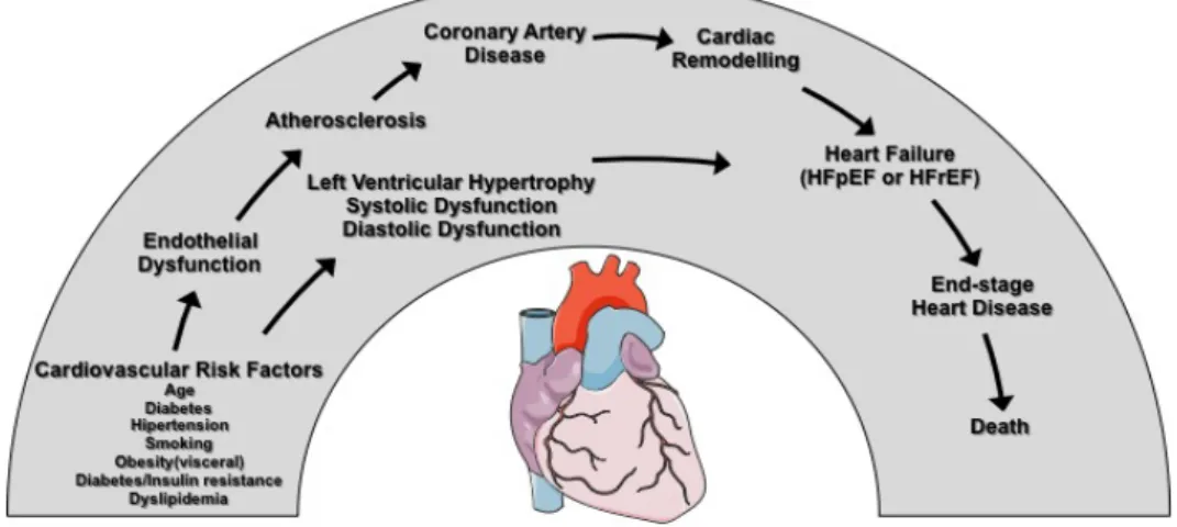
(32) CHAPTER I | INTRODUCTION. 2. DIASTOLIC DYSFUNCTION ACROSS THE CARDIOVASCULAR CONTINUUM In 1991, a panel of experts representing the full spectrum of cardiovascular disease generated an hypothesis that framed cardiovascular disease as a chain of events that were initiated by several related and unrelated risk factors, and progressing through several pathophysiological pathways and processes until the development of end-stage heart disease (6), as detailed in figure 1. More importantly, this model acknowledged that a therapeutic intervention anywhere along this chain of events could disrupt the pathophysiological process and therefore, confer “cardioprotection” and prevent end-stage cardiovascular disease (7). In the meantime, the concept of the cardiovascular continuum has been validated in several critical studies at both the mechanistic and clinical level (7) (8). It is now known that the events leading to disease progression overlap and intertwine, and do not always occur as a sequence of discrete, tandem incidents. Although the original concept focused on the traditional risk factors, and the consequences of coronary artery disease, during the last years the concept of cardiovascular continuum has expanded to include other areas such as heart failure, cerebrovascular disease, peripheral vascular disease, and renal disease (7).. Figure 1. The modern concept of the cardiovascular continuum.. HFpEF: Heart Failure Preserved Ejection Fraction; HFrEF: Heart Failure reduced ejection fraction. Adapted from Dzau et al, Circulation. 2006 (7). In this project we aimed to evaluate the role of diastolic function in individuals and patients in different steps of the cardiovascular continuum. LVDD is frequent, showing increasing prevalence throughout the cardiovascular continuum. In the community, subclinical LVDD affects between 20-30 % of the general population (9) (10) (11) (12), depending on the criteria used for its definition and the general characteristics. RICARDO FONTES-CARVALHO. of the studied population. In several phases of the cardiovascular continuum, the evaluation of diastolic function is also clinically relevant providing significant prognostic information. Even in asymptomatic individuals, subclinical diastolic dysfunction is a marker of cardiac remodeling. It is considered an intermediate step in the cardiovascular continuum, being associated with the progression to heart failure and adverse outcomes (13). Also, in latter phases of the cardiovascular continuum (such as in patients after myocardial infarction or with heart failure) diastolic dysfunction is an important prognostic marker (1) (14). 30.
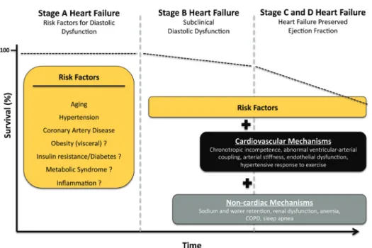
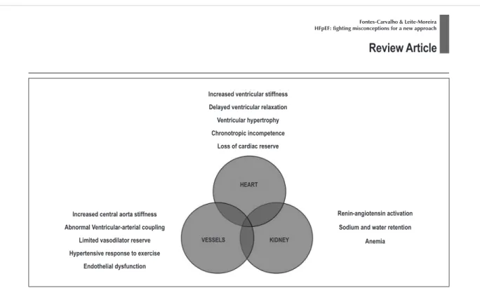
(33) CHAPTER I | INTRODUCTION. 3. THE IMPACT OF DIASTOLIC DYSFUNCTION: PROGNOSTIC IMPLICATIONS 3.1. Diastolic Dysfunction and Heart Failure Risk. In heart failure (HF) progression, alterations in myocardial structure and function appear before the onset of symptoms, a process known as myocardial remodeling (15). Heart failure guidelines define the classification according to the ACCF/AHA stages of heart failure (Stages A, B C and D), recognizing that both risk factors and abnormalities of cardiac structure and function are associated with the onset of clinical heart failure (16) (17). These stages are progressive and inviolate, and once a patient moves to a higher stage, regression to an earlier stage of heart failure is not possible. Prospective studies have shown that asymptomatic diastolic dysfunction is associated with the development of symptomatic heart failure and increased long-term mortality (9) (18) (19). Therefore, an individual with diastolic dysfunction, but without symptoms of heart failure, is classified has having a stage B in the ACCF/AHA classification of heart failure (20), as shown in figure 2.. Figure 2. Risk factors for subclinical diastolic dysfunction and mechanisms involved in the progression from subclinical diastolic dysfunction to HFpEF. Several risk factors can contribute to the development of subclinical diastolic dysfunction (stage B Heart Failure). At a latter stage, other cardiac and extra-cardiac mechanisms are responsible for the progression from subclinical diastolic dysfunction to clinical heart failure (stages C and D). It is known that the survival decreases significantly once symptoms appear (stages C and D of heart failure) but the duration and survival of stages A and B is not well established.. 31.
(34) CHAPTER I | INTRODUCTION. 3. The Impact of Diastolic Dysfunction: prognostic implications. 3.2. Diastolic Dysfunction in the Pathophysiology of Heart Failure with Preserved Ejection Fraction. (HFpEF) Heart failure with Preserved Ejection Fraction (HFpEF), also known as diastolic heart failure, accounts for approximately 50% of heart failure patients (21) (22), but its prevalence is increasing (21). LVDD is present in nearly all of HFpEF patients (23) (24) and, therefore, current consensus guidelines recommend that evidence of abnormal LV diastolic function is required for the diagnosis of HFpEF (17) (25). Nevertheless, the pathophysiology of HFpEF is complex, and involves several other cardiac and noncardiac factors, including loss of chronotropic reserve, ventriculo-vascular coupling mismatch, changes in systemic and pulmonary vascular function, impaired left atrium function, water and sodium retention and numerous comorbidities (2) (26) (27), as summarized in figure 2. Furthermore, most patients with heart failure, irrespective of ejection fraction, have abnormalities in both systolic and diastolic dysfunction (3). For a more detailed overview on the role of diastolic dysfunction in the pathophysiology of heart failure with preserved ejection, two review articles were included at the end of this first chapter (4) (26).. 3.3. The Impact of Diastolic Function on Exercise Capacity and Quality of Life. Beyond its prognostic impact and association with adverse clinical outcome, LVDD is also associated with impaired quality of life (11), probably due to reduced exercise capacity. The evaluation of the main determinants of reduced exercise capacity is complex because it is caused by simultaneous changes in both cardiac and non-cardiac factors. Nevertheless, several studies have shown a poor relation between exercise capacity and systolic function, especially when assessed by ejection fraction (28). On the contrary, in small and selected groups of patients, LVDD was an important and independent determinant of reduced exercise capacity, namely in patients with systolic heart failure (29-31), heart failure preserved ejection fraction (32) and in patients referred for exercise echocardiography (28). Several pathophysiological mechanisms can explain the association between worse diastolic function and reduced exercise performance. In patients with LVDD, impaired relaxation and increased filling pressures cause a reduction in left ventricular filling, thus decreasing the stroke volume response to exercise (2). Moreover, increased end-diastolic pressure leads to increased pulmonary capillary wedge pressure, which negatively affects the gas-exchange response during exercise and causes the sensation of exertion dyspnea (33).. RICARDO FONTES-CARVALHO. After myocardial infarction, exercise capacity is an important predictor of cardiovascular outcomes,. 32. decreased quality of life and disability (33) (34). However, in these patients, the information on the relative contribution of diastolic and systolic function as predictors of reduced functional capacity is sparse, especially using modern echocardiographic parameters. Therefore, in this project we also aimed to evaluate the impact of diastolic function as a predictor of reduced functional capacity after myocardial infarction..
(35) CHAPTER I | INTRODUCTION. 3. The Impact of Diastolic Dysfunction: prognostic implications. Recent studies have shown that exercise training can be a new intervention to improve diastolic function. In selected groups of patients with HFpEF the combination of endurance and resistance training significantly enhanced diastolic function and improved exercise capacity and quality of life (32). Thus, we aimed to evaluate whether a structured program of exercise training could improve diastolic function in patients after myocardial infarction.. 33.
(36) CHAPTER I | INTRODUCTION. 4. THE EVALUATION OF LEFT VENTRICULAR DIASTOLIC FUNCTION The evaluation of diastolic function can be complex and challenging. It involves the characterization of myocardial relaxation, of ventricular stiffness and of pressure changes during diastole. All these events can be measured invasively using several hemodynamic parameters(2). However, in routine clinical practice the assessment of diastolic function should be made non-invasively by echocardiography. In the last two decades, the echocardiographic evaluation of diastolic function has been difficult due to lack of consensus on the echocardiographic definition of LVDD (35) and to the limitations of the traditional echocardiographic parameters, especially those derived from mitral inflow velocities analysis(3). More recently, new echocardiographic techniques for the evaluation of diastolic function have appeared and the European Association of Echocardiography and the American Society of Echocardiography published new consensus recommendations for left ventricular diastolic function assessment(3). This document strongly advise the systematic use of tissue Doppler-derived early mitral annulus velocity (E’ wave) and E/E’ ratio (ratio between the E wave velocity of the diastolic mitral inflow and the E’ velocity of tissue Doppler) for the evaluation of diastolic function. It is known that the E’ velocity is a preload-independent index of LV relaxation (36) (37), whereas the E/E’ ratio can be used to estimate increased left ventricle filling pressures (3) (38) (39). Moreover, using an integrated analysis of E’ velocities with other echocardiographic parameters of diastolic function evaluation (such as E/A ratio, E-wave deceleration time, pulmonary flow analysis and left atrium volume), patients can be categorized in 4 grades of diastolic dysfunction: 1) normal diastolic function; 2) grade I or mild LVDD; 3) grade II or moderate LVDD and 4) grade III or severe LVDD(3). Recently, a new grade of LVDD has been proposed – grade IA – which is an intermediate step between grades I and II of LVDD, and has been proposed to be associated with increased risk of cardiovascular events (40). In this study, the new grade IA of diastolic dysfunction was frequent, affecting almost 18% of a selected population of patients from a tertiary center showing a high prevalence of cardiovascular disease and cancer. Until now, no further information is available regarding the prevalence and clinical characteristics of this new grade of diastolic dysfunction in the general population, a gap that we tried to evaluate in this project. Another approach for the evaluation of left ventricular diastolic function can be the assessment of left atrium (LA) volumes and function. Several studies have shown a significant correlation between LA remodeling and LVDD(41). Indeed, it is stated that, whereas Doppler velocities and time measurements. RICARDO FONTES-CARVALHO. reflect diastolic properties at the time of measurement, LA volumes can be considered “chronic markers”. 34. of diastolic dysfunction and of long-term increased filling pressures (3) (35) (41). Left atrium function encompasses 3 phases – reservoir, conduit and active contraction phases – that can actively modulate left ventricular function, especially diastolic function (42). Therefore, the interaction between the LA and the LV, also known as the atrium-ventricular coupling, can directly influence global cardiac function, cardiac output and exercise capacity (43). There is a growing interest in studying the role of.
(37) CHAPTER I | INTRODUCTION. 4. The Evaluation of left Ventricular Diastolic Function. LA volumes and several indexes of LA function – especially using novel speckle tracking echocardiographic analysis (44) (45) – as markers of diastolic dysfunction and determinants of exercise capacity. Twodimensional speckle tracking is a new echocardiographic tool that tracks the speckle pattern, frame by frame, to calculate strain and strain rate. Strain represents myocardial deformation, whereas strain rate represents the speed at which myocardial deformation occurs(46). Figure 3 illustrates the variation of LA volumes and longitudinal strain curves across the cardiac cycle and gives an overlook of the three phases of LA function.. Figure 3. The evaluation of left atrium function, volumes and longitudinal strain by speckle tracking analysis. The left atrium plays an important role in the regulation of global cardiac function. It acts as a reservoir during systole, as a conduit during early diastole and as an active blood pump during late diastole. The yellow line shows the variation of LA volumes across the cardiac cycle. The black line depicts the curve of LA longitudinal strain, showing the peak longitudinal atrial strain (PLAS) and the peak atrial contraction strain (PACS). In the future, the evaluation of diastolic function will continue to evolve with the introduction of new echocardiographic techniques, such as the evaluation of LV twist and torsion (3), 3D myocardial strain (47) and left atrium analysis (48). Diastolic function can also be evaluated by cardiac magnetic resonance using volumetric filling curves analysis, phase-contrast imaging, tagging and strain-encoded imaging (49). A new promising technique is the use of post-contrast T1 mapping, which is a marker of diffuse myocardial fibrosis showing a good correlation with LV stiffness (50). A complete review on the different methods for the evaluation of diastolic function is beyond the scope of this document. However, at the end of this chapter, one of the review articles provides additional information on the advantages and limitations of the different echocardiographic parameters for the evaluation of diastolic function (26). 35.
(38) CHAPTER I | INTRODUCTION. 5. THE DETERMINANTS OF DIASTOLIC DYSFUNCTION: WHAT IS KNOWN AND WHAT IS UNKNOWN The early identification and correction of the main determinants of subclinical diastolic dysfunction can be important to reduce the risk of progression to overt heart failure (16) (20). This can be especially important for the prevention of HFpEF, a disease where no therapy or intervention has shown to significantly change the prognosis(51).. 5.1. Established Determinants of Diastolic Dysfunction. Aging, hypertension and coronary artery disease are well-known determinants of left ventricular diastolic function(9) (10) (11) (12) (52). The major determinant of LVDD is age(13) (11) with almost 70% of individuals older than 75 years having some degree of diastolic dysfunction(9). Several studies have also shown a strong association between diastolic dysfunction and hypertension(9) (11) (53). Because myocardial relaxation is an active process that depends on ATP production, LVDD is frequent in patients with coronary artery disease and, when present, is associated with worse outcome(54).. 5.2. Obesity as a New Determinant of Diastolic Dysfunction: Understanding “Obesity. Cardiomyopathy” Obesity has reached global epidemic proportions worldwide(55), being nowadays a public health concern. Obesity is associated with increased risk of death (56) and of numerous comorbidities, including hypertension, type II diabetes mellitus, dyslipidemia, obstructive sleep apnea, certain cancers, and atherosclerotic cardiovascular disease(57). Beyond these well-known maladaptive effects, more recent studies have shown that obesity can also directly induce changes in cardiac structure and function, particularly left ventricular hypertrophy and subclinical diastolic dysfunction (58) (59). Because obesity is also independently associated with heart failure risk, it is hypothesized that diastolic dysfunction is an important pathophysiological link between obesity and heart failure (60). These observations have given a new insight into the recognition of “obesity cardiomyopathy” as a new entity, as stated in the recent heart failure guidelines(16). Data from the Framingham Heart Study showed that for every 1Kg/m2 increase in body mass index, the risk of heart failure increased 5% in men and 7% in women, with graded increases in the risk of heart failure across all body mass index categories(60). Therefore, given the high prevalence of. RICARDO FONTES-CARVALHO. obesity worldwide and the increased risk of heart failure in obese individuals, there is considerable interest. 36. in understanding the mechanisms involved in the association between obesity, diastolic dysfunction and heart failure(16). In the general population, increased body mass index has been recently associated with subclinical diastolic dysfunction(58). However, it is widely known that body mass index is an imperfect measure of.
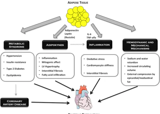
(39) CHAPTER I | INTRODUCTION. 5. The Determinants of Diastolic Dysfunction: what is known and what is unknown. body fat, being influenced by muscle mass, body water content and others. Furthermore, the relative role of central versus total or subcutaneous adiposity in inducing changes in cardiac structure and function needs further clarification. Visceral fat is the metabolically most active organ, secreting adipokines and contributing to a systemic pro-inflammatory state that can affect the cardiovascular system and diastolic function(61). Interestingly, several studies have shown that waist circumference – which is more closely related with excess visceral fat (62) – is a stronger determinant of myocardial infarction and cardiovascular disease and death, when compared to body mass index or subcutaneous fat (63) (56). Therefore, in this project we aimed to evaluate the relative role of adipose tissue distribution, especially the role of central (and visceral) versus peripheral (and subcutaneous) fat, as determinants of LVDD. The heart is also covered by fat, the epicardial adipose tissue(64). Epicardial fat contacts directly with the heart and the coronary arteries, without any mechanical barrier separating it from the cardiomyocytes and vessels, and sharing the same blood supply (65). Because epicardial fat produces several pro-inflammatory and pro-atherogenic cytokines (66, 67) it is postulated that it can directly influence the heart. Indeed, several studies have recognized epicardial fat as an independent determinant of the development and progression of coronary artery disease(68) (69) (70) (71) (72). Thus, it is expected that epicardial adipose tissue can also directly influence myocardial structure and function(73) (74) (75) (76), especially diastolic function, by local/paracrine mechanisms. The evaluation of the effect of epicardial fat on left ventricular diastolic function was another aim of this work. The pathophysiological mechanisms involved in the association between obesity and LVDD are poorly understood. Figure 4 illustrates several of these potential mechanisms (77). First, the effect could be indirect, because obesity is associated with other cardiovascular risk factors that can cause LVDD, such as hypertension(57). However, previous studies have shown that the association between increased adiposity and diastolic function is independent of these traditional risk factors(58). Alternatively, obesity can directly affect myocardial structure and function by several mechanisms, such as by increasing circulating volume and cardiac output(78), by inducing a systemic pro-inflamatory state(61) (79) or through the secretion of several adipokines (80, 81). In this project we aimed to evaluate the relative importance of the direct (adiposity mediated) and indirect (blood-pressure- and inflammation-mediated) mechanisms involved in the association between increased adiposity and diastolic function. As previously stated, the adipose tissue is an endocrine organ that produces several substances, called adipokines. In experimental studies, some of these adipokines (such as leptin, adiponectin and resistin) have been shown to directly induce left ventricle remodeling and myocardial dysfunction (66, 82). However, few data are available on the effect of adipokines as determinants of diastolic function in the general population.. 37.
(40) CHAPTER I | INTRODUCTION. 5. The Determinants of Diastolic Dysfunction: what is known and what is unknown. Figure 4. Potential (direct and indirect) pathophysiological mechanisms involved in the association between increased adiposity and diastolic dysfunction.. 5.3. Diastolic Function in the Diabetic Continuum: the Role of Insulin Resistance, Metabolic. Syndrome and Diabetes on Diastolic Dysfunction Although controversial, several studies have shown that diabetes can affect cardiac structure and function, independently of changes in blood pressure or coronary artery disease, a condition called diabetic cardiomyopathy (83) (84). In humans, LVDD is considered the earliest manifestation of myocardial involvement in type 2 diabetes mellitus (85) and a key component of diabetic cardiomyopathy (83). More recent data have shown that changes in diastolic function precede the onset of diabetes, being already present in pre-diabetic patients (86) (87), suggesting that LVDD is not caused by sustained hyperglycemia or to a “glucotoxic” effect. Insulin resistance can be one of the main pathophysiologic mechanisms involved in this diabetic cardiomyopathy because insulin resistance induces changes in myocardial substrate utilization (88) (83), increases myocardial interstitial fibrosis (89), activates the sympathetic nervous system (90) and impairs ventricular-vascular coupling by increasing arterial stiffness (91) (92).. RICARDO FONTES-CARVALHO. Metabolic syndrome, or insulin resistance syndrome, is a cluster of cardiovascular risk factors with. 38. a growing prevalence worldwide (93) that have been shown to act synergistically to increase the risk of adverse cardiovascular events (94). Interestingly, individuals with metabolic syndrome have increased prevalence of LVDD (95) (96) that frequently has a subclinical course (97). Few population-based studies have evaluated LVDD in these different stages of the diabetic continuum, especially the role of insulin resistance..
(41) CHAPTER I | INTRODUCTION. 6. REVIEW ARTICLE “Heart Failure with Preserved Ejection Fraction: Fighting Misconceptions for a New Approach”. Review Article Heart Failure with Preserved Ejection Fraction: Fighting Misconceptions for a New Approach Ricardo Fontes-Carvalho1,2 and Adelino Leite-Moreira1,3 Serviço de Fisiologia da Faculdade de Medicina do Porto1; Serviço de Cardiologia do Centro Hospitalar de Vila Nova de Gaia2; Centro de Cirurgia Torácica do Hospital de São João3, Porto - Portugal. Abstract Over the last decades, heart failure with preserved ejection fraction (HFpEF) has received less attention by the medical and scientific communities, which led to the emergence of a number of misconceptions concerning its characteristics, diagnostic and therapeutic approach. In recent years, new studies have changed the concepts traditionally associated with HFpEF, contributing to a new view towards this disease. This review is intended to discuss the latest evidence on HFpEF and to fight the main misconceptions associated with it in order to improve its diagnostic and therapeutic approach. Today we have several data showing that HFpEF is a condition that requires a different clinical approach from that used in systolic heart failure (SHF). HFpEF is no longer seen as a “benign” disease because it is associated with a poor prognosis and high prevalence. Its pathophysiology is complex and not fully clarified. In addition to diastolic dysfunction, we now know that other cardiac and extracardiac factors are also involved in its onset and progression. Using recent consensus guidelines we have objective criteria for its diagnosis, especially by using the new echocardiographic parameters for assessing diastolic function, including the E/e’ ratio obtained by tissue Doppler. Finally, treatment of HFpEF remains unknown, because no therapeutic strategy has been shown to improve HFpEF prognosis. Thus, in this review we will also discuss the potentially new therapeutic targets for HFpEF.. Introduction Heart failure (HF) represents a major and growing public health problem, affecting 2% - 3% of adults in developed countries1. Patients with heart failure are classically divided into two groups: those with HF with preserved ejection fraction. Keywords Heart failure/therapy; stroke volume; ventricular function; demographic aging. Mailing address: Ricardo Fontes-Carvalho • Serviço de Fisiologia da Faculdade de Medicina do Porto Al. Prof. Hernâni Monteiro 4200 - 319 Porto - Portugal E-mail: fontes.carvalho@gmail.com, ricardo@med.up.pt Manuscript received December 01, 2009; revised manuscript received February 17, 2010; accepted March 24, 2010.. (HFpEF), also called diastolic HF (DHF) and those with HF and reduced ejection fraction (HFrEF), better known as systolic HF (SHF)1. In recent decades, HFpEF has received much less attention from medical and scientific communities, a situation that is finally starting to change. Such lack of attention resulted in the gradual emergence, within the medical community, of a series of misconceptions and dogmas concerning the epidemiology, diagnosis, pathophysiology and treatment of HFpEF. With this review, we intend to explore and tackle the major misconceptions associated with HFpEF. We will discuss the latest evidence concerning HFpEF, providing a new view on this complex syndrome, in order to improve its clinical and therapeutic approach.. Frequent misconceptions in heart failure with preserved ejection fraction Misconception 1: HFpEF is a benign condition Until recently, HFpEF had been considered an essentially “benign” disease associated with a better prognosis. Epidemiological studies have shown that the prognosis for these patients is as bad as those who have systolic HF (SHF)2,3. Patients with HFpEF have mortality rates of 29% after one year (versus 32% in patients with systolic HF), and 65% after five years (versus 68%)3. The morbidity of HFpEF is also very high, requiring frequent admissions and a significant consumption of resources4,5. Once admitted due to HF, these patients have a high rate of readmission of 50% after one year5. Equally worrying is the evidence showing that the survival of patients with HFpEF has not been improving in recent decades, unlike what has been observed in patients with systolic HF6. Such observation is probably related to the fact that the management and treatment of these patients are not producing the desired effects, probably due to various misconceptions concerning HFpEF. Misconception 2: diastolic HF is an uncommon syndrome A second misconception in HFpEF is to think that this is a clinical condition that is less common than the SHF. This is quite the opposite! We know today that HFpEF is responsible for about 50% of all patients admitted with HF, a proportion that increases with age2,4,6-8. Moreover, in the last two decades the proportion of patients with HFpEF increased. 504. 39.
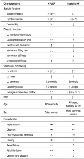
(42) CHAPTER I | INTRODUCTION. 6. Review Article | “Heart Failure with Preserved Ejection Fraction: Fighting Misconceptions for a New Approach”. Fontes-Carvalho & Leite-Moreira HFpEF: fighting misconceptions for a new approach. Review Article from 38% to 54% out of cases of HF6, a proportion that will continue to rise due to the progressive aging of population and expected increase in the prevalence of hypertension, obesity and diabetes. Misconception 3: diastolic HF and systolic HF are the same condition The classical separation of HF in HFpEF is questioned by several authors who argue that these relate to the same condition, albeit with different phenotypes9,10.. RICARDO FONTES-CARVALHO. Characteristics. HFpEF. Systolic HF. Systolic function Ejection fraction. N (or ↑). ↓↓. Ejection volume. N (or ↓). ↓ (or N). N. ↓. Contractility Diastolic function. However, there are many demographic, epidemiological, histological, molecular and structural arguments, as well as some relating to ventricular function and even therapeutic effectiveness, which seem to clearly indicate that these two conditions are quite different (Table 1)6,11.. LV telediastolic pressure. ↑↑. ↑. Constant relaxation time. ↑↑. ↑. Relative wall thickness1. ↑. ↓. Ventricular filling rate. ↓↓. ↓. Regarding the characteristics of the population, patients with HFpEF are older, often female, and have a high prevalence of hypertension, diabetes and obesity, as well as various comorbidities such as atrial fibrillation, renal failure and anemia2-4,7,12,13 (Table 1).. Ventricular stiffness. ↑↑. ↓. Myocardial stiffness. ↑. N. N (or ↓). ↑↑. The hearts of patients with systolic HF and HFpEF also have significant differences in terms of structure and ventricular function (Table 1). The hearts of patients with SHF present an eccentric ventricular modeling with increased diastolic volumes and the main anomaly occurs in LV systolic properties14 (Figure 1). By contrast, patients with HFpEF present as concentric remodeling, the volumes are normal or even reduced, and the main change occurs in the diastolic properties, with delayed relaxation and/or increased ventricular stiffness14-16 (Figure 1 and Table 1).. 40. Table 1 – Comparison of characteristics of patients with systolic HF and HFpEF. Other recently published studies have also shown differences at histological and molecular level. For example, analysis of endomyocardial biopsies revealed that cardiomyocytes of patients with HFpEF are structurally different, with larger diameters, greater stiffness and increased density of myofilaments, compared to patients with ICS17. Significant differences were also discovered at the molecular level. Titin is a molecule found inside the sarcomere which, given its elastic properties, is the main determinant of the stiffness of cardiomyocytes. It was found that in patients with HFpEF there is a change in the expression of the isoforms of this molecule - with increased expression of the stiffer isoform - or its degree of phosphorylation, which contributes to the increase in ventricular stiffness observed in these patients17,18. Patients with HFpEF and systolic HF also have significant differences in fibrosis and extracellular collagen matrix, due to distinct patterns of extracellular matrix metalloproteinases (MMP) and tissue inhibitors of such metalloproteinases (TIMP) activation. While in HFpEF there is a decreased degradation of extracellular matrix (resulting in increased ventricular stiffness), in dilated cardiomyopathy there is an increased matrix degradation19,20. In the HFpEF, diastolic dysfunction ca occur due to changes in the passive properties of the ventricle - particularly increased ventricular stiffness - or due to alterations in myocardial relaxation. The delay in myocardial realaxation seen in patients with HFpEF is caused by changes in calcium kinetics, especially by reduced activity. Ventricular remodeling LV volume LV mass. ↑. ↑. LV geometry. Concentric. Eccentric. Cardiomyocytes. ↑ Diameter. ↑ Length. ↑↑. ↓ (or N or ↑). Collagen extracellular matrix BNP. ↑. ↑↑. Age. Often elderly. All ages, typically 50-70. Sex. Often women. More common in men. Hypertension. +++. ++. Diabetes. +++. ++. +. +++. Obesity. +++. +. Renal failure. ++. 0. Atrial fibrillation. ++. +. Chronic lung disease. ++. 0. Comorbidities. Prior myocardial infarction. Abbreviations: LV - left ventricle, N - normal; ↑ - increased; ↓ - decreased; BNP brain natriuretic peptide; The relative wall thickness describes the left ventricular geometry and is defined as the ratio between the left ventricular thickness and the left ventricular cavity diameter.. of SERCA2, the main protein responsible for the reuptake of calcium back into the sarcoplasmic reticulum21. Finally, strong arguments related to the response to pharmacological therapy justify the separation of these two conditions. Few clinical trials performed to date on HFpEF reveal that these patients do not respond as well to therapy commonly used in systolic HF, suggesting that different pathophysiological mechanisms operate in these two conditions. These differences mean that the therapeutic approach to HFpEF must be different from that used in systolic HF, as prescribed in the guidelines for heart failure1,22.. Arq Bras Cardiol 2011;96(6):504-514. 505.
Imagem
+7

Documentos relacionados
Neste trabalho o objetivo central foi a ampliação e adequação do procedimento e programa computacional baseado no programa comercial MSC.PATRAN, para a geração automática de modelos
É nesta mudança, abruptamente solicitada e muitas das vezes legislada, que nos vão impondo, neste contexto de sociedades sem emprego; a ordem para a flexibilização como
Assim, todas as pessoas, independente de sexo, orientação sexual ou identidade de gênero, têm direito de gozar da proteção assegurada pelo regime internacional
Porém, ao analisar as 11 dissertações e uma tese a que tivemos acesso na íntegra, verifi camos que entre as obras mais citadas estão: Processo de escolarização do Ensino
Deste modo ampliamos a noção de “texto” para além do escrito e do verbal e buscamos aliar esse olhar a uma perspectiva que relaciona Ciência, Tecnologia e Sociedade
Peça de mão de alta rotação pneumática com sistema Push Button (botão para remoção de broca), podendo apresentar passagem dupla de ar e acoplamento para engate rápido
Extinction with social support is blocked by the protein synthesis inhibitors anisomycin and rapamycin and by the inhibitor of gene expression 5,6-dichloro-1- β-
•Para não perder a garantia e para evitar problemas técnicos, não consertar o aparelho em casa.. Quando precisar, o usuário deverá levá-lo à Assistência Técnica
