Universidade de Lisboa
Faculdade de Medicina Dentária
In vitro microtensile bond strength of a
Universal Adhesive System to dentin using
Etch-and-Rinse and Etch-and-Dry technique
Sara Palmares Fernandes
Dissertação
Mestrado Integrado em Medicina Dentária
Universidade de Lisboa
Faculdade de Medicina Dentária
In vitro microtensile bond strength of a
Universal Adhesive System to dentin using
Etch-and-Rinse and Etch-and-Dry technique
Sara Palmares Fernandes
Dissertação orientada pela Dra. Ana Pequeno
e co-orientada pela Dra. Ana Catarina Coito
Mestrado Integrado em Medicina Dentária
i
“Para ser grande sê inteiro: nada Teu exagera ou exclui. Sê todo em cada coisa; Põe quanto és
No mínimo que fazes. Assim em cada lago, a lua toda
Brilha, porque alta vive.”
iii
Agradecimentos
À minha orientadora, Dra. Ana Pequeno, pelo constante apoio, paciência e disponibilidade que sempre demonstrou perante as minhas dúvidas e nas inúmeras vezes que reviu a minha tese. Por me ter introduzido no mundo da Adesão Dentária e da Microtração e partilhado comigo todo o seu conhecimento.
À minha co-orientadora, Dra. Catarina Coito, por me ter transmitido o gosto pela investigação, pela constante disponibilidade e entusiasmo contagiante nas horas laboratoriais.
Ao Departamento de Dentisteria Operatória por toda a ajuda prestada e pela forma alegre com que brindaram todas as horas laboratoriais. Ao Prof. Dr. Alexandre Cavalheiro, mentor deste projeto.
Ao Prof. Dr. Luís Pires Lopes pela disponibilização das instalações, materiais e equipamentos do Laboratório de Biomateriais da Faculdade de Medicina Dentária da Universidade de Lisboa.
Aos meus queridos amigos, Joana Costa, Inês Marcos, Margarida Simões, Daniela Rocha, Ruben Pereira e David Braz, pela sua amizade durante estes cinco anos e por terem partilhado comigo os bons e os maus momentos.
Aos meus amigos de infância, Ricardo Santos, Pedro Ribeiro, Marta Didier e Nadja Leite, por estarem presentes em todos os momentos da minha vida.
À minha família por tudo, e em especial aos meus Pais e Irmã, pelo amor incondicional, motivação e apoio em todas as etapas da minha vida.
v
Table of Contents
Abstract ... xi
Resumo ... xiii
I. Introduction ... 1
1. Classification of contemporary adhesives ... 2
1.1 Total-etch or Etch-and-rinse adhesives ... 2
1.2 Self-etch or Etch-and-dry adhesives ... 3
2. Microtensile bond strength test ... 5
II. Materials and Methods ... 7
1. Type of study ... 8
2. Design of the study ... 8
3. Teeth preparation ... 8
4. Distribution and treatment of the crown segments ... 11
5. Specimens preparation for the microtensile tests... 13
6. Microtensile tests ... 14 7. Statistical Analysis ... 15 III. Results ... 17 IV. Discussion ... 21 V. Conclusions ... 29 VI. Appendices ... I APPENDIX 1 ... II APPENDIX 2 ... III APPENDIX 3 ... IV APPENDIX 4 ... V APPENDIX 5 ... VI VII. References... VII
vii
List of tables and figures
Table 1 - Tests of Normality of the µTBS in MPa for the two different
experimental groups tested. ... 15
Table 2 - Test for Equality of Variances of the µTBS in MPa. ... 15
Table 3 - Descriptive statistics of the µTBS in MPa for the two experimental groups tested. ... 18
Table 4 - t-test for Equality of Means (p≤0,05). ... 18
Table 5 - Number of specimens according to the failure mode and premature failures of the two different experimental groups tested. ... 19
Table 6 - Number of specimens according to the failure mode and premature failures of all experimental groups tested. ... 20
Table 7 - Materials, Manufacturer, Composition and Batch Numbers. ... II Table 8 - Application mode of Scotchbond Universal Adhesive according to the manufacturer’s instructions. ... III Table 9 - Application mode of Scotchbond Universal Adhesive according to the detailed manufacturer’s instructions. ... IV Table 10 - Number of sticks obtained in each tooth. ... VI Figure 1 - Diamond wafering blade. ... 8
Figure 2 - Isomet 1000 Precision Saw, hard tissue microtome. ... 8
Figure 3 - Tooth attached to an acrylic holder with sticky wax. ... 9
Figure 4 - Roots cutting 1-2 mm below CEJ. ... 9
Figure 5 - Exposure of the pulp chamber after roots cut. ... 9
Figure 6 - Removal of the pulp tissues. ... 9
Figure 7 - Filling of the pulp chamber with cyanoacrylate glue. ... 9
Figure 8 - The crown fixed to the acrylic holder. ... 10
Figure 9 - Removal of the occlusal enamel and superficial dentin. ... 10
Figure 10 - Mid coronal dentin surface. ... 10
Figure 11 - Lunn Major, mechanical grinder. ... 10
Figure 12 - Scotchbond Universal Adhesive and Scotchbond Universal Etchant. ... 12
viii
Figure 14 - Resin composite build-up. ... 12
Figure 15 - The teeth after being sectioned in both “x” and “y” directions. ... 13
Figure 16 - The sticks obtained after the final cut. ... 13
Figure 17 - The specimen attached to a Geraldelli’s jig. ... 14
Figure 18 - Instron 4502, universal testing machine. ... 14
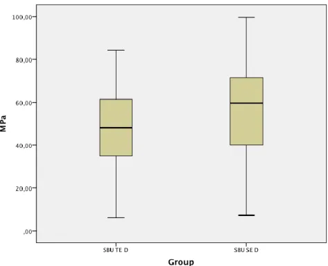
Figure 19 - Box-whisker plots of the µTBS in MPa for the two different experimental groups tested. ... 18
Figure 20 - Distribution of the specimens according to the failure mode of the two different experimental groups tested. ... 19
Figure 21 - Distribution of the specimens according to the failure mode of all experimental groups tested. ... 20 Figure 22 - Cyanoacrylate glue. ... V Figure 23 - Halogen light-activation unit. ... V Figure 24 - Incubator. ... V Figure 25 - Radiometer. ... V Figure 26 - Digital caliper. ... V Figure 27 - Stereomicroscope. ... V
ix
Abbreviates
ED ER HAp 10-MDP 4-MET MMP-2 MMP-8 MMP-9 PA Phenyl-P Ptf SBU SE TE UA µTBS Etch-and-dry Etch-and-rinse Hydroxyapatite10-methacryloyloxydecyl dihydrogen phosphate 4-methacryloyloxyethyl trimellitic acid
Matrix metalloproteinase-2 Matrix metalloproteinase-8 Matrix metalloproteinase-9 Polyalkenoic acid copolymer
2-(methacryloyloxyethyl)phenyl hydrogenphosphate Pre-testing failures Scotchbond Universal Self-etch Total-etch Universal adhesives Microtensile bond strength
xi
Abstract
Objectives: The purpose of the present study is to compare the immediate microtensile bond strengths (µTBS) of one universal adhesive applied to dentine according to the etch-and-rinse and the self-etch technique. The null hypothesis tested was that the bonding effectiveness to dentin was not affected when the adhesive was applied either following an etch-and-rinse or etch-and-dry technique.
Methods: Six recently extracted human third molars, intact and without macroscopic evidence of caries or restorations, were assigned to two groups according to the etching strategy: Group SBU TE D – Scotchbond Universal Adhesive (3M ESPE) applied as a 2-step etch-and-rinse adhesive on moist dentin; Group SBU SE D - Scotchbond Universal Adhesive applied as a 1-step self-etch adhesive on dentin. After resin composite build-ups were performed, the teeth were stored in distilled water in an incubator for 24 hours at 37°C. Specimens were sectioned with a slow-speed diamond saw under water irrigation to obtain bonded sticks that were tested to failure in a universal testing machine at a crosshead speed of 1mm/minute. The statistical analysis of the results was performed with SPSS. A paired-sample t-test was performed when the assumption of normality was valid.
Results: The mean µTBS to dentin of the Scotchbond Universal Adhesive applied following the etch-and-dry approach (SBU SE D) was statistically higher than that when the SBU was applied following the etch-and-rinse approach (SBU TE D) (p = 0,008).
Conclusions: Within the limitations of the present laboratory study, it may be concluded that improved bonding effectiveness of SBU on dentin is obtained when the adhesive is applied with the self-etch approach. The etch-and-rinse approach tested (wet dentin) resulted in immediate bond strength statistically lower than the self-etch group.
Keywords: Dental adhesives, universal adhesives, dentin bonding, microtensile
xiii
Resumo
Durante as últimas décadas, a adesão dentária tem adquirido uma importância cada vez maior. O principal desafio dos sistemas adesivos amelodentinários é providenciar uma adesão eficaz a tecidos duros de diferente natureza. Desde os trabalhos de Buonocore, a adesão ao esmalte permaneceu simples e consistente ao longo dos anos. Pelo contrário, a adesão à dentina revelou-se imprevisível devido às suas características morfológicas, levando à sucessiva modificação dos sistemas adesivos.
O mecanismo base da adesão, tanto ao esmalte, como à dentina, é essencialmente um processo que envolve a substituição da porção mineralizada destes tecidos pelos monómeros da resina adesiva, que se unem micromecanicamente às porosidades criadas pelo condicionamento ácido. As fragilidades inerentes a este mecanismo, em especial na dentina, devem-se à complexidade da técnica e à sua sensibilidade. Consequentemente, a exigência por sistemas adesivos mais simples, com um menor número de passos e uma técnica menos sensível, é cada vez maior, levando os fabricantes a desenvolverem novos adesivos num curto período de tempo.
Apesar dos sistemas adesivos mais simplificados apresentarem uma longevidade comprometida tanto in vitro, como in vivo, comparativamente com aqueles que se baseiam em diversos passos, alguns fabricantes introduziram recentemente uma classe de adesivos mais versátil, desenvolvida para simplificar e reduzir o tempo de aplicação clínico. Estes novos adesivos podem ser aplicados segundo a técnica etch-and-rinse (2 passos) ou etch-and-dry (1 passo). Para os adesivos etch-and-rinse, a força adesiva provém da formação da camada híbrida. Contrariamente, para os adesivos etch-and-dry, a adesão química tem mostrado ter um papel mais importante. Os adesivos mild self-etch têm como mecanismo de adesão uma combinação entre a adesão química à hidroxiapatite e a adesão micromecânica.
Objetivos: Um único sistema adesivo que possa ser aplicado de diferentes formas e que permita ao clínico escolher qual a estratégia adesiva mais adequada para a cavidade preparada, é desejável. Estes novos adesivos são chamados de Adesivos Universais, ou
“Multi-purpose” ou “Multi-mode”. Devido ao fato destes adesivos serem novos no
mercado e do respetivo conceito ser recente, a informação existente na literatura sobre o desempenho in vitro e in vivo desta nova categoria de adesivos é escassa. Por esta razão, o objetivo deste estudo laboratorial é comparar as forças de adesão à dentina de um
xiv
adesivo universal, segundo a estratégia etch-and-rinse e etch-and-dry, através de testes de microtração (µTBS) (Scotchbond Universal Adhesive, 3M ESPE, Neuss, Germany). A hipótese nula deste estudo é: não há diferenças nas forças de adesão à microtração quando o adesivo é aplicado segundo a técnica etch-and-rinse ou etch-and-dry.
Materiais e métodos: Uma amostra conveniente composta por seis terceiros molares humanos recentemente extraídos, intactos e sem evidência macroscópica de cárie ou restaurações (n=6), foi distribuída aleatoriamente em dois grupos, segundo a estratégia de adesão: Grupo SBU TE D - Scotchbond Universal Adhesive aplicado como um adesivo etch-and-rinse de 2 passos (em dentina húmida); Grupo SBU SE D - Scotchbond Universal Adhesive aplicado como um adesivo etch-and-dry de 1 passo.
Após a preparação dos dentes e, com o objetivo de formar uma smear layer semelhante à que é obtida em situações clínicas, a superfície dos dentes foi polida com papel abrasivo de carbureto de silício (SiC) de grão 600, durante 60 segundos sob refrigeração com água, numa máquina polidora (Lunn Major, Struers, Denmark). Procedeu-se de seguida à aplicação do sistema adesivo de acordo com a distribuição nos respetivos grupos experimentais. Em todas as amostras o adesivo foi fotopolimerizado durante 10 segundos com o fotopolimerizador ELIPAR S10 (3M ESPE Seefeld, Germany) com intensidade de 600 mW/cm2, controlado periodicamente por um
radiómetro (Curing Radiometre P/N 10503, USA). De seguida foi aplicada a resina composta ENAMEL Plus HRi (Micerium S.p.A. Avegno (GE) Italy) em camadas de aproximadamente 2 mm e fotopolimerizadas entre si durante 20 segundos, de acordo com as instruções do fabricante. As faces vestibular, lingual, mesial e distal foram polimerizadas adicionalmente por mais 10 segundos cada. A face exterior da resina composta de todos os espécimes foi pintada com tinta à prova de água, com o objetivo de excluir do estudo os palitos nos quais a adesão foi feita em esmalte. Os dentes foram armazenados em água destilada numa estufa de incubação durante 24h a 37ºC e registado o dia e a hora da reconstrução.
Posteriormente, foram efetuados cortes no eixo do “x” e do “y” com um disco de diamante, a baixa rotação e sob refrigeração com água, num micrótomo de tecidos duros (Isomet 1000 Precision Saw, Buehler Ltd., Lake Buff, IL, USA), com o objetivo de obter palitos com uma área de, aproximadamente, 0,7 mm2. Cada palito foi colado individualmente num suporte de Geraldelli´s, com cola de cianoacrilato, e testados um a um. Foram sujeitos a forças de tração numa máquina de Teste Universal, a uma velocidade de 1 mm/minuto até ocorrer fratura. Mediu-se a secção de cada espécimen
xv
fraturado com uma craveira digital e determinou-se a área em milímetros quadrados (mm2). A área de superfície de cada palito e a sua resistência à fratura, medida em
KiloNewtons (KN), foram registadas e, a partir delas, calculadas as forças de adesão em MegaPascais (MPa). Cada fratura foi observada ao estereomicroscópio (Nikon, Japan) com uma ampliação de 10x, para se caracterizar o tipo de fratura ocorrida (coesiva, adesiva ou mista). Quando a fratura ocorreu exclusivamente na dentina foi denominada como coesiva de dentina (CD) e quando ocorreu exclusivamente na resina composta foi classificada como coesiva de compósito (CC). Quando a fratura ocorreu na interface dentina-adesivo, denominou-se de adesiva (A) e, quando atingiu tanto a dentina como a resina composta, foi denominada como mista (M).
A análise estatística dos resultados foi realizada através do teste paramétrico emparelhado t-test, após se ter verificado que a amostra seguia uma distribuição normal (os testes de Kolmogorov-Smirnov e Shapiro-Wilk foram usados para avaliar se os resultados seguiam uma distribuição normal; o teste de Levene foi usado para determinar a igualdade de variâncias). O intervalo de confiança definido foi de 95%. O número de palitos fraturados durante a sua preparação (palitos descolados) foi registado, no entanto, esses valores não foram considerados para a análise estatística.
Resultados: Foram testados 140 palitos, 68 correspondentes ao grupo SBU TE D e 72 correspondentes ao grupo SBU SE D. As forças de adesão quando o SBU foi aplicado segundo a técnica self-etch (SBU SE D) (56,9 ± 2,5 MPa) foram superiores às forças de adesão quando o SBU foi aplicado segundo a técnica total-etch (SBU TE D) (48,0 ± 2,1 MPa). A análise estatística com o t-test determinou a existência de diferenças estatisticamente significativas entre estes grupos (p=0,008). Após a observação do tipo de fratura ocorrida, verificou-se que, no total dos dois grupos, 49,3% foram fraturas adesivas, 7,8% mistas, 17,9% coesivas de dentina e 25% coesivas de compósito.
Conclusões: Tendo em conta as limitações deste estudo laboratorial, pode-se concluir que o SBU apresenta uma melhor eficácia na dentina quando aplicado segundo a técnica self-etch. A técnica total-etch (em dentina húmida) resultou em forças adesivas imediatas estatisticamente inferiores ao grupo self-etch.
Estudos futuros poderão avaliar o efeito a longo prazo do armazenamento em água no desempenho deste adesivo. Além disso, estudos em diferentes substratos, como por exemplo, em dentina cariada, em associação com estudos in vivo são necessários para avaliar o desempenho clínico desta nova classe de adesivos, para que possam ser utilizados futuramente com maior conhecimento.
xvi
Palavras-chave: Adesivos dentários, adesivos universais, adesão à dentina, forças
1
2
During the last decades, restorative concepts have been continually changing, and adhesive technology has become steadily more important. Adhesion to enamel was followed by adhesion to dentin. Initially simply designed “bonding agents” gradually evolved to multi-step “adhesive systems” with rather complicated application procedures (Swift et al., 1995; Van Meerbeek et al., 1998). The main challenge for dental adhesives is to provide an equally effective bond to two hard dental tissues of different nature. Bonding to enamel has been proven to be durable. Bonding to dentin is far more intricate and can apparently only be achieved when more complicated and time-consuming application procedures are followed (Swift et al., 1995; Perdigao et al., 2000; Van Meerbeek et al., 2011). Consequently, today’s adhesives are often regarded as technique-sensitive with the smallest error in the clinical application procedure being penalized either by rapid debonding or early marginal degradation (De Munck et al., 2005a; Breschi
et al., 2008; Van Meerbeek et al., 2011). As a consequence, the demand for simpler, more
user-friendly and less technique-sensitive adhesives remains high, urging manufacturers into developing new adhesives at a rapid pace.
1. Classification of contemporary adhesives
The bonding mechanism of adhesive systems basically involves the replacement of minerals removed from the hard dental tissue by resin monomers, in such a way that a polymer becomes micro-mechanically interlocked to the dental substrate (Nakabayashi
et al., 1982). However, the adhesive systems available on the market can be classified
into two categories, in function of how the adhesive interacts with the smear layer: etch-and-rinse (ER) or total-etch (TE) and etch-and-dry (ED) or self-etch (SE) adhesives (Van Meerbeek et al., 1998; De Munck et al., 2005a).
1.1 Total-etch or Etch-and-rinse adhesives
When using the ER strategy, the first step involves the application of phosphoric acid gel to both dental substrates, which allows removal of the smear layer, exposure of the collagen fibrils in dentin, and increase in surface area and surface energy in the enamel substrate (Nunes et al., 2001; Perdigao & Lopes, 2001). This conditioning step is followed by a priming step and application of the adhesive resin, resulting in a three-step procedure. Simplified two-step ER adhesives combine the primer and adhesive resin into one
3
application (Van Meerbeek et al., 1998; Van Meerbeek et al., 2003; De Munck et al., 2005a).
Irrespective of the number of steps, the main disadvantage of the ER system, mainly two-step version, is that there is risk of collagen fibre collapse during the process of demineralized dentin drying, which leads to a decrease in bond strength (Pashley et al., 1992; Swift et al., 1995; Hashimoto et al., 2000; Perdigao et al., 2002; De Munck et al., 2009). The collagen collapse is prevented by keeping demineralized dentin moist, which is a difficult task to perform clinically. In fact, adequate moisture depends on both the solvent used in the material and on the clinician’s interpretation of the manufacturer’s directions (Pashley et al., 2011; Munoz et al., 2013).
The incomplete impregnation of collagen fibers by the adhesive monomers can lead to the degradation of the exposed collagen fibrils within the hybrid layers by endogenous proteases enzymes present in dentin (colagenase MMP-8 and gelatinases MMP-2 and -9) (De Munck et al., 2005a; Pashley et al., 2011; Mazzoni et al., 2013). The need to protect them against the degrading mechanisms present in the oral cavity environment (Perdigao & Lopes, 2001; De Munck et al., 2005a), led to the development of the second category, an adhesive using the self-etch strategy (Pashley et al., 2011; Munoz et al., 2013).
1.2 Self-etch or Etch-and-dry adhesives
In the ED strategy, there is no need to apply a preliminary phosphoric acid gel on dental substrates. Dentin demineralization and priming occur simultaneously, based on the use of non-rinse acidic monomers dissolved in an aqueous solution (Van Meerbeek et
al., 2003; De Munck et al., 2005a). The goal is to incorporate the smear layer into the
hybrid layer (Tay & Pashley, 2001). This approach has been claimed to be user-friendly (shorter application time, less steps) and less technique-sensitive (no wet-bonding, simple drying) (Pashley & Tay, 2001; Tay & Pashley, 2001; Van Meerbeek et al., 2003; Yoshida
et al., 2004). Another important advantage of the self-etch is that the infiltration of resin
occurs simultaneously with the self-etching process to the same depth, by which the risk of discrepancy between both processes is low or non-existent. Therefore, collapse of air-dried, demineralized collagen is prevented (Tay & Pashley, 2001; Van Meerbeek et al., 2003). This should be attributed to their less aggressive and thus more superficial interaction with dentin (Van Meerbeek et al., 2011).
4
Even if all ED adhesives systems rely on the same bonding mechanism, they differ from each other in many aspects, such as acidic resin monomer composition, water content and acidity. Thus, these adhesives may be further classified according to the pH of the adhesive solution as mild (pH>2), intermediately strong (1<pH<2) or strong (pH≤1). Indeed the adhesive acidity influences the ability of the system to interact with the underlying enamel and dentin (Van Meerbeek et al., 2003). The high acidity of strong self-etch adhesives results in rather deep demineralization effects. Consequently, the underlying bonding mechanism is primarily diffusion-based, similar to the etch-and-rinse approach (Van Meerbeek et al., 2003; Yoshida et al., 2004). Mild self-etch systems demineralize dentin only to a depth of 1 µm. This superficial demineralization occurs only partially, keeping residual hydroxyapatite still attached to exposed collagen. Specific carboxyl or phosphate groups of functional monomers (4-MET, phenyl-P and 10-MDP) can then chemically interact with this residual hydroxyapatite (Yoshida et al., 2000; Van Meerbeek et al., 2003; Yoshida et al., 2004). This chemical bond seems to be important in stabilizing the adhesive interface on dentin over time (Van Meerbeek et al., 2011). Nevertheless, sufficient surface-porosity is created to obtain micromechanical interlocking through hybridization. The thickness of the hybrid layer is, however, much smaller than that produced by the strong self-etch or etch-and-rinse approach (Van Meerbeek et al., 2003).
Self-etch adhesives can come as “two-step” or “self-etching primers” and “one-step” adhesives, depending on whether a self-etching primer and adhesive resin are separately provided or combined into one single solution. One-step adhesives can be further divided into two-component one-step self-etch adhesive (by separating active ingredients), and single-component one-step self-etch adhesives, that are considered the only true “one-bottle” or “all-in-one” adhesives (Van Meerbeek et al., 2003; Van Meerbeek et al., 2011).
“All-in-one” must today be regarded as the most simplified adhesive protocol that can clinically be employed to bond composite to tooth enamel and dentin, combining all bonding steps into one single application step. Concerns remain regarding both their short- and long-term bonding effectiveness (Frankenberger et al., 2001). In terms of immediate performance these simplified one-step adhesives showed promising results (De Munck et al., 2003). However, the long-term performance is inferior in terms of bond durability, in particular when compared to the gold-standard three-step etch-and-rinse approach (Breschi et al., 2008; De Munck et al., 2012; Marchesi et al., 2013).
5
2. Microtensile bond strength test
The popularity of laboratory studies in the field of adhesive dentistry may in part be ascribed to the rapid evolution of dental adhesive technology and the resultant high turnover of adhesive systems, which often tempts manufacturers to release a successor product on the market even before its precursor has been clinically evaluated, at least in the long term (Van Meerbeek et al., 2003). Laboratory tests can evaluate the effect of a single variable, while keeping all other variables constant (Van Meerbeek et al., 2003; De Munck et al., 2005a).
In order to measure the bonding effectiveness of adhesives to enamel and dentin, diverse methodologies can today be used. The bond strength can be measured statically (macro-shear, macro-tensile, micro-shear, micro-tensile) or dynamically (fatigue test) (Van Meerbeek et al., 2010).
Sano et al. (1994) developed the microtensile bond strength (µTBS) test. This methodology has been accepted as a versatile and reliable in vitro static test to quantify the bonding effectiveness and stability of adhesive biomaterials bonded to tooth structure. It appears to be able to discriminate adhesives better on their bonding performance than a traditional shear bond strength approach (Armstrong et al., 1998; Schreiner et al., 1998; Pashley et al., 1999; El Zohairy et al., 2010; Poitevin et al., 2010; Van Meerbeek et al., 2010). Pashley et al. (1995) have stated a number of potential advantages for this methodology: more adhesive failures and fewer cohesive failures; higher interfacial bond strengths can be measured; the ability to measure regional bond strengths; means and variances can be calculated for single teeth; permits testing of bonds to irregular surfaces; permits testing of very small areas; and facilitates examination of the failed bonds by scanning electron microscopy, since the surface area is approximately 1 mm2. Disadvantages of µTBS test include: labor intensive and technically demanding; difficult to measure bond strengths <5 MPa, requires special equipment; samples are so small they dehydrate and damaged easily (Pashley et al., 1995; Armstrong et al., 1998; Pashley et
al., 1999).
Despite the importance of laboratory studies attempting to predict the clinical performance of biomaterials, clinical trials remain the ultimate way to collect scientific evidence on the clinical effectiveness of restorative treatment (Swift et al., 1995; Van Meerbeek et al., 2003; De Munck et al., 2005a).
6
In spite of compromised in vitro and clinical longevity associated with ultrasimplified adhesives compared with that of adhesives that rely on several steps, some manufacturers have introduced recently more versatile adhesive systems, developed to simplify and shorten the application time, making the clinical procedure more user-friendly. These new adhesives can be applied either with the etch-and-rinse (two-step) or the etch-and-dry (one-step) technique. For etch-and-rinse adhesives, bond strength is derived both from resin tag and hybrid layer formation, with resin tags being responsible for an estimated one third of the bond strengths (Van Meerbeek et al., 2003; De Munck
et al., 2005a). Otherwise, for etch-and-dry adhesives, chemical bonding has been shown
to play a more important role. Mild self-etch adhesives rely on a combination of chemical adhesion to hydroxyapatite with some micromechanical interlocking that does not depend much on resin-tag formation (Van Meerbeek et al., 2003; Yoshida et al., 2004).
Hence, an adhesive that can be applied both ways and thus enables the practitioner to decide for a specific adhesive protocol, considered to be the most suited for the cavity prepared, would be very desirable. These new materials are called “Universal”, “Multi-purpose” or “Multi-mode” adhesives (Hanabusa et al., 2012; Perdigao et al., 2012; Yoshida et al., 2012; Marchesi et al., 2013; Mena-Serrano et al., 2013; Munoz et al., 2013; Perdigao et al., 2013a; Perdigao et al., 2013b; Taschner et al., 2013; de Goes et al., 2014; Munoz et al., 2014; Perdigao & Loguercio, 2014; Pongprueksa et al., 2014; Wagner
et al., 2014).
These new adhesives and the respective concept behind them are novel. Therefore, there is little information in the literature about the performance of this new class of adhesives. Only few ultramorphological and bond strength studies, and short-term clinical studies have been published. More in vitro and clinical studies are needed to back the use of these new materials.
For this reason, the purpose of the present work is to compare the immediate microtensile bond strengths (µTBS) of one universal adhesive applied to dentin according to the etch-and-rinse and the self-etch strategies (Scotchbond Universal Adhesive, 3M ESPE, Neuss, Germany). The following null hypothesis was tested in this study: (1) There were no differences in dentin microtensile bond strength when the adhesive was applied either following an etch-and-rinse or etch-and-dry technique.
7
8
1. Type of study
Experimental in vitro study with the purpose of evaluating the microtensile bond strength of a universal adhesive system applied to dentin according to: 1) the etch-and-rinse strategy, or 2) the etch-and-dry strategy.
2. Design of the study
A convenient sample of six recently extracted human third molars, intact and without macroscopic evidence of caries or restorations, was used in this study. Before preparation, the teeth were randomly selected from a group of teeth, firstly stored in 0,5% Chloramine T (Sigma Chemical Co., St Louis, MO, USA) at 4ºC for one week and then, left in distilled water at 4ºC, according to the ISO TR 11405 standard, no more than three months. All teeth were cleaned under running water using a periodontal scaler before preparation.
3. Teeth preparation
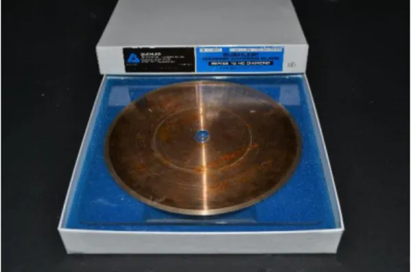
From each tooth, a crown segment was obtained exposing middle dentin by sectioning the crowns with two cuts, a few millimeters apart, parallel to the occlusal surface, with a precision diamond disk at low speed (Diamond Wafering Blade - 10,2cmx0,3mm - Series 15HC, Buehler Ltd., Lake Bluff, IL, USA) (Figure 1), on a hard tissue microtome (Isomet 1000 Precision Saw, Buehler Ltd. Ltd., Lake Buff, IL, USA) (Figure 2) under constant distilled water irrigation, in the following way:
Figure 1 - Diamond wafering blade.
Figure 2 - Isomet 1000 Precision Saw, hard tissue microtome.
9
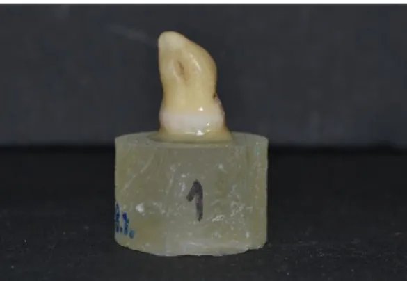
1. The teeth crowns were attached to an acrylic holder with sticky wax, perpendicular to the long axis of the tooth (Figure 3);
2. The first cut was made parallel to the occlusal surface 1-2 mm below the cementoenamel junction to remove the roots (Figures 4 and 5);
3. The crowns were detached from the acrylic holders and the pulp tissues were removed from the pulp chamber with a dentin curette (Figure 6). The pulp chamber was then filled with cyanoacrylate glue (737 Black Magic Toughened adhesive, Permabond, Hampshire, UK) (Figure 7) and the crowns were fixed with the same glue to the acrylic holders, by the sectioning surface (Figure 8).
Figure 3 - Tooth attached to an acrylic holder with sticky wax.
Figure 4 - Roots cutting 1-2 mm below CEJ. Figure 5 - Exposure of the pulp chamber after roots cut.
Figure 7 - Filling of the pulp chamber with cyanoacrylate glue.
10
4. Mid-coronal dentin surfaces were obtained by removing the occlusal enamel and superficial dentin of the molar crowns, perpendicular to the long axis of tooth (Figures 9 and 10).
5. With the purpose of creating a uniform smear layer obtained in similar conditions to those occurring in clinic situations, the dentin surface was polished with 600-grit silica-carbide (SiC) sandpaper (Ultra-Prep, Buehler Ltd., Lake Bluff, IL, USA) on a mechanical grinder (Lunn Major, Struers, Denmark) (Figure 11), according to the ISO TR 11405 standard, during 60 seconds under water irrigation (Pashley et al., 1988).
Figure 8 - The crown fixed to the acrylic holder.
Figure 9 - Removal of the occlusal enamel and superficial dentin.
Figure 10 - Mid coronal dentin surface.
11
4. Distribution and treatment of the crown segments
The six crown segments were randomly assigned to one of the two adhesive groups. The order in which the crown segments were treated was random, to avoid possible bias due to any particular sequence of treatment. All the treatment procedures were performed by the same operator in the way that is followed described:
Group A – Scotchbond Universal Adhesive (3M ESPE, Neuss, Germany) (Figure 12) as per manufacturer’s instructions – Self-etch (Etch-and-Dry) technique on dentin (SBU SE D):
Adhesive application
1. The occlusal surface was rinsed with water. The excess of water was removed from the dentin surface using a moist cotton pellet, so that the surface remained shiny and visibly moist.
2. The adhesive was applied, using a disposable microbrush, to the entire dentin surface, scrubbing lightly for 20 seconds.
3. The surface was then gently air-dried until it ceases to show any movement and the solvent was evaporated completely, forming a homogenous and slightly shiny film. Beginning with a soft blow of air from a distance of approximately 10 cm, the air pressure was increased while decreasing distance, finishing at a distance of approximately 1-2 mm from the surface at maximum air pressure.
4. Finally, the surface was polymerized for 10 seconds.
Group B – Scotchbond Universal Adhesive (3M ESPE, Neuss, Germany) as per manufacturer’s instructions – Total-etch (Etch-and-Rinse) technique on dentin (SBU TE D):
Conditioning step
1. The phosphoric acid etching gel was applied to the entire dentin surface, during 15 seconds. The occlusal surface was rinsed with water. The excess of water was removed from the dentin surface using a moist cotton pellet, so that the surface remained shiny and visibly moist.
12 Adhesive application
The adhesive was applied as for the self-etch mode.
Resin composite build-ups were performed using a resin composite, ENAMEL Plus HRi (Micerium S.p.A. Avegno (GE) Italy) (Figures 13 and 14), shade UD4, in three increments of 2 mm each. Each increment was light cured for 20 seconds, according to the manufacturer's instructions, until reaching a height of 6 mm. Additional light polymerization was performed on facial, lingual, mesial and distal surfaces of the composite build-up for 10 seconds each.
All light curing was performed with a light intensity of 600 mW/cm2 using a
halogen light-activation unit (ELIPAR S10, 3M ESPE Seefeld, Germany), with the 13 mm light guide held 1-2 mm from the treatment surface. The output of the curing light was periodically verified at >600 mW/cm2 with a radiometer (Curing Radiometer P/N
10503, USA) throughout the procedure.
Figure 12 - Scotchbond Universal Adhesive and Scotchbond Universal Etchant.
Figure 14 - Resin composite build-up. Figure 13 - Resin composite ENAMEL Plus HRi.
13
5. Specimens preparation for the microtensile tests
All teeth were painted with different colors with waterproof ink. The exterior surface of the resin composite was painted in order to identify, and then, exclude from the study, the sticks in which the adhesion was made to enamel.
The teeth were stored in distilled water in an incubator for 24 hours at 37 °C, according to the ISO TR 11405 standard. Date and time of the restoration was registered. Posteriorly, the teeth were longitudinally sectioned in both “x” and “y” directions with a slow-speed diamond disk (Diamond Wafering Blade - 10,2cmx0,3mm - Series 15HC, Buehler Ltd., Lake Bluff, IL, USA) (Figure 15) under water irrigation, using a hard tissues microtome (Isomet Precision Saw, Buehler Ltd. Ltd., Lake Buff, IL, EUA), to obtain sticks with a cross-sectional area of approximately 0,7 mm2.
A final cut is made at the base of the root, perpendicular to the long axis of the tooth, to separate the sticks from the acrylic supports (Figure 16).
Debonded or lost sticks are registered. Debonded sticks were those separated in the adhesive interface during the cutting procedure. Lost sticks were those which were lost or fractured during test preparation.
The obtained sticks were kept in distilled water for a maximum of 24 hours, until the conclusion of the microtensile tests.
Figure 16 - The sticks obtained after the final cut. Figure 15 - The teeth after being sectioned in both “x”
14
6. Microtensile tests
The specimens were individually attached to a stainless-steel grooved Geraldelli´s jig with cyanoacrylate glue (737 Black Magic Toughened adhesive, Permabond, Hampshire, UK) (Figure 17) and then submitted to a tension load using a universal testing machine (Instron 4502 Series, Serial no. H3307, Instron Corporation, Canton, MA, USA) (Figure 18), at a crosshead speed of 1mm/min until fracture occurred.
A digital caliper (Ficher Darex, France) was used to measure the sides of the bonding interface and calculate the bonding area in mm2. The load at fracture (KN) and the bonding surface area of the specimens were registered and µTBS calculated in MPa, by dividing the force imposed at time of fracture by the bonding area (mm2).
The failure modes were analyzed under a stereomicroscope (Nikon, Japan) at 10X magnification and classified as: adhesive (A, failure occurring at the dentin-adhesive interface); cohesive in dentin (CD, when the failure occurred in dentin); cohesive in composite (CC, when the failure occurred in composite); and mixed (M, failure with composite and dentin at the interface).
Figure 18 - Instron 4502, universal testing machine. Figure 17 - The specimen attached to a Geraldelli’s jig.
15
7. Statistical Analysis
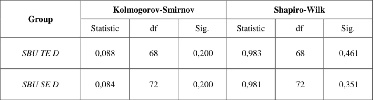
The statistical analysis of the results was performed through descriptive and inference methods. A paired-sample t-test was performed when the assumption of normality was valid: the Kolmogorov-Smirnov and Shapiro-Wilk Tests were used to assess whether the data followed a normal distribution (Table 1); the Levene’s Tests was computed to determine if the assumption of equal variances was valid (Table 2). Significance was set at a 95% confidence level.
The number of prematurely debonded sticks (pretesting failures that occurred during specimen preparation) in each test group was recorded, but these values were not included in the statistical analysis (Perdigao et al., 2006).
Group
Kolmogorov-Smirnov Shapiro-Wilk
Statistic df Sig. Statistic df Sig.
SBU TE D 0,088 68 0,200 0,983 68 0,461
SBU SE D 0,084 72 0,200 0,981 72 0,351
Table 1 - Tests of Normality of the µTBS in MPa for the two different experimental groups tested.
Levene’s Test F Sig. Equal variances assumed 3,334 0,070 Equal variances not assumed
17
18
The number of microtensile sticks (N), mean and standard error (SE), standard deviation (SD), minimum (Min), maximum (Max) are shown in Table 3.
The highest mean µTBS was obtained with SBU SE D (56,9 ± 2,5 MPa) and the lowest was obtained with SBU TE D (48,0 ± 2,1 MPa) (Figure 20).
After observing the normality of the data distribution and the equality of the variances, data from µTBS were analyzed using a paired-sample t-test (Table 4).
Group N Mean ± SE SD Min Max
SBU TE D 68 48,0 ± 2,1 17,6 6,1 84,4
SBU SE D 72 56,9 ± 2,5 21,2 7,2 99,6
Table 3 - Descriptive statistics of the µTBS in MPa for the two experimental groups tested.
t-test for Equality of Means
t df Sig. (2-tailed) Mean Difference
Equal variances assumed -2,673 138 0,008 -8,84586
Equal variances not assumed -2,687 135,753 0,008 -8,84586
Table 4 - t-test for Equality of Means (p≤0,05).
Figure 19 - Box-whisker plots of the µTBS in MPa for the two different experimental groups tested. The median µTBS is represented by the central line. The box represents the interquartile range.
19
The mean µTBS to dentin of the Scotchbond Universal Adhesive applied following the etch-and-dry approach (SBU SE D) was statistically higher than that when the SBU was applied following the etch-and-rinse approach (SBU TE D) (p = 0,008).
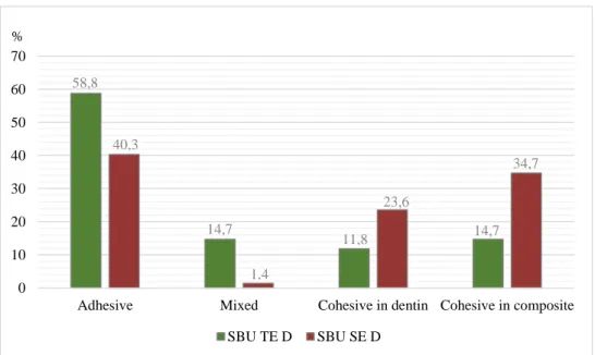
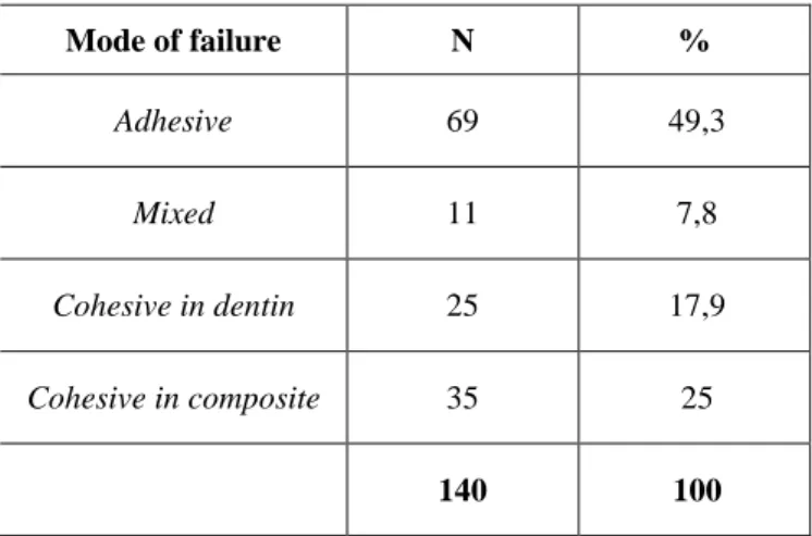
Failure mode distribution of the debonded specimens is shown in Table 5 and 6, Figures 20 and 21. Fracture analysis revealed that the most predominant failure pattern was “adhesive” in both groups tested. Cohesive failures in composite and dentin were observed in 25% and 17,9% of the specimens, respectively. Mixed failures were observed in few specimens (7,8%).
Group
Mode of failure N
Adhesive Mixed Cohesive in
dentin Cohesive in composite Total Pretesting failures SBU TE D 40 10 8 10 68 4 SBU SE D 29 1 17 25 72 14
Table 5 - Number of specimens according to the failure mode and premature failures of the two different experimental groups tested.
Figure 20 - Distribution of the specimens according to the failure mode of the two different experimental groups tested.
0 10 20 30 40 50 60 70
Adhesive Mixed Cohesive in dentin Cohesive in composite
% SBU TE D SBU SE D 58,8 40,3 14,7 1,4 11,8 23,6 14,7 34,7
20 Mode of failure N % Adhesive 69 49,3 Mixed 11 7,8 Cohesive in dentin 25 17,9 Cohesive in composite 35 25 140 100
Table 6 - Number of specimens according to the failure mode and premature failures of all experimental groups tested.
0 5 10 15 20 25 30 35 40 45 50
Adhesive Mixed Cohesive in
dentin
Cohesive in composite %
Figure 21 - Distribution of the specimens according to the failure mode of all experimental groups tested.
49,3
7,8
17,9
21
22
As the bond strength was measured upon 24 hours water storage, it should be considered as “immediate” bond strength. Universal adhesives represent the last generation of adhesives in the market. They are designed under the “all-in-one” concept of already existing one-step self-etch adhesives, but incorporating the versatility of adapting them to the clinical situation, by application under different adhesive approach.
The null hypothesis was rejected, as there were statistical differences in dentin µTBS among the different adhesion strategies for SBU: when SBU was applied following an etch-and-dry approach (SBU SE D) resulted in statistical higher µTBS than when applied following an etch-and-rinse approach (SBU TE D) (p=0,008).
A possible reason for the higher performance of SBU when applied in the SE mode (SBU SE) is that the SBU bonding ability is two-fold, relying on a combination of chemical adhesion to hydroxyapatite (HAp) with some micromechanical interlocking that does not depend much on resin-tag formation (Van Meerbeek et al., 2003; Yoshida et al., 2004). In fact, the pH of Scotchbond Universal Adhesive is 2,7, which is within the range of the mild self-etch adhesives (Van Meerbeek et al., 2003). Mild self-etch adhesives demineralise dentin only partially, keeping residual hydroxyapatite still attached to collagen. The abundant presence of HAp remaining around the collagen provides natural protection of the collagen, but also provides the potential for chemical interaction with HAp (Tay & Pashley, 2001; Van Meerbeek et al., 2003; Yoshida et al., 2004; De Munck
et al., 2005b). Molecules like 10-MDP and also polyalkenoic acid copolymer (PA) are
present in SBU composition (Appendix 1) and will chemically bond to Ca of HAp, forming stable calcium-phosphate and calcium-carboxylate salts, respectively, along with only a limited surface-decalcification effect (Yoshida et al., 2000; Fukuda et al., 2003; Yoshida et al., 2004; Van Meerbeek et al., 2011; Perdigao et al., 2012). Depending on the stability of the resulting calcium-monomer complex in adhesive suspension, this ionic bond may either decompose and demineralize the tooth surface, or remain stable and chemically bond with calcium (Yoshida et al., 2000; Yoshida et al., 2004). The monomer 10-MDP appears to not only interact more intensively with HAp, but also be hydrolytically more stable, as compared with other functional monomers as 4-MET and phenyl-P (Yoshida et al., 2004; Van Landuyt et al., 2007; Van Landuyt et al., 2008). The high chemical affinity of 10-MDP for HAp was found to involve the formation of a self-assembled nano-layered structure of 10-MDP molecules, so-called “nanolayering”. Such nanolayering is typical of 10-MDP (could not be detected for the functional monomer 4-MET and phenyl-P) and may explain the high long-term stability of 10-MDP-based
self-23
etch adhesives (rather than its immediate bond strength), which has previously been proven both in laboratory and clinical research (Peumans et al., 2010; Yoshihara et al., 2010; Yoshihara et al., 2011; Yoshida et al., 2012). Beyond that, the partial dissolution of the dentin surface is sufficient to dissolve the smear layer and to provide some micro-mechanical interlocking. The thickness of the hybrid layer is, however, much smaller than that produced by the strong self-etch or etch-and-rinse adhesives, but has been proven to be minor in importance with regard to actual bonding effectiveness (Van Meerbeek et al., 2003; De Munck et al., 2005b; Van Meerbeek et al., 2011).
The lower µTBS obtained with SBU applied in the ER mode are in contrast with other studies of the same universal adhesive in the literature, reporting that phosphoric-acid etching of dentin prior to self-etch adhesive application (SBU TE) had no negative effect on microtensile bond strengths when compared to SBU SE (Perdigao et al., 2012; Marchesi et al. 2013; Munoz et al., 2013; Munoz et al., 2014; Wagner et al., 2014). It is known that smear layer constitutes a true physical barrier against the penetration of resin monomers, making the hybrid layer formation extremely difficult (Kenshima et al., 2005; Kenshima et al., 2006). After preliminary etching with phosphoric acid, the smear layer is removed and superficial dentin is demineralized. This seems to increase adhesive infiltration, allowing the creation of a well impregnated hybrid layer, when compared to their respective SE counterparts (Hanabusa et al., 2012; Perdigao et al., 2012; Taschner
et al., 2012; Munoz et al., 2013; Wagner et al., 2014). However, our results revealed that
the group in which dentin was etched with phosphoric-acid prior to the application of a self-etch adhesive (SBU TE D) obtained inferior bond strengths, compared with its application on smear layer-covered dentin (SBU SE D). Phosphoric acid gels used with etch-and-rinse adhesives are more acidic than the phosphate or carboxylate monomers found in the composition of self-etch adhesives (Van Meerbeek et al., 2003). Incomplete infiltration of the deeply demineralized collagen network by resin monomers was the main reason reported for this decrease in bond strength, due to the fact that phosphoric acid can decalcify human dentin more deeply (up to 3-6 um) than a (mild) self-etch adhesive is designed to infiltrate (Van Landuyt et al., 2006a; Van Landuyt et al., 2006b; Van Meerbeek et al., 2011; Hanabusa et al., 2012). In fact, the more resin that infiltrates acid-etched matrices, the higher are the resin-dentin bond strengths (Nishitani et al., 2006). Such poorly resin-infiltrated zones and thus unprotected collagen fibrils are readily exposed to oral fluids. Hydrolytic breakdown, potentially accelerated by acids and enzymes produced by bacteria, and/or by enzymes present in dentin and activated by the
24
adhesive procedure, is expected to jeopardize the long-term stability of such porous etch-and-rinse hybrid layers (Breschi et al., 2008; Pashley et al., 2011). Previous studies in the literature (Van Landuyt et al., 2006a; Van Landuyt et al., 2006b), also reported a decreasing in the bond strength after phosphoric-acid etching of dentin prior to the application of a self-etch adhesive.
We could presume that the lower µTBS obtained with SBU applied in the ER mode can also be attributed to the variability in the amount of moisture left by the operator in each dentin surface, before adhesive application. Over-drying and over-wetting are considered as two extremes (Tay et al., 1998). In both situations will occur defects in the interface which could weak the resin-dentin adhesion: water excess leads to a decrease in adhesion as a result of the primer dilution and reduced conversion degree of the monomer (Jacobsen & Soderholm, 1995); over-drying dentin could rise the risk of collagen fibre collapse (Pashley et al., 1992; Swift et al., 1995). Between these two extremes is a “window of opportunity” wherein optimal hybridization and tubular seal may be achieved (Tay et al., 1998). However, although most manufacturers of adhesive systems (including Scotchbond Universal Adhesive manufacturer) recommend the application of their materials to moist dentin and the adhesive application has been the same in all tested groups, it seems clear that the wet-bonding technique is difficult to standardize and sensitive to errors, since it is very difficult to determine the amount of water that should be kept on dentin surface before the adhesive application (Frankenberger et al., 2000).
Since such adhesives combine the primer and the adhesive in the same solution, both hydrophilic and hydrophobic monomers are blended, with a relatively high concentration of solvent required to keep them in solution (Van Landuyt et al., 2005). Consequently, increasing the risk of forming a hybrid layer without full involvement of the exposed collagen by adhesive monomers (Van Meerbeek et al., 2001). This could be another possible explanation for the lower µTBS obtained with SBU applied in the ER strategy.
In the present study, the most predominant failure pattern was adhesive in both SBU TE and SBU SE groups (58,8 and 40,3%, respectively) (figure 20). This is in accordance with previous studies in literature, reporting that more adhesive failures than cohesive failures are expected when performing a microtensile bond strength test (Pashley et al., 1995; Schreiner et al., 1998). Indeed, an accurate assessment of the strength of an adhesive material is best determined when the failure occurs within the
25
material itself and does not involve dentin or composite (Sano et al., 1994; Ghassemieh, 2008).
Although the high number of adhesive failures obtained in this study, the number of cohesive failures in composite were higher than was expected for this kind of methodology (Pashley et al., 1995; Schreiner et al., 1998): in 25% of the specimens the fracture occurred exclusively in composite (Figure 21). Similar results were obtained previously suggesting that the curing time of the resin composite recommended by the manufacturer (20 seconds) was inferior to the ideal (Silva, 2008; Pequeno, 2009). In our study, the curing time of the resin composite recommended by the manufacturer was also 20 seconds. Proença et al. (2007) realized an in vitro study with ER and SE adhesives, however the resin composite was cured twice over (40 seconds) than the recommended time, and obtained less cohesive failures in composite. A previous study of Perdigao et
al. (2006) obtained similar results for a curing time of 40 seconds. Although additional
light polymerization was performed on facial, lingual, mesial and distal surfaces of the composite build-up for 10 seconds each, besides the 20 seconds recommended by the manufacturer, a high number of cohesive failures in composite were still observed. We could speculate that the additional light polymerization did not achieve the more central layers of the composite, taking into account that they had already been polymerized. Thus, we could presume that the additional light polymerization was useless in terms of preventing cohesive failures.
Cohesive failures in dentin were observed in 17,9% of the specimens (figure 21). Such cohesive failures in dentin do not mean that the resin-dentin bonds are uniformly stronger than the intrinsic strength of dentin, but that the manner in which the bond is stressed is so non-uniform that is concentrated or focused at one highly localized region where it opens a crack in dentin that then fails (Pashley et al., 1995; Armstrong et al., 1998; Pashley et al., 1999). Moreover this type of failure is usually present when the sample has considerable damage or defect on the dentin side, which is hard to control (Phrukkanon et al., 1998; Ghassemieh, 2008). For these reasons, this kind of failure pattern is somewhat expected and they should not be considered in the evaluation of the bond strengths (Ghassemieh, 2008).
Different types of imperfections and variations that might occur in the specimens can change the pattern of progression of failure, such as the misalignment of the axis of loading with specimen, the thickness of adhesive and uneven adhesion application (Ghassemieh, 2008).Thus, the specimens preparation for the microtensile tests requires
26
extremely careful manipulation (Cardoso et al., 2001; De Munck et al., 2005a), otherwise it may induce defects at the interface that may lead to lower bond strength values or even pre-testing failures (De Munck et al., 2005a).
Pre-testing failures (ptf) were recorded, but not included in the statistical analysis. Correct interpretation of pre-testing failures with regard to the calculation of the average µTBS is currently a matter of debate. There are studies that considered the ptf as (1) 0 MPa, which actually penalizes the adhesive too severely (as there was a certain bond strength), (2) others that excluded the ptf from the average µTBS calculation and (3) others that a ptf received the lowest µTBS measured within the respective group (Mine
et al., 2009). It is important to highlight that the non-inclusion of ptf in this study could
have generated overestimated strength bond values (Wagner et al., 2014), especially in the SE group, which obtained a high number of pre-testing failures, in spite of the higher µTBS obtained. It is worth mentioning that these ptf occurred predominantly in only one tooth (Appendix 5), which was probably due to an error in adhesive application or the result of the entrapment of air bubbles in the adhesive layer.
The most used dentin preparation methods before bonding are preparation by either carbide or diamond bur or by silicon-carbide (SiC) paper (De Munck et al., 2012). In this study, a thin artificial smear layer was created by means of grinding with 600-grit SiC paper, according to the ISO TR 11405 standard. Clinically, the smear layer created using a medium-grit diamond bur (average particle size of 70 µm) is rougher than the smear layer produced by using 600-grit wet paper used in in vitro studies. Because increased smear-layer thickness and greater surface roughness correspond to lower SE adhesive impregnation, bond strength obtained after using a 600-grit paper (average particle size of 14,5 µm) may be overestimated due to enhanced adhesive penetration (Oliveira et al., 2003; Cardoso et al., 2008; Taschner et al., 2010), and thus, explaining the higher µTBS obtained when SBU was applied in SE mode.
Taking into consideration that different bonding strategies (ER and SE) are influenced by chemical bonding and by micromechanical interlocking in different degrees, variation in regional characteristics of the substrate may also affects the two bonding strategies differently. However, there is some controversy as to whether sticks obtained from the peripheral area of the restored tooth result in decreased or increased µTBS (Sezinando et al., 2012). Whereas one study reported lower µTBS for peripheral specimens than for centrally located specimens (Loguercio et al., 2005), another study
27
reported that µTBS to “periphery” dentin is higher than for the “central” specimens (De Munck et al., 2011). In our study we used sticks from the entire interface, whereas Hanabusa (2012) only used nine central dentin sticks from each tooth to reduce substrate-regional variability. With this in mind, we should have divided the bonded sticks from each tooth in peripheral sticks and central sticks to avoid any bias in the µTBS distribution caused by regional differences (Sarr et al., 2010; Hanabusa et al., 2012). In fact, water takes some time to diffuse from the external surface into the inner bonded interface, and thus, the resin-dentin interface at the periphery of the restoration (peripheral sticks) is more susceptible to water degradation than that located centrally (Loguercio et al., 2005). When performing microtensile tests, the stress is applied to a bonded surface area of 0,25-1 mm2 (Sano et al., 1994). The specimens used in this study had a cross-sectional area of approximately 0,7 mm2, which corresponds to the bonding area size recommended by Phrukkanon and others (1998). An inverse relationship between dentin µTBS and cross-sectional area was found (Sano et al., 1994; Phrukkanon et al., 1998). The reason for the increase in bond strength with decreases in bonded surface area is probably due to the presence of more defects (air bubbles, phase separations, surface roughness, nonuniform film thickness and angle of application of load) at the bonded interface or within the substrate of larger specimens. When the specimen is loaded, stress concentration occurs at the defects and initiates crack formation (Van Noort et al., 1989; Sano et al., 1994; Pashley et al., 1995; Ghassemieh, 2008). The reduced bonding area of the sticks in this study could somehow explain the higher bond strengths obtained relatively to other studies, in the same conditions, but with sticks with a larger bonding area (Marchesi et al., 2013; Munoz et al. 2013; Munoz et al., 2014).
In spite of using a small bonding area, standard deviations were higher than expected, particularly for the SE group (figure 19). Apart from the bond strength, the variability is also an indicator for the performance of an adhesive. A low variability may indicate a good accuracy and low technique sensitivity of the adhesive. Variability in the bond strength results between different samples may be caused by several factors, such as the variability between teeth, variability in the application procedure, irregularities in the composite (such as air bubbles), the presence of voids in the adhesive (Phrukkanon et
al., 1998; Pashley et al., 1999; Perdigao et al., 2002). For these reasons, future studies
should increase the number of teeth included in the sample, in an attempt to reduce the standard deviation.
28
It should be mentioned that we have detailed the manufacturer’s instructions (Appendix 3). Due to commercial constrains, frequently manufacturers of adhesive systems when formulating the application instructions of their products, choose a solution between what would be the ideal instructions to maximize the results of a particular adhesive, and what would they think it would be acceptable for the majority of the clinicians. In fact, it was not possible to evaporate the solvent completely from the surface with a gentle stream of air over the liquid for only 5 seconds. Due to that reason the surface was gently air-dried until it ceases to show any movement and the solvent was evaporated completely, forming a homogenous and slightly shiny film. After the careful application of the primers, it is necessary to evaporate the solvent carefully. If this procedure is not respected, the solvents, which are used as transporters and facilitators of the resin infiltration, may prevent the formation of a perfectly infiltrated, sealed, fully polymerized hybrid layer (Pashley & Carvalho, 1997), and might result in accelerated bond degradation due to the lower polymerization conversion at the bonding interface (Ikeda et al., 2008). Previous studies reported the importance of solvent/water evaporation with respect to dentin adhesion (Cavalheiro et al., 2006; Cavalheiro, 2008).
One of the limitations of this study has to do with the lack of long-term water storage. With this in mind, future studies should analyze the effect of long-term water storage on the in vitro performance of the universal adhesive SBU.
The in vitro nature of this study does not allow direct extrapolation of the results to an in vivo situation, so whether the same results would be obtained in vivo should be the object of further investigation. Laboratory results do not always correlate with clinical performance. They should be regarded as screening tools, whose value lies only in their ability to discriminate between a multitude of adhesive systems and thus allowing researchers to focus on the more promising materials. Ultimately, the fact remains that the true performance of a material cannot be determined without rigorous clinical testing (El Zohairy et al., 2010; Van Meerbeek et al., 2010). It also should be made clear that tests used to evaluate bond strength of dental adhesives are not without their limitations. Another limitation of this study are that caries-free teeth were used. Specimens with any sign of caries were excluded because these lesions are seen as pathways for fluid escape and because there was a need for standardization. However, in a clinical scenario, the majority of the adhesive procedures are performed on teeth affected by caries lesions. Yet, it is known that caries significantly change the dentin permeability (Tagami et al., 1992; Ceballos et al., 2003; Scholtanus et al., 2010).
29
30
Within the limitations of the present laboratory study, it may be concluded that improved bonding effectiveness of SBU on dentin is obtained when the adhesive is applied with the self-etch approach. The etch-and-rinse approach tested (wet dentin) resulted in immediate bond strength statistically lower than the self-etch group.
Due to the fact that these new adhesives and the respective concept behind them are novel, there is little information in the literature about the in vitro and in vivo performance of this new class of adhesives. Future studies should analyze the effect of long-term water storage on the in vitro performance of the universal adhesive SBU. Besides that, further studies on different substrates, such as carious dentin, in association with in vivo tests are needed to assess the long-term clinical behavior of this new class of adhesives and to back the use of these new materials.
Clinical significance: Improved bonding effectiveness of the tested universal adhesive system on dentin may be obtained if the adhesive is applied with the self-etch approach.
I
II
APPENDIX 1
Materials, Manufacturers, Components and Batch Numbers
Material Manufacturer Composition Batch Number
Scotchbond Universal Adhesive 3M ESPE Neuss, Germany Bis-GMA (15-25 vol%) HEMA (15-25 vol%) 10-MDP (5-15%) Ethanol (10-15 vol%) Water (10-15 vol%)
Silane treated silica (5-15 vol%) 2-Propenoic acid, 2-Methyl-, reaction products with 1,10-Decanediol and phosphorous oxide (1-10 vol%)
Copolymer of acrylic and itaconic acid (1-5 vol%)
Dimethylaminobenzoat (<2 vol%) Camphorquinone (<2 vol%) (Dimethylamino)ethyl methacrylate (<2 vol%)
Methyl ethyl ketone (<0,5 vol%)
Lot 540368 Exp 12/2015 Scotchbond Universal Etchant 3M ESPE Neuss, Germany Water (50-65 vol%)
Phosphoric acid (30-40 vol%) Synthetic amorphous silica, fumed, crystalline free (5-10 vol%)
Polyethylene glycol (1-5 vol%) Aluminum oxide (<2 vol%)
Lot 537103 Exp 11/2015 Enamel Plus HRi UD4 Micerium S.P.A. Avegno, Italy 1,4-Butandioldimethacrylate Urethandimethacrylate Bis-GMA Lot 2010006016 Exp 06/2015 Lot 2012000921 Exp 12/2018
III
APPENDIX 2
Application mode of Scotchbond Universal Adhesive
according to the manufacturer’s instructions
(information supplied in the safety data sheets and material instructions)
1) Tooth preparation
Remove loose preparation debris by spraying with water, and lightly air dry the cavity in only 2-3 bursts of water-free and oil-free air, or use cotton pellets to dry it off. Do not overdry.
The cavity should be just dry enough that the surface has a slightly glossy appearance. Overdrying can lead to post-operative sensitivity.
2) Procedure for direct restorations with light-curing composites
Adhesive System Technique Instructions for use
Scotchbond Universal
Adhesive
Etch-and-dry
1. Use the disposable applicator to apply the adhesive to the entire tooth structure and rub it in for 20 seconds;
2. If necessary, rewet disposable applicator during treatment;
3. Subsequently direct a gentle stream of air
over the liquid for about 5 seconds until it no
longer moves and the solvent has evaporated completely.
4. Harden the adhesive with a commonly used curing light for 10 seconds.
Etch-and-rinse
1. Apply a commonly used phosphoric acid etching gel (about 35%), e.g., Scotchbond Universal Etchant, to the prepared and unprepared (if present) tooth structure (enamel and dentin) and allow to react for 15 seconds. Rinse thoroughly with water and dry with water-free and oil-water-free air or with cotton pellets. Do not overdry;
2. Apply adhesive as for the self-etch mode.