Abstract
Submitted: August 19, 2016 0RGL¿FDWLRQ6HSWHPEHU Accepted: October 2, 2016
The effect of silver nanoparticles
on composite shear bond strength
to dentin with different adhesion
protocols
In Dentistry, restorative materials and oral bacteria are believed to be responsible for restoration failure. To make long-lasting restorations, antibacterial agents should be made. Inorganic nanoparticles and their nano composites are applied as good antibacterial agents. Objective: The purpose of this study was to investigate the effect of silver nanoparticles on composite shear bond strength using one etch and rinse and one self-etch adhesive systems. Material and Methods: Silver nanoparticles were prepared. Transmission electron microscope and X-ray diffraction were used to characterize the structure of the particles. Nanoparticles were applied on exposed dentin and then different adhesives and composites were applied. All samples were tested by universal testing machine and shear bond strength was assesed. Results: Particles with average diameter of about 20 nm and spherical shape were found. Moreover, it was shown that pretreatment by silver nanoparticles enhanced shear bond strength in both etch and rinse, and in self-etch adhesive systems p0.0. &onclusions: &onsidering the positive antibacterial effects of silver nanoparticles, using them is recommended in restorative dentistry. It seems that silver nanoparticles could have positive effects on bond strength of both etch-and-rinse and self-etch adhesive systems. The best results of silver nanoparticles have been achieved with Adper Single Bond and before acid etching.
Ke yw or ds: Silver. Nanoparticles. Shear strength. Dentin-bonding agents. Fatemeh KOOHPEIMA1
Mohammad Javad MOKHTARI2
Samaneh KHALAFI1
http://dx.doi.org/10.1590/1678-7757-2016-0391
1Shiraz University of Medical Sciences, School of Dentistry, Biomaterial Research Center, Department
of Operative Dentistry, Shiraz, Iran.
2Islamic Azad University, Zarghan Branch, Young Researchers and Elite Club, Zarghan, Iran.
Introduction
Silver nanoparticles (NPs) have called the
researcher’s attention because of their unique properties, for example, their optical, antimicrobial,
and electrical properties15. The antimicrobial,
antifungal and antiviral action of silver or silver
compounds depends on the level of released bioactive silver ions (Ag+) and its availability to interact with
bacterial or fungal cell membranes14. Silver NPs have
the potential to serve as a bactericidal agent because
of the inherent antimicrobial inÀuences of silver ion. NPs are insoluble particles that are smaller than 100
nm in diameter25. They bene¿t smaller particles that
demonstrated stronger antibacterial activity compared to larger particles due to their higher surface area and,
thus, enhanced interaction with organic and inorganic
molecules. However, many properties of metal NPs
are still unknown27. The potency of the antibacterial
effects corresponds to the nanoparticle size. Smaller
particles have more antibacterial properties because
of the equivalent silver mass content23. Silver NPs
adhere and penetrate the bacterial cell wall, thereby causing structural changes in the cell membrane, such
asin cell membrane permeability and cell apoptosis.
There is a formation of pits on the cell surface, and
there is accumulation of NPs on the cell surface9.
Silver NPs may have potential toxicities at some
concentrations and can produce many health problems
if used inappropriately. Metal NPs have been recently
applied in Dentistry because of their bactericidal and bacteriostatic effects11.
Caries preventive measures with the purpose of
reducing demineralization should be independent of
the patient’s compliance5. These preventive measures
consist of antimicrobial bonding agents, antibacterial
mouth rinses, and remineralizing agents adjacent to
oral appliances. It has been demonstrated that the
presence of silver in dental restorative materials is effective against caries producing bacteria, such as
streptococci and lactobacillus19. It can also be effective
as an antibacterial additive to dental restorations. We do not know exactly whether the incorporation
of silver NPs would interfere with bond strength of
dental restorative materials16,19. Until now, only few
studies have determined the effect of silver NPs on composite shear bond strength (SBS). Adhesion
mechanism is based on the penetration of resin
molecules into enamel and dentin22. Bond strength
of dental composites to dentin is one of the main
criteria in clinical durability of composite restoration.
Various bonding systems have been introduced in order
to ful¿ll a reliable bond to tooth structure based on two
main methods: the etch-and-rinse and the self-etching
adhesive systems7. Smear layer removal and formation of
collagen ¿bril layer by means of acid conditioner to form
hybrid layer are the main adhesion mechanisms in
etch-and-rinse systems3. The self-etching adhesive system is
subdivided into two groups: two-step and one-step
self-etching. Self-etching adhesive systems employ an acidic
monomer as conditioner. In self-etch adhesives, acidic
functional monomers react to the mineral content of tooth
surface24. Self-etch adhesives are less time-consuming
and technique-sensitive. Etch-and-rinse adhesive systems
are still considered as a golden standard among bonding
systems. However, dentists have a tendency to use
adhesive systems with a simpli¿ed application procedure12.
Therefore, in this study we examined whether an
additional pretreatment with silver NPs provides any
supplementary effect on bond strength of self-etch
commercial adhesive (Clear¿l SE Bond) and etch-and-rinse (Adper Single Bond) using standard SBS
methodology.
Material and methods
Preparation and characterization of silver NPs
Portions of 1 g/L AgNO3 with sodium dodecyl sulfate(SDS) (Merck) were mixed in an aqueous solution
under a N2 atmosphere. SDS molecules were applied to disperse the silver NPs and also to stabilize the
formation of shaped silver NPs. Deposition was carried
out at different reaction times. Oxygen was removed
from water by nitrogen bubbling and the electrolyte was combined under a nitrogen atmosphere. SDS
was added at the 40 g/L level to avoid aggregation.
The products were washed with distilled water and collected by centrifugation at 15,000 rpm for 10
min (Hettich Universal 320, Tuttlingen, Germany). A
transmission electron microscope (TEM) (JEM-1011,
JEOL, Japan) was used to determine size, shape, and size distribution of silver NPs. Silver NPs were prepared
by placing a drop of working solution on a TEM grid and
dryed for TEM analysis. X-ray diffraction (XRD) was
used as a secondary or complementary technique to determine particle size by analyzing diffraction peaks.
X-ray powder diffractometer (DX-1000X, Dandong
Fangyuan, China)17.
Specimen preparation
Ninety extracted, noncarious human premolar
teeth were cleaned and stored in 0.1% thymol
solution for one week. The teeth were prepared using
a diamond bur (4138 KG Sorensen, Barueri, Brazil) and a high-speed handpiece under water coolant up
to the nearmost half of the DEJ to the pulp, which is
the proposed location for measuring bond strength of composites, so that no pulp exposure occur in the
preparation site. Then the exposed super¿cial dentin
surface was polished using silicon carbide paper (600
grit) under water coolant to standardize the smear layer. The teeth were rinsed with distilled water to
remove any debris and then mounted in acrylic resin
(2×3×5 cm) and randomly divided into six groups
(n=15).
The specimens’ preparation was based on the type
of employed adhesive system and tested silver NPs
as follows:
Group 1: Acid etching + Adper single Bond + Composite Z250
Group 2: Silver NPs + Acid etching + Adper single
Bond + Composite Z250
Group 3: Acid etching + Silver NPs + Adper single Bond + Composite Z250
Group 4: Clear¿l SE Bond Primer + Clear¿l SE Bond
+ Composite Z250
Group 5: Silver NPs + Clear¿l SE Bond Primer + Clear¿l SE Bond + Composite Z250
Group 6: Clear¿l SE Bond Primer + Silver NPs +
Clear¿l SE Bond + Composite Z250
In all groups tested the application of silver NPs were followed by a 60-second rinse with water.
Adhesive systems were applied following the
manufacturer’s instructions (Figure 1). Subsequent
to the application of adhesive on dentin surface, a
resin composite block (3M ESPE Filtek Z250, St Paul Minnesota, USA) was built up over the bonded dentin
with the aid of a TeÀon mold (2 mm height×5 mm
diameter) at room temperature, followed by
light-curing for 40 seconds in vertical position on composite surface (600 mW/cm2)4,26.
SBS measurement
Specimens were stored in distilled water at 37°C for one week and then they were tested in shear mode
using a rod attached to the universal testing machine
(Zwick/Roell Z020, Stuttgart, Germany) at a
cross-head speed of 0.5 mm/min until the bond fractured and the shear force were evaluated. The force was
measured in Newtons (N). SBS values were calculated
by converting Newtons into megapascals (MPa)4.
Statistical analysis
Statistical analysis was performed using SPSS
18.0.0 software (SPSS Inc, Chicago, USA). The
statistically signi¿cant difference among groups was
determined using analysis of variance (ANOVA) and Tukey test with the level of signi¿cance at p=0.05.
Results
TEM and XRD analysis
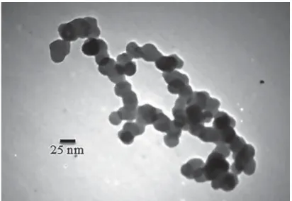
The size and morphology of particles were assessed
using a TEM (Figure 2). TEM image shows silver
particles with average diameter of about 20 nm and with spherical shape.
A typical XRD pattern of the as-prepared silver NPs
is shown in Figure 3. All peaks in the XRD pattern can be indexed as a face centered cubic (fcc) structure
Adhesive/ Composite System Composition Application Mode
Adper Single Bond 3M ESPE, USA Adhesive: Bis-GMA; HEMA; cimethacrylatas; polyalkanoic acid copolymer; initiators; water;
and ethanol
Acid etch (15 s); rinse (15 s); air-dry (30 s); GHQWLQUHZHWWHGZLWKZDWHUȝOVRQH coat of adhesive; air-dry (10 s); light cure (10
s) &OHDU¿O6(%RQG.XUDUD\0HGLFDO,QF
Japan
Primer: MDP; HEMA; hydrophilic dimethacrylate; camphorquinone; water
Adhesive: MDP; HEMA; Bis-GMA; hydrophobic dimethacrylate; N, N diethanol p-toluidine; camphorquinone bond; silanated
colloidal silica
air-dry the dentin surface; two coats of primer with slight agitation (20 s); air-dry (20 s); one coat of the adhesive with slight agitation (20
s); light cure (10 s)
Z250 3M ESPE, USA Organic Matrix: BisGMA; UDMA; BisEMA; camphorquinone (initiator) Filler: Zirconia/silica
layering technique after applying adhesive systems
(JCPDS, ¿le no. 4- 0783). XRD pattern shows the
presence of diffraction peaks corresponding to (111), (200), and (220) planes. Mean particle size
determined using the Scherrer method on the main
(111) diffraction peak of the X-ray diffraction pattern is 20±2 nm, agreeing with the size of isolated NPs
observed in TEM.
SBS measurement
Table 1 and Figure 4 demonstrated mean SBS values and standard deviation of the two adhesive
systems at the three tested conditions. Maximum
and minimum SBS in the presence of silver NPs were
observed in group 2 (25.42±3.36, p=0.000) and group 6 (17.95±3.31, p=0.05), respectively.
Discussion
Nanotechnology is making advances in many
¿elds, specially in Dentistry. In this technology, the speci¿c material is converted into nanometric sizes for showing new properties21. Antibacterial properties
of silver NPs and their compounds have been used in
various medical and recently dental branches16. Due to
the potential of microorganisms to stick to composite resins and adhesives compared to other restorative
materials, the present study made an attempt to
evaluate the effect of silver NPs on composite SBS
with different adhesion protocols. In this study, etch-and-rinse adhesive with the application of silver NPs
before acid etching showed better results. The use of
silver NPs also had no adverse effect on bond strength
in other groups.
Silver is a safe bactericidal metal because it
is biocompatible to animal cells but is very toxic
to bacteria16. Smaller silver NPs (<100 nm) can
interact very closely with microbes. They provide a larger surface area for antimicrobial activity because
of greater ratio of surface-to-volume than larger
particles1. A variety of preparation methods have
been demonstrated for the synthesis of silver NPs; some of them are laser ablation, electron irradiation,
gamma irradiation, microwave processing, chemical
reduction, photochemical methods, and biological
synthetic methods. The most common method for the preparation of silver NPs is chemical reduction
that produce stable, colloidal dispersions in water or
organic solvents15. In Dentistry, two mechanisms are
applied for bacterial reduction: a) combining dental Figure 2- Transmission electron microscope (TEM) image of spherical silver nanoparticles (NPs) and their particle size distributions. TEM
image shows silver particles with average diameter of about 20 nm and spherical shape
Figure 3- X-ray diffraction (XRD) pattern of silver nanoparticles
materials with NPs; and b) coating surfaces with NPs to prevent microbial adhesion13. In this study, we used
the second mechanism and for the ¿rst time silver
NPs were applied with etch-and-rinse and self-etch
adhesive systems as pretreatment. The rationale was
to achieve bene¿ts of antibacterial properties and
perhaps positive effects on bond strength.
Although discoloration and color changing to a tone of gray are common problems in all materials
containing silver, especially composite resins, in the
present study we did not see silver spots by visual eye
check on any tooth, but we did not check it with the microscope either. It is showed in the previous study
that using low concentrations of metal NPs can prevent
severe discoloration of composite resins. Therefore,
according to previous studies we chose concentrations of 1% silver NPs to impart the less detrimental effect
of these NPs on the color of composite resins16.
From this study it can be concluded that the
bonding effectiveness of Clear¿l SE Bond can be improved by selectively use of silver NPs on the walls
of the cavity. An antibacterial bonding agent at the
tooth-restoration interface is very important because
usually there are bacteria in the prepared tooth cavity. In previous studies, primer and adhesives that contain
silver NPs could kill the residual bacteria8,18. Bacteria
at the interface of the tooth-restoration margins could
harm the dental pulp and also affect bond strength.
The primer could be an important vehicle to deliver
antimicrobial agents such as silver NPs to kill bacteria in the tooth cavity because it has direct contact
with dentin10,30. Silver NPs has a strong antibacterial
activity, low cytoxicity and acceptable biocompatibility with human cells, and a long-term antibacterial
effect by means of sustained silver ion release6. The
development of new self-etch adhesives offers some
promising opportunities. Unlike with etch and rinse adhesives, not all hydroxyapatites are removed from
the hybrid layer in dentin as the demineralization by
mild self-etch adhesives occurs. Depth and extent
of demineralization by mild self-etch adhesives is limited compared to etch-and-rinse20,28. Researches
have shown that functional monomers in self-etch
adhesives can chemically interact with hydroxyapatite
within a clinically acceptable time7, and this chemical
interaction has been hypothesized to be improved by
possible in¿ltration of silver NPs into dentinal tubules
and perhaps it could provide better resistance against
degradation by prevention of micro- and nanoleakage. The present study demonstrated that bond strength Figure 4- Means and standard deviations of shear bond strength (SBS) for the groups
Column 1 Column 2 Column 3 Column 4 Column 5
Group Code n Mean±SD [MPa] 6LJQL¿FDQFH3
33
1 A 15 17.10±3.18 A,B***
2 B 15 25.42±3.36 B,A*** B,C**
3 C 15 20.34±2.50 C,B**
4 D 15 14.34±1.75 D,E* D,F*
5 E 15 18.08±3.28 E,D*
6 F 15 17.95±3.31 F,D*
of etch-and-rinse adhesive, (Adper Single Bond) was
higher than self-etch (Clear¿l SE Bond) adhesive, using
silver NPs, under SBS test. Therefore, it seems that the effect of etching in the etch-and-rinse adhesive
on cavity surfaces has an important role in producing
high bond strength. In a previous study, 250 ppm and
500 ppm of silver NPs with a size smaller than 5 nm in combination with nanosized silica particles were added
to self mixed experimental composite adhesives. They
found that silver NPs did not signi¿cantly affect SBS among adhesives2. However, we added silver NPs
with different protocols. Bonding agents seemed to
have in¿ltrated and wetted the dentin surface well,
after silver NPs application. It might perhaps change the etching pattern of phosphoric acid and form the
long resin tags from well-¿lled dentinal tubules. They
also found that an experimental composite adhesive
containing silica nano¿llers and silver NPs can help prevent enamel demineralization around bracket
surfaces without compromising physical properties2.
Yoshida, et al.29 (1999) demonstrated that a resin
composite incorporated with silver NPs had a long-term effect against St rept ococcus m ut ans. In addition,
they showed that a resin composite that has ¿llers
containing Ag ions had antibacterial effects on oral
streptococci with no adverse effect on adhesive mechanical properties, allowing thus their use in
restorative treatments29.
The present study measured dentin bond strength
after one week of water-aging. More studies should be conducted to investigate the long-term bond
durability of adhesive systems after long-term
water-aging to investigate the effects of silver ion release
and its effects on dental hard tissue remineralization, antibacterial characteristics, and the durability of the
resin-dentin bond. In our in vit r o study, we did not
investigate the release of silver NPs into oral cavity and saliva, but these studies should be considered. It
seems good to emphasize that more researches are
needed to con¿rm these results, in which other types of
adhesives and other methods that can produce similar conditions of degradation of the adhesive interface in
the oral cavity should be performed. Furthermore, the
results of this study were developed in vit ro, therefore,
clinical investigations are required to establish these
¿ndings and provide clinical recommendations.
Conclusion
Considering the positive antibacterial effects of
silver NPs, using them is recommended in restorative Dentistry. It seems that silver NPs could have positive
effects on bond strength of both etch-and-rinse and
self-etch adhesive systems. The best results of silver
NPs have been achieved with Adper Single Bond and before acid etching.
Acknowledgements
The authors thank the Vice-Chancellor of Shiraz
University of Medical Sciences for supporting this research (Grant 8995), which is based on the thesis
by Dr Samaneh Khala¿.
References
1- Agnihotri S, Mukherji S, Mukherji S. Size-controlled silver nanoparticles synthesized over the range 5-100 nm using the
same protocol and their antibacterial efficacy. RSC Advances. 2014;4(8):3974-83.
2- Ahn SJ, Lee SJ, Kook JK, Lim BS. Experimental antimicrobial orthodontic adhesives using nano¿llers and silver nanoparticles. Dent
Mater. 2009;25(2):206-13.
3- Al-Agha EI, Alagha MI. Nanoleakage of class V resin restorations
using two nano¿lled adhesive systems. J Int Oral Health. 2015;7(7):6-11.
4- Alavi S, Koohpeima F, Motamedi M, Heidari S. The effect of different light curing units and composite thicknesses on the shear bond strength
of composite to dentin. J Isl Dent Assoc Iran. 2013;25(1):251-5. 5- Bergstrand F, Twetman S. A review on prevention and treatment
of post-orthodontic white spot lesions - evidence-based methods and emerging technologies. Open Dent J. 2011;5(1):158-62.
6- Bondarenko O, Ivask A, Käkinen A, Kurvet I, Kahru A. Particle-cell contact enhances antibacterial activity of silver nanoparticles. PLoS One. 2013;8(5):e64060.
7- Cardoso MV, Almeida Neves A, Mine A, Coutinho E, Van Landuyt K, De Munck J, et al. Current aspects on bonding effectiveness and stability
in adhesive dentistry. Aust Dent J. 2011;56(s1):31-44.
8- Chen C, Weir MD, Cheng L, Lin NJ, Lin-Gibson S, Chow LC,
et al. Antibacterial activity and ion release of bonding agent containing amorphous calcium phosphate nanoparticles. Dent Mater.
2014;30(8):891-901.
9- Chen M, Yang Z, Wu H, Pan X, Xie X, Wu C. Antimicrobial activity
and the mechanism of silver nanoparticle thermosensitive gel. Int J Nanomedicine. 2011;6:2873-7.
10- Cheng L, Zhang K, Melo MA, Weir MD, Zhou X, Xu HH. Anti-bio¿lm dentin primer with quaternary ammonium and silver nanoparticles. J
Dent Res. 2012;91(6):598-604.
11- Corrêa JM, Mori M, Sanches HL, Cruz AD, Poiate E, Poiate IA. Silver
nanoparticles in dental biomaterials. Int J Biomater. 2015;2015. doi: 10.1155/2015/485275.
12- Giannini M, Makishi P, Ayres AP, Vermelho PM, Fronza BM, Nikaido T, et al. Self-etch adhesive systems: a literature review. Braz Dent J.
2015;26(1):3-10.
13- Hamouda IM. Current perspectives of nanoparticles in medical and
14- Hsueh YH, Lin KS, Ke WJ, Hsieh CT, Chiang CL, Tzou DY, et al. The
antimicrobial properties of silver nanoparticles in Bacillus subt ilis are mediated by released Ag+ ions. PloS One. 2015;10(12):e0144306.
15- Iravani S, Korbekandi H, Mirmohammadi S, Zolfaghari B. Synthesis of silver nanoparticles: chemical, physical and biological methods. Res
Pharm Sci. 2014;9(6):385-406.
16- Kasraei S, Azarsina M. Addition of silver nanoparticles reduces the
wettability of methacrylate and silorane-based composites. Braz Oral Res. 2012;26(6):505-10.
17- Long NN, Doanh SC, Trung BQ. Preparation of silver nanoparticles by pulse sonoelectrochemical method and studying their characteristics.
J Phys Conf Ser. 2009:012077.
18- Melo MA, Cheng L, Zhang K, Weir MD, Rodrigues LKA, Xu HH. Novel
dental adhesives containing nanoparticles of silver and amorphous calcium phosphate. Dent Mater. 2013;29(2):199-210.
19- Melo MA, Guedes SF, Xu HH, Rodrigues LK. Nanotechnology-based restorative materials for dental caries management. Trends Biotechnol.
2013;31(8):459-67.
20- Mortazavi V, Khademi A, Khosravi K, Fathi M, Ebrahimi–Chaharom
M, Shahnaseri S, et al. Effect of MTAD on the shear bond strength of self-etch adhesives to dentin. Dent Res J (Isfahan). 2012;9(1):24-30.
21- Ozak ST, Ozkan P. Nanotechnology and dentistry. Eur J Dent. 2013;7(1):145-51.
22- Perdigão J, Frankenberger R, Rosa BT, Breschi L. New trends in dentin/enamel adhesion. Am J Dent. 2000;13(Spec No):25D-30D.
23- Reidy B, Haase A, Luch A, Dawson KA, Lynch I. Mechanisms of
silver nanoparticle release, transformation and toxicity: a critical review of current knowledge and recommendations for future studies and
applications. Materials. 2013;6(6):2295-350.
24- Sabatini C. Effect of phosphoric acid etching on the shear bond
strength of two self-etch adhesives. J Appl Oral Sci. 2013;21(1):56-62. 25- Seil JT, Webster TJ. Antimicrobial applications of nanotechnology:
methods and literature. Int J Nanomedicine. 2012;7:2767-81. 26- Sharafeddin F, Nouri H, Koohpeima F. The effect of temperature on
shear bond strength of Clear¿l SE Bond and Adper Single Bond adhesive systems to dentin. J Dent (Shiraz). 2015;16(1):10-6.
27- Teske SS, Detweiler CS. The biomechanisms of metal and metal-oxide nanoparticles’ interactions with cells. Int J Environ Res Public
Health. 2015;12(2):1112-34.
28- Van Landuyt KL, Kanumilli P, De Munck J, Peumans M, Lambrechts
P, Van Meerbeek B. Bond strength of a mild self-etch adhesive with and without prior acid-etching. J Dent. 2006;34(1):77-85.
29- Yoshida K, Tanagawa M, Matsumoto S, Yamada T, Atsuta M. Antibacterial activity of resin composites with silver-containing materials. EurJ Oral Sci. 1999;107(4):290-6.
30- Zhang K, Melo MAS, Cheng L, Weir MD, Bai Y, Xu HH. Effect of quaternary ammonium and silver nanoparticle-containing adhesives