Improved antimelanogenesis and antioxidant effects
of polysaccharide from
Cuscuta chinensis
Lam
seeds after enzymatic hydrolysis
Zi-Jun Liu, Ya-Lan Wang, Qi-Ling Li and Liu Yang
School of Traditional Chinese Medicine, Southern Medical University, Guangzhou, China
Abstract
Cuscuta chinensis polysaccharide (CPS) was extracted using hot water and enzymatically hydrolyzed C. chinensis poly-saccharide (ECPS) was produced by the mannase enzymatic hydrolysis process. The purpose of this research was to investigate the antimelanogenic activity of ECPS and CPS in B16F10 melanoma cells. The in vitroantioxidant activity was assessed by their ferric iron reducing power and DPPH free radical scavenging activities. The molecular mass distribution of polysaccharides was determined using SEC-MALLS-RI. CPS was successfully enzymatically degraded using mannase and the weighted average molecular weights of CPS and ECPS were 434.6 kDa and 211.7 kDa. The results of biological activity assays suggested that the enzymatically hydrolyzed polysaccharide had superior antimelanogenic activity and antioxidant effect than the original polysaccharide. ECPS exhibited antimelanogenic activity by down-regulating the expression of tyrosinase, MITF, and TRP-1 without cytotoxic effects in B16F10 melanoma cells. In conclusion, ECPS have the potential to become a skin whitening product.
Key words: Cuscuta chinensis polysaccharide; Antimelanogenic activity; Enzymatic hydrolysis polysaccharide; Antioxidant activity
Introduction
Melanin is the L-tyrosine transformation end-product, which is the major determinant of hair and skin color and plays a vital role in protection against ultraviolet radiation injury (1). However, accumulation of melanin might be involved in abnormal pigmentation and result in hyperpigmentation of skin, melasma, solar melanosis, and ephelides (2). Bio-synthesis of melanin involves a sequence of enzymatic and oxidative reactions and tyrosinase plays an important role in the process (3). The tyrosinase-related protein (TRP-1) facilitates the formation of DHICA oxidase in melanin bio-synthetic pathway (4). Intracellular microphthalmia-associated transcription factor (MITF) is an important transcription regulator of melanin biosynthesis genes. MITF also partic-ipates in regulation of melanocyte pigmentation, proliferation, and differentiation (5).a-MSH-melanocortin 1 receptor signal-ing occurs in melanogenic specific enzymes, including TRP-1; tyrosinase is also regulated by the MITF (5). Many skin whitening agents exert the anti-melanogenic effects by regulation of tyrosinase expression or inhibitory effects on tyrosinase activity. Moreover, the intracellular antioxi-dant level and free radical production also have an effect on melanin content (6). Therefore, tyrosinase inhibitors
and antioxidant compounds are often selected as skin whitening agents.Cuscuta chinensisLam., called TuSiZi in Chinese, is a traditional Chinese medicine generally used as a functional food and known to enhance reproductive system ability (7). In recent years, some reports have indi-cated its use to treat freckles and vitiligo (8). Other reports have shown that it exerts a positive effect on skin protec-tion (9), and induces the inhibiprotec-tion of tyrosinase activity (10). Polysaccharides are the main constituents from the water extract ofC. chinensis Lam. seed, which are con-sidered to have anti-apoptosis (11) and immunological activities (12). Previous analytical results have indicated that C. chinensis Lam. polysaccharide is composed of fructose, mannose, xylose, and arabinose; mannose is the main sugar component (13). Many researchers have demonstrated that the viscosity (14), molecular weight (Mw) distributions (15), and monosaccharide proportion (16) of polysaccharides have a great effect on their bioactivity. Moreover, recent research has shown that degraded polysaccharides with low Mw exhibit higher antioxidant and tyrosinase-inhibiting activities than the original poly-saccharide (17). Thus, the production of a low Mw
Correspondence: Liu Yang:<[email protected]>
polysaccharide fromC. chinensisLam. seed is necessary to improve its bioactivity. Among the different degradation processes, the major advantages of enzymatic degrada-tion are the substrate specificity, high selectivity, and mild conditions, which produce hydrolysates with well-defined structures (18).
Based on these pharmacological studies, we specu-lated that C. chinensispolysaccharide (CPS) and enzy-matically hydrolyzedC. chinensispolysaccharide (ECPS) might be effective botanical drugs for the improvement of hyperpigmentation. Mannase was used to obtain low Mw ECPS from seed. In addition, the antimelanogenesis and antioxidant activities of polysaccharides with different Mw were estimated, and the relationship between bioactivities and Mw of polysaccharides were investigated.
Material and methods
Reagents
Chemicals for enzyme and antioxidant activities were purchased from Sigma Co. (USA). All other reagents and chemicals were purchased from Aladdin (China).
Preparation of CPS and ECPS
The medicinal materials of Cuscuta chinensis Lam seeds were provided from Guang Dong Feng Chun Phar-maceutical CO., LTD (China). About 500 g dry materials were powdered, and soaked with 1200 mL 80% ethanol for 24 h under room temperature to remove lipids, oligo-saccharides, and colored materials. The pretreated samples were filtrated with cloth, and then the dried residue was extracted with 3000 mL water at 90°C for three times. The aqueous extracts were separated from the residue by centrifugation (4000 g for 5 min at 22°C) and then concentrated at 70°C under vacuum; the condensate was precipitated with 60% ethanol at 3°C for 24 h. Finally, the precipitate was deproteinated by the Sevag method, dialyzed with 3500 Da membrane, lyophilized, and then labeledC. chinensispolysaccharide (CPS).
The enzymatically hydrolyzed C. chinensis polysac-charide (ECPS) was obtained by hydrolysis with mannase (0.1% in sodium acetate buffer) in a mannase to substrate ratio of 5:1 (v/w) at 60°C, pH 4.5 for 6 h. Thereafter, the catalysis reaction was terminated in boiling water for 10 min. The reaction solution was centrifuged at 10,000g
for 15 min (4°C), the supernatant was collected for dialysis at 3°C for 3 days with a 3500 Da membrane to remove the small molecular substances, and was lyophilized.
The carbohydrate content was tested by the phenol-sulfuric acid method with glucose as the standard sub-stance of a calibration curve.
SEC-MALLS-RI measurement
Size exclusion chromatography (Waters, USA) combined with multi-angle laser light scattering detector (Wyatt, USA) and a refractive index detector (Waters, USA) (SEC-MALLS-RI)
were used to detect weighted average molecular weights of polysaccharide. SEC-MALLS-RI was carried out with Phenomenex Polysep-GFC-Linear column (8 mm300 mm);
samples (2 mg/mL) were dissolved with mobile phase, which consisted of 0.1 M sodium chloride. The injection volume was 100mL andflow was set at 0.7 mL/min.
Mushroom tyrosinase inhibition assay
Mushroom tyrosinase inhibition (19) was performed as previously reported with modifications. Briefly, 25mL of Kojic acid (positive control) or sample solutions (25mL of 10 mM L-tyrosine, 25mL of 0.5 mM L-DOPA, and 875mL of 50 mM phosphate buffer (pH 6.5) solution) were mixed. Then, 38 mL of 2100 U/mL mushroom tyrosinase was added and vortexed. After 0.5-h incubation at 37°C, the absorbance was measured with a microplate reader at 475 nm (Thermo Fisher, USA). The inhibition percent of tyrosinase activity was calculated by the following formula: % tyrosinase inhibition = [(A-control–A-sample) / A-control]
100, where A-controlrepresents the absorbance at 475 nm without sample and A-samplerepresents the absorbance at 475 nm with sample.
Cell culture and viability assay
Murine B16F10 melanoma cells were purchased from Biochemistry and Cell Biology (China). Cells were main-tained in Dulbecco’s Modified Eagle Medium (DMEM) supplemented with 10% fetal bovine serum (FBS), 100mg/mL streptomycin, and 100 IU/mL penicillin at 37°C in a
humidi-fied circumstance containing 5% CO2. Cells were seeded on culture plates and supplemented with different con-centrations of samples and a-melanocyte stimulating hormone (a-MSH) for 72 h to measure the intracellular tyrosinase activity and quantitate melanin contents.
The 3-(4,5-dimethylthiazol-2-yl)-2,5-diphenyltetrazolium bromide (MTT) assay was carried out to test cell viability (20). Briefly, 96-well plates were seeded with murine B16F10 melanoma cells. A volume 50mL of 2 mg/ml MTT was transferred into each well after treatment with 100mL of different sample concentrations for 24 h. After 4-h incubation, the reaction was terminated and the dimethyl sulfoxide was added to dissolve the insoluble resultant. Absorbance was measured at 590 nm with the microplate reader.
Measurement of melanin content
The detection of melanin content was carried out with the slightly modified method (21). After washing with iced PBS, melanoma cells (2 104cells per well)
Intracellular tyrosinase activity assay
Intracellular tyrosinase activity assay was carried out according to previous literature with minor modification (22). Briefly, melanoma cells were lysed with lysis buffer (1 mM PMSF, 1% Triton X-100, 20 mM sodium phosphate) by freeze-thawing. After centrifugation of the lysate at 15,000gfor 10 min (4°C), the protein content of the super-natant was determined by a bicinchoninic acid (BCA) assay. The supernatant protein (10 mg) was transferred into 100 mL of the reaction mixture (0.1% L-DOPA and 0.1 M phosphate buffer). After 60 min incubation at 37°C, tyrosinase activity was measured with the microplate reader at 450 nm. All the experiments were carried out in triplicate.
Ferric iron reducing power
The ferric iron reducing power assay was performed according to previously published method with minor
modi-fications (23). The different concentrations of samples (2 mL) or Vc (a positive control) were mixed with 2 mL potassium ferricyanide (1%, W/V) and 2 mL phosphate buffer (0.2 M, pH 6.8). After incubation at 50°C for 30 min, 2 mL trichloroacetic acid (10%, W/V) was transferred into the reaction mixture and centrifuged at 4000 g for 15 min (22°C). The supernatant (2 mL) was mixed with the mixture containing 2 mL distilled water and 0.4 mL FeCl3 (0.1%, W/V). After 10 min incubation at 37°C, the absorb-ance was measured with the microplate reader at 700 nm.
DPPH radical-scavenging activity assay
The DPPH-scavenging activity assay was carried out as previously reported with some modifications (24). Briefly, 2 mL of the sample were added to 2 mL 0.1 mM DPPH solution and vortexed. After 30 min incubation in the dark, the absorbance was measured with the micro-plate reader at 517 nm.
Protein expression analysis by western blot
After treatment with different concentrations of ECPS for 72 h, the cells were washed with PBS and lysed in RIPA buffer (150 mM NaCl in 50 mM pH 8.0 Tris-HCl, 0.5% sodium deoxycholate, 1.0% nonidet P-40, and 0.1% sodium dodecyl sulfate). After centrifugation at 10,000 g
for 25 min (4°C), the supernatant of lysates was collected. The proteins were subjected to 12% SDS-PAGE and then transferred to polyvinylidene difluoride membrane. Block-ing was carried out in Tris-buffered saline with Tween-20 and 2% skim milk powder (TBST), and then incubated for 12 h at 4°C. The primary antibodies used were:
anti-b-actin (1:5000), anti-TRP-1 (1:500), anti-tyrosinase (1:500), and anti-MITF (1:1000). The primary antibodies were removed and the membranes were cleaned twice with TBST. After that, membranes with horseradish peroxidase-conjugated secondary antibody (Santa Cruz, USA) were incubated for 60 min at room temperature. The protein bands were washed with TBST again and visualized with ECL kit
(Amersham Pharmacia Biotech, USA) using the UVP imaging system (UVP, USA).
Statistical analysis
All results are reported as means±SD and the
experi-ments were replicated three times. Comparisons between groups were estimated using ANOVA followed by Dunnett’s test. Single comparisons between two groups were made by Student’st-test. All statistical analyses were made using SPSS software (version 16.0). Po0.05 was usually
con-sidered to be statistically significant.
Results
Mw and total polysaccharides of ECPS and CPS
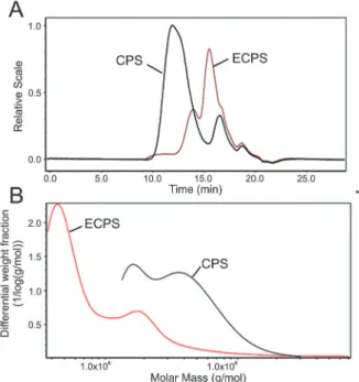
The total polysaccharide contents of ECPS and CPS measured by phenol-sulfuric acid assay were 89.17 and 90.26%, respectively. Meanwhile, the Mw of ECPS and CPS were measured by SEC-MALLS-RI. The Mw of ECPS was 211.7 kDa, which was lower than CPS (434.6 kDa). Figure 1A shows the relative intensity (RI) for ECPS and CPS; after enzymatic hydrolysis by mannase, the peak retention time of ECPS was longer than that of CPS. As displayed in Figure 1B, the differential weight fractions of polysaccharides were portrayed as the function of molar mass for samples. The molar mass distribution of poly-saccharide changed significantly by enzymatic hydrolysis. The differential weight fraction of ECPS in the low Mw
region increased, which suggested that the CPS was enzymatically degraded into low Mw polysaccharide.
Antioxidant activities of polysaccharides
The DPPH free-radical scavenging abilities of ECPS and CPS are reported in Figure 2A. The free-radical scavenging activities of polysaccharide samples and Vc exhibited a dose-dependent activity. In the current study, the free-radical scavenging ability of CPS was lower than that of ECPS. However, both exhibited lower free-radical scavenging effect than the positive sample. The IC50 values of ECPS and CPS were 0.39 and 0.51 mg/mL, respectively. As displayed in Figure 2B, the total antiox-idant activity can be assessed by testing the ferric iron reducing power. The concentrations varied from 0.1 to 1 mg/mL; both polysaccharide samples and Vc presented antioxidant activity in a dose-dependent manner. More-over, the absorbance value of ECPS was always higher than that of CPS at the same concentration.
Effect of ECPS and CPS on mushroom tyrosinase activity and cell viability
As shown in Figure 2C, the tyrosinase inhibitory activity of polysaccharides (0.1B1 mg/mL) presented
a dose-dependent relationship. Moreover, the inhibitory effect of ECPS was always higher than that of CPS at
the same concentration. The MTT assay was performed to assess the cytotoxic effects of ECPS and CPS in B16F10 melanoma cells. As displayed in Figure 2D, there were no significant changes in B16F10 cell viability with different concentrations (0B320mg/mL) of ECPS and CPS. Based
on these results, we used these concentration ranges in further research.
Effect of ECPS and CPS on intracellular tyrosinase activity and melanin contents
To compare the effects of ECPS and CPS on the activity of intracellular tyrosinase and melanogenesis in B16F10 melanoma cell model, the inhibitory potency of ECPS and CPS on melanin content and tyrosinase activity in a-MSH-stimulated B16F10 cells were examined. As shown in Figure 3, melanin content and tyrosinase activity of B16F10 cells were significantly increased when com-pared to the unstimulated B16F10 cells (Po0.01). At
con-centrations of 40 mg/mL (ECPS) and 160 mg/mL (CPS), the increase of melanin contents could be mitigated in a dose-dependent manner (Po0.01 and Po0.05). Similarly,
treatment with ECPS (40 mg/mL) and CPS (160 mg/mL) suppressed the tyrosinase activity of B16F10 cells (Po
0.01 and Po0.05). Moreover, ECPS exhibited higher
tyro-sinase inhibitory activity on melanogenesis than CPS. ECPS (160 and 320 mg/mL) exerted antimelanogenesis
effect comparable to the positive control (Kojic acid), which is widely used as skin whitening bioactive compound.
Effect of ECPS on tyrosinase, MITF, and TRP-1 protein levels in B16F10 cells
As shown in Figure 4, the ECPS significantly decreased tyrosinase, MITF, and TRP-1 protein expression levels in B16F10 cells in a dose-dependent manner (Po0.05 and
Po0.01). These results show that ECPS inhibited the
expression of tyrosinase by down-regulating protein expres-sion of TRP-1 and MITF.
Discussion
The natural polysaccharides from C. chinensis have received attention attributed to the good effects on
tyrosinase inhibition, free radical scavenging, and skin protection (25–27). However, little research has focused on
the antimelanogenesis activity of enzymatic modification of polysaccharides. Previous research has demonstrated that degraded polysaccharides by enzymatic hydrolysis process exhibited superior free radical scavenging effect (28). More-over, the biological activities of polysaccharides are closely related to their Mw distributions. Theoretically, low Mw poly-saccharides are more active than high Mw polypoly-saccharides due to their high penetration property on cell membranes (29,30). However, the antimelanogenesis effect of ECPS on B16F10 cells had not yet been studied. The low Mw polysaccharide was prepared by enzymatic hydrolysis with mannase.
Oxidative stress can produce excessive free radicals and lead to oxidative injury. Previous studies have proven
Figure 3. Effects of enzymatically hydrolyzedCuscuta chinensispolysaccharides (ECPS) and Cuscuta chinensispolysaccharides (CPS) on B16F10 cells. Melanin content and tyrosinase (TYR) activity of melanoma cells were measured after ECPS (AandC) and CPS (BandD) treatment. Kojic acid (160mg/mL) was used as positive control. Data are reported as means±SD.#P
that skin disease is closely related to accumulation of free radicals (31). Moreover, excessive free radicals play a vital role in suppressing melanogenesis of melanoma cells and growth of melanocytes (32). Tyrosinase is a multi-functional oxidant enzyme that contains bronze and is vital in promoting melanin biosynthesis (33). However, skin pigmentation and various skin diseases are closely related to the accumulation of melanin and cause a serious esthetic problem (34).
Active ingredients with antioxidant and anti-tyrosinase abilities can exert skin protection and inhibit melanogen-esis (35). Our results have demonstrated that the lower Mw of enzymatically modified polysaccharides exhibited superior antioxidant and anti-tyrosinase activities than ori-ginal polysaccharidesin vitro. The improvement is attrib-uted to the greater surface area and better water solubility, which was consistent with a previous study (17) that showed that the degraded polysaccharide fromSargassum fusiforme
possesses superior anti-tyrosinase activity and antioxidant activity than the original polysaccharide.
Normal melanocytes lie at the junction of the epidermis and dermis of the skin and generate melanin, which is transferred to keratinocytes (36). In the present study, the murine B16F10 melanoma cells were used because they possess melanogenic mechanism, are known to have intracellular tyrosinase, and can generate melanin, which are related toa-MSH stimulation and melanogenesis (37).
Tyrosinase activity, melanin content, and cell viability were the in vitro assays used to screen antimelanogenesis in present study. CPS and ECPS exhibited a dose-dependent inhibitory effect on tyrosinase activity and melanin synthesis in B16F10 cells. ECPS showed a stronger anti-melanin synthesis and anti-tyrosinase effect.
Tyrosinase-related protein-1 (TRP-1) and tyrosinase play a vital role in melanin biosynthesis and melanogen-esis pathways (38). MITF is a cellular transcription factor of the tyrosinase gene, which takes part in melanogenesis. Usually, the activation of TRP-1 and tyrosinase enhances MITF protein expression and causes the increase of melanin synthesis (39). Thus, skin whitening agents may have the property of inhibiting the signaling pathway involved in the activation of TYP-1 or tyrosinase. There-fore, we investigated the effects of ECPS on TRP-1, cellular tyrosinase and MITF protein expressions to study the mechanisms underlying the inhibition of tyrosinase activity and melanogenesis. The results of western bolt assay showed that ECPS suppressed the expression of TRP-1, tyrosinase, and MITF in B16F10 cells and implied that ECPS decreased melanogenesis by down-regulating tyrosinase, MITF, and TRP-1 expression in B16F10 melanoma cells. The result was in accordance with a previous study showing that the aqueous extract fromCuscuta japonica
seed significantly inhibited a-MSH-induced melanin syn-thesis and tyrosinase activity by suppressing p38 MAPK
phosphorylation, inhibiting cAMP levels, and subsequently decreasing the expression of TRP and MITF (40).
In summary, the enzymatically modified polysaccha-ride possessed superior antioxidant and antimelanogenic effects than the original polysaccharide. Furthermore, this antimelanogenic effect of ECPS was mediated by the suppression of TRP-1, tyrosinase, and MITF expression in
murine B16F10 cells. ECPS can be applicable for use in thefields of cosmetic and medicine products.
Acknowledgements
This work was supported by the National Natural Science Foundation of China (grant No. 81373640).
References
1. Riley PA. Melanogenesis and melanoma. Pigment Cell Research 2003; 16: 548–552, doi: 10.1034/j.1600-0749. 2003.00069.x.
2. Ortonne JP, Bissett DL. Latest insights into skin hyperpig-mentation.J Investig Dermatol Symp Proc2008; 13: 10–14, doi: 10.1038/jidsymp.2008.7.
3. Arung ET, Kuspradini H, Kusuma IW, Shimizu K, Kondo R. Validation ofEupatorium triplinerveVahl leaves, a skin care herb from East Kalimantan, Using a Melanin Biosynthesis Assay.J Acupunct Meridian Stud2012; 5: 87–92, doi: 10.1016/ j.jams.2012.01.003.
4. Kobayashi T, Urabe K, Winder A, Jiménez-Cervantes C, Imokawa G, Brewington T, et al. Tyrosinase related protein 1 (TRP1) functions as a DHICA oxidase in melanin biosynth-esis.EMBO J1994; 13: 5818–5825.
5. Costin GE, Hearing VJ. Human skin pigmentation: melano-cytes modulate skin color in response to stress.FASEB J 2007; 21: 976–994, doi: 10.1096/fj.06-6649rev.
6. Galván I, Alonso-Alvarez C. An Intracellular antioxidant determines the expression of a melanin-based signal in a bird.PLoS One2008; 3: e3335, doi: 10.1371/journal.pone. 0003335.
7. Yang J, Wang Y, Bao Y, Guo J. The totalflavones from Semen cuscutaereverse the reduction of testosterone level and the expression of androgen receptor gene in kidney-yang deficient mice.J Ethnopharmacol2008; 119: 166–171, doi: 10.1016/j.jep.2008.06.027.
8. Donnapee S, Li J, Yang X, Ge AH, Donkor PO, Gao XM, et al.Cuscuta chinensisLam.: A systematic review on ethno-pharmacology, phytochemistry and pharmacology of an impor-tant traditional herbal medicine.J Ethnopharmacol2014; 157: 292–308, doi: 10.1016/j.jep.2014.09.032.
9. Nisa M, Akbar S, Tariq M, Hussain Z. Effect ofCuscuta chinensiswater extract on 7,12-dimethylbenz[a]anthracene-induced skin papillomas and carcinomas in mice.J Ethnophar-macol1986; 18: 21–31, doi: 10.1016/0378-8741(86)90040-1. 10. Wang TJ, An J, Chen XH, Deng QD, Yang L. Assessment
of Cuscuta chinensis seeds’ effect on melanogenesis: Comparison of water and ethanol fractionsin vitroandin vivo. J Ethnopharmacol 2014; 154: 240–248, doi: 10.1016/j.jep. 2014.04.016.
11. Sun SL, Guo L, Ren YC, Wang B, Li RH, Qi YS, et al. Anti-apoptosis effect of polysaccharide isolated from the seeds of Cuscuta chinensisLam on cardiomyocytes in aging rats. Mol Biol Rep 2014; 41: 6117–6124, doi: 10.1007/s11033-014-3490-1.
12. Wang Z, Fang JN, Ge DL, Li XY. Chemical characterization and immunological activities of an acidic polysaccharide isolated from the seeds of Cuscuta chinensis Lam. Acta Pharmacol Sin2000; 21: 1136–1140.
13. Yang S, Xu X, Xu H, Xu S, Lin Q, Jia Z, et al. Purification, characterization and biological effect of reversing the kidney-yang deficiency of polysaccharides from Semen cuscutae. Carbohydr Polym2017; 175: 249–256, doi: 10.1016/j.carbpol. 2017.07.077.
14. Katayama S, Nishio T, Kishimura H, Saeki H. Immunomo-dulatory properties of highly viscous polysaccharide extract from the Gagome alga (Kjellmaniella crassifolia). Plant Food Humn Nutr2012; 67: 76–81, doi: 10.1007/s11130-011-0271-z.
15. Pengzhan Y, Ning L, Xiguang L, Gefei Z, Quanbin Z, Pengcheng L. Antihyperlipidemic effects of different molecular weight sulfated polysaccharides fromUlva pertusa (Chloro-phyta). Pharmacol Res 2003; 48: 543–549, doi: 10.1016/ S1043-6618(03)00215-9.
16. Jiang Y, Qi X, Gao K, Liu W, Li N, Cheng N, et al. Relationship between molecular weight, monosaccharide composition and immunobiologic activity of Astragalus poly-saccharides.Glycoconj J2016; 33: 755–761, doi: 10.1007/ s10719-016-9669-z.
17. Chen BJ, Shi MJ, Cui S, Hao SX, Hider RC, Zhou T. Improved antioxidant and anti-tyrosinase activity of poly-saccharide fromSargassum fusiformeby degradation.Int J Biol Macromol2016; 92: 715–722, doi: 10.1016/j.ijbiomac. 2016.07.082.
18. McCleary BV. Enzymatic modification of plant polysacchar-ides.Int J Biol Macromol1986; 8: 349–354, doi: 10.1016/ 0141-8130(86)90054-1.
19. Baurin N, Arnoult E, Scior T, Do QT, Bernard P. Preliminary screening of some tropical plants for anti-tyrosinase activity. J Ethnopharmacol2002; 82: 155–158, doi: 10.1016/S0378-8741(02)00174-5.
20. Mosmann T. Rapid colorimetric assay for cellular growth and survival: Application to proliferation and cytotoxicity assays. J Immunol Methods1983; 65: 55–63, doi: 10.1016/0022-1759(83)90303-4.
21. Hosoi J, Abe E, Suda T, Kuroki T. Regulation of melanin synthesis of B16 mouse melanoma cells by 1 alpha, 25-dihydroxyvitamin D3 and retinoic acid. Cancer Res 1985; 45: 1474–1478.
22. Wang HM, Chen CY, Wen ZH. Identifying melanogenesis inhibitors fromCinnamomum subaveniumwithin vitroand in vivoscreening systems by targeting the human tyrosinase. Exp Dermatol2011; 20: 242–248, doi: 10.1111/j.1600-0625. 2010.01161.x.
24. Parejo I, Codina C, Petrakis C, Kefalas P. Evaluation of scavenging activity assessed by Co(II)/EDTA-induced luminol chemiluminescence and DPPH(2,2-diphenyl-1-picrylhydrazyl) free radical assay.J Pharmacol Toxicol Methods2000; 44: 507–512, doi: 10.1016/S1056-8719(01)00110-1.
25. Rout S, Banerjee R. Free radical scavenging, anti-glycation and tyrosinase inhibition properties of a polysaccharide frac-tion isolated from the rind fromPunica granatum.Bioresour Technol2007; 98: 3159–3163, doi: 10.1016/j.biortech.2006. 10.011.
26. Yu P and Sun H. Purification of a fucoidan from kelp polysaccharide and its inhibitory kinetics for tyrosinase. Carbohydrate Polymers2014; 99: 278-283, doi: 10.1016/ j.carbpol.2013.08.033.
27. Wei X, Liu Y, Xiao J, Wang Y. Protective effects of tea polysaccharides and polyphenols on skin.J Agric Food Chem 2009; 57: 7757–7762, doi: 10.1021/jf901340f.
28. Xu J, Xu LL, Zhou QW, Hao SX, Zhou T, Xie HJ. Enhanced in vitro antioxidant activity of polysaccharides from Enter-omorpha prolifera by enzymatic degradation. J Food Bio-chem2016; 40: 275–283, doi: 10.1111/jfbc.12218. 29. Zhou J, Hu N, Wu Yl, Pan Yj, Sun CR. Preliminary studies
on the chemical characterization and antioxidant proper-ties of acidic polysaccharides from Sargassum fusiforme. J Zhejiang Univ Sci B2008; 9: 721–727, doi: 10.1631/jzus. B0820025.
30. Wu Q, Zheng C, Ning ZX, Yang B. Modification of low molecular weight polysaccharides fromTremella fuciformis and their antioxidant activityin vitro.Int J Mol Sci2007; 8: 670–679, doi: 10.3390/i8070670.
31. Yasui H, Sakurai H. Age-dependent generation of reactive oxygen species in the skin of live hairless rats exposed to UVA light.Exp Dermatol2003; 12: 655–661, doi: 10.1034/ j.1600-0625.2003.00033.x.
32. Yamakoshi J, Otsuka F, Sano A, Tokutake S, Saito M, Kikuchi M, et al. Lightening effect on ultraviolet-induced
pigmentation of guinea pig skin by oral administration of a proanthocyanidin-rich extract from grape seeds.Pigment Cell Res 2003; 16: 629–638, doi: 10.1046/j.1600-0749. 2003.00093.x.
33. Strothkamp KG, Jolley RL, Mason HS. Quaternary structure of mushroom tyrosinase.Biochem Biophys Res Commun 1976; 70: 519–524, doi: 10.1016/0006-291X(76)91077-9. 34. Parvez S, Kang M, Chung HS, Bae H. Naturally occurring
tyrosinase inhibitors: mechanism and applications in skin health, cosmetics and agriculture industries.Phytother Res 2007; 21: 805–816, doi: 10.1002/ptr.2184.
35. Perluigi M, De Marco F, Foppoli C, Coccia R, Blarzino C, Luisa Marcante M, et al. Tyrosinase protects human melano-cytes from ROS-generating compounds.Biochem Biophys Res Commun 2003; 305: 250–256, doi: 10.1016/S0006-291X(03)00751-4.
36. Hirobe T. How are proliferation and differentiation of mela-nocytes regulated?Pigment Cell Melanoma Res2011; 24: 462–478, doi: 10.1111/j.1755-148X.2011.00845.x.
37. Buscà R, Ballotti R. Cyclic AMP a key messenger in the regulation of skin pigmentation.Pigment Cell Res2000; 13: 60–69, doi: 10.1034/j.1600-0749.2000.130203.x.
38. Slominski A, Tobin DJ, Shibahara S, Wortsman J. Melanin pigmentation in mammalian skin and its hormonal regulation. Physiol Rev 2004; 84: 1155–1228, doi: 10.1152/physrev. 00044.2003.
39. Shibahara S, Yasumoto K-I, Amae S, Udono T, Watanabe K-I, Saito H, et al. Regulation of pigment cell-specific gene expression by MITF.Pigment Cell Res2000; 13: 98-102, doi: 10.1034/j.1600-0749.13.s8.18.x.