Vol.58, n.2: pp. 198-207, March-April 2015 http://dx.doi.org/10.1590/S1516-8913201400173
ISSN 1516-8913 Printed in Brazil
BRAZILIAN ARCHIVES OF BIOLOGY AND TECHNOLOGY
A N I N T E R N A T I O N A L J O U R N A L
Antimicrobial, Antioxidant and Cytotoxic Activity of Marine
Streptomyces parvulus
VITJS11 Crude Extract
S. Jemimah Naine, C. Subathra Devi
*, V. Mohanasrinivasan and Vaishnavi. B.
School of Biosciences and Technology; VIT University; Vellore, Tamil Nadu - India
ABSTRACT
The main aim of the study was to evaluate the bioactive properties of ethyl acetate crude extract of Streptomyces parvulus VITJS11 with a view to assess their therapeutic potential. The biological activity of ethyl acetate extract was tested against fungal and bacterial pathogens. The free radical scavenging potential of the crude extract was determined by DPPH assay. The chemo preventive properties of S. parvulus VITJS11 ethyl acetate extract was examined by MTT assay on HepG2 cells. The morphological, physiological and the biochemical properties of the strain S. parvulus VITJS11 was confirmed by conventional methods. Genotypic characterization was done using 16S r-DNA partial gene amplification and sequencing. The authenticity of the crude chemical constitutes were determined by the GC-MS. The ethyl acetate extract of VITJS11 showed maximum antifungal activity against three Aspergillus species and prominent antibacterial activity against two Gram positive and Gram negative bacteria at 20 mg/mL. The antioxidant potential of the crude extract exhibited strong reducing power activity at 5mg/ mL with 85% inhibition and the cytotoxic effect was found with IC50 of 500µg/ mL on HepG2 cell lines. The GC-MS analysis and the chromatogram patterns revealed 16 peaks, indicating the presence of bioactive constituents, which included several important organic compounds, namely 9-(2',2’ -dimethylpropanoilhydrazono)-3,6-dichloro-2,7-Bis-[2-(diethylamino)-ethoxy]fluorine (23.1) Dotriacontylpentafluoropropionate,(25.0) Octadecanoic acid, (20.0); Trans-2-methyl-4-n-butylthiane, S, S-dioxide.(19.0). The results showed the benefit of ethyl acetate extract from S. parvulus VITJS11 in treating microbial infections and indicated their broad spectrum of activity with beneficial virtues for therapeutic use.
Key words: Marine actinomycetes, Streptomyces parvulus VITJS11, antimicrobial activity, antioxidants,
cytotoxicity, bioactive compounds
*Author for correspondence: csubathradevi@vit.ac.in
INTRODUCTION
The development of drug resistance by the pathogens is due to the concurrent usage of
existing antibiotics. The search for new, safer,
broad-spectrum antibiotic with greater potency has been progressing slowly (Gupte et al. 2002). Hence, to combat these drug resistant pathogens, new antibiotics are in need and should be developed. There is no satisfactory drug as of now to control the occurrence of cancer and it is known to be the leading cause of death worldwide
actinomycetes, Streptomyces species are one of the largest sources of bioactive natural products
and they are taxonomically diverse and
biologically active. The potential contribution of
marine Streptomyces in the discovery of new
bioactive molecules is increasingly challenging
and the study of marine Streptomyces and their
potential role in the production of metabolites is becoming an interesting new topic for research. Several investigations have shown an increasing number of biologically active and structurally unique compounds from it (Hardt et al. 2000; Marsgy et al. 2001). Indian coastal region is an ideal location for harvesting many valuable species producing bioactive compounds with numerous complex and novel chemical entities. Recently, much attention has been directed toward biologically active compounds from natural products as they exhibit fewer side effects in comparison to orthodox medical drugs. However, the potential beneficial effects of these natural compounds need to be confirmed in large, rigorous trials. These structures can be chemically modified and improved through knowledge of the structure-activity relationship, mechanism of action, drug metabolism, molecular modelling and combinatorial chemistry studies. The discovery of new active metabolites must be followed by adequate biological testing (Jemimah et al. 2014). Hence, the aim of this study was to investigate the
bioactive potential of S. parvulus VITJS11crude
extracts from marine environment of Bay of Bengal, South coast of India for its antifungal, antibacterial, antioxidant and cytotoxic potential.
MATERIALS AND METHODS
Sample collection and isolation
Marine soil samples were collected from the south East coast of India, Ramanathapuram- Sethu Karai (Lat.9°50"N and 78°10'E) at the depth of 10-100 cm at littoral zone and the collected samples were stored at 4°C.The isolation was performed on selective media such as actinomycetes isolation
agar (AIA), Kuster’s agars, Bennett agar, Starch
casein agar along with 25% marine water and 25% marine soil extract for effective isolation (Balagurunathan et al. 2001). All the plates were
incubated at 30oC for 1 - 2 weeks. Emerging
isolates were sub cultured on ISP2 agar and stored under refrigeration. The morphological and cultural characteristics were determined on
various ISP medium (Newman et al. 2003; Ravikumar et al. 2008).
Cross streak method
The S. parvulus VITJS11 was cross streaked on the modified nutrient glucose agar plates near the periphery against wide range of Gram-positive and Gram-negative bacteria at right angle and incubated for three days (Alexander et al. 1977; Brock et al. 1994).The zone of inhibition was measured after two days of incubation.
Scanning electron microscopy
Streptomyces parvulus VITJS11 was grown on starch casein agar at 27°C for seven days. The spore chain morphology and spore surface ornamentation was evaluated by scanning electron microscopy analysis.
Fermentation and extraction of antibacterial compound
The inoculum of S. parvulus VITJS11 was
prepared on starch casein broth at a seed concentration of 100 mL in a 250 mL Erlenmeyer flask (pH 7.2) incubating at room temperature for seven days. For the isolation of bioactive compounds, extraction process was carried out successively with using ethyl acetate solvent with increasing polarity. The crude extract powder was weighed and studied for its bioactivity (Remya et al. 2007).
Test organisms
The pathogens, including Aspergillus niger
MTCC No:872; A. fumigatus MTCC No:8877; A.
flavus MTCC No:8790;Pseudomonas aeruginosa
(MTCC No: 4676), Staphylococcus aureus
(MTCC No: 7405), Salmonella typhi (MTCC No:
1167), Escherichia coli (MTCC No: 1588), were
obtained from the Microbial culture collection, IMTECH, Chandigarh, INDIA.
Determination of antifungal activity
The antifungal activity was assessed by agar-well diffusion assay. The culture medium of sabouraud dextrose broth was inoculated with the fungal
strains and was adjusted to 1 x 10⁸ CFU/mL. Then
100 µL of the inoculum was placed on the sabouraud dextrose agar and the wells were filled
with 100 µL of 20 mg/mL S. parvulus VITJS11
crude extract. The plates were incubated at 27oC
Agar-well diffusion assay
The in vitro antibacterial activity of all the solvent
extracts of S. parvulus VITJS11 was determined
by agar- well diffusion method (Pandey et al.
2004). Log phase bacterial cultures of
108 CFU/mL were used. One hundred microlitre of
various concentrations ranging 1.0 – 20 mg/mL of
crude extract of S. parvulus VITJS11 were tested.
Diameter of the zone was measured and expressed in mm.
Free radical Scavenging Activity
The antioxidant activity was determined by DPPH scavenging assay (Khalaf et al. 2008). Various concentration (0.1, 0.5, 1.0, 3.0 and 5.0 mg/mL) of S. parvulus VITJS11 crude extract was taken in separate tubes. Ascorbic acid was used as reference compound (0.1, 0.5, 1.0, 3.0 and 5.0 mg/mL). A freshly prepared solution of 0.002 % DPPH (2, 2, Diphenyl-2-Picryl hydrazyl) in methanol was added to each tube containing different concentrations of extracts (2.0 mL). The samples were incubated in dark at 37 °C for 20 min and read at 515 nm. The data were expressed as the percent decrease in the absorbance compared to the control. The percentage inhibition of radical scavenging activity was calculated.
Maintenance of cell cultures
The HepG2 cells were obtained from NCSS, Pune and cultured in RPMI-1640 medium on 10 cm
tissue culture dishes (Greiner Bio-one™,
Germany) supplemented with 10% heat
inactivated fetal bovine serum. Cells were
incubated in humidified incubator with 5% CO2 at
37°C, and sub cultured when confluence was reached up to 80% (Freshney et al. 1982).
Cell viability assay
The 3-(4, 5-dimethylthiazol-2-yl)-2-5-diphenyl tetrazolium bromide) MTT assay, was used to assess the viability of the cells (Mosmann et al.
1983). The HepG2 cells (5x10³)were seeded per
well into 96-well plates containing 100 µL
DMEM medium with 10% FBS incubated at 37◦C
for 24 h, Then, the cells were treated with 100 µL
of the S. parvulus VITJS11 crude extract in
triplicates with different concentrations ranging
from 50 to 1000 μg/mL. Doxorubicin (5µg / mL)
was used as internal positive control and 100 µL of DMEM was used as negative control; wells without any cells were considered as blank. After
incubating in humidified incubator with 5% CO2
at 37°C for 48 h, 20 µL of 5 mg/mL MTT diluted in PBS were added to each well and incubated for 4 h. One hundred microlitre of 10% SDS in, 0.01M HCl solution was added to each well to dissolve the formazan crystals formed. The plates were covered with aluminium foil and kept in an incubator for 12 h for dissolution of the formed formazan crystals. Amount of formazan was determined measuring the absorbance at 560 nm using a micro plate reader.
Molecular Characterization
Total genomic DNA was isolated using the phenol chloroform method (Nathan et al. 2004). PCR amplification of 16S r-DNA was carried out using
the primers FC27 (5’_ to
3’_AGAGTTTGATCCTGGCTCAG) and
RC1492 (5’_to3’_TACGGCTACCTT
GTTACGACTT) (Rainey et al. 1996). The PCR
product was detected by agarose gel
electrophoresis. Sequencing was performed using big dye terminator cycle sequencing kit (Applied Bio Systems, USA). The sequence was subjected to homology search using BLAST programme of
the National Centre for Biotechnology
Information (NCBI) and the sequence data was submitted to the GenBank database under the accession number (KC961640).
Phylogenetic analysis
The acquired sequences was used for a gene homology search, with the 16S r-DNA sequences available in the public databases from BLAST and identified to the generic level (Altschul et al. 1997; Benson et al. 1999). Using the CLUSTAL-X Multiple Sequence Alignment Program (Strasburg, France), the 16S r-DNA sequences of the strains were aligned with sequences of related organisms obtained from GenBank and a phylogenetic tree was constructed via the neighbor-joining method using the EvolView program (Huangkai et al. 2012). To validate the reproducibility of the branching pattern, a bootstrap analysis was performed.
GC-MS analysis
calculated by comparing its average peak area to the total areas. (Jemimah et al. 2013)
RESULTS AND DISCUSSION
Recently, numerous pathogens have developed resistance due to the indiscriminate use of commercial antimicrobial drugs. Hence, there is an urge to search the novel drugs against such
pathogens. It has been envisaged that marine environment are extreme source to provide exciting new bioactive compounds. In the present study, an attempt was made to identify the
bioactive potential of S. parvulus VITJS11. The
morphology, biochemical and taxonomical
characterization of S. parvulus VITJS11
represented the features of Streptomycetaceae
family belonging to genera Streptomyces (Table 1).
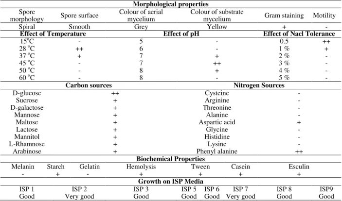
Table 1 - Morphological properties, biochemical, cultural and process parameters of Streptomyces parvulus
VITJS11.
Morphological properties
Spore
morphology Spore surface
Colour of aerial mycelium
Colour of substrate
mycelium Gram staining Motility
Spiral Smooth Grey Yellow + -
Effect of Temperature Effect of pH Effect of Nacl Tolerance
15oC - 5 - 0.5 ++
28 oC ++ 6 - 1 % +
37 oC + 7 + 2 % -
45 oC - 7 ++ 3 % -
50 oC - 8 + 4 % -
60 oC - 8 - 5 % -
Carbon sources Nitrogen Sources
D-glucose ++ Cysteine -
Sucrose + Arginine -
D-galactose + Threonine -
Mannose + Alanine -
Maltose + Aspartic acid +
Lactose + Glycine -
Mannitol + Histidine -
L-Rhamnose + Lysine -
Arabinose + Phenyl alanine ++
Biochemical Properties
Melanin Starch Gelatin Hemolysis Tween Casein Esculin
- + - + + + +
Growth on ISP Media
ISP 1 ISP 2 ISP 3 ISP 5 ISP 6 ISP 7 ISP 8 ISP9
Good Very good Good Good Good Very good Good Good
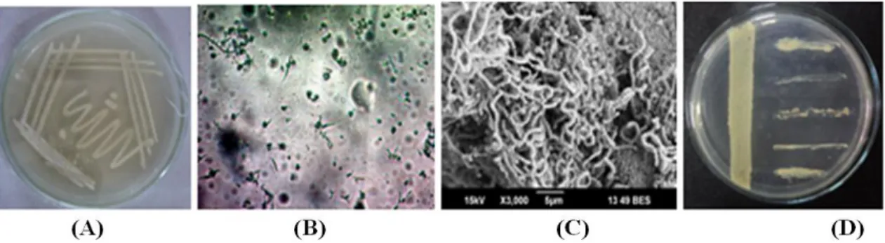
The strain was studied on the basis of its morphology and recognized by their presence of aerial hyphae with grey coloration on starch casein medium. The frequent production of spreading substrate mycelia was observed and the colonies were found darkened during sporulation with spirals. The colonies with powdery appearance had a characteristic feature with smooth surface. The
ornamentation of spore surface of S. parvulus
VITJS11 was observed under light microscopy. Scanning electron microscopy (magnification, 6,000) showed aerial mycelium bearing spores with 0.7 µm (Fig. 1).
The physiological characteristics of the S. parvulus
VITJS11 was compared with Streptomyces species
given in the key of Nonomura. The strain VITJS11
showed more similarity to the reference strain, S.
Figure 1 -Streptomyces parvulus VITJS11- (A) Pure culture plate of on starch casein agar; (B) Light microscopy at 40X; (C) Scanning electron microscopy; (D) Cross streak assay of the strain against the bacterial pathogens.
Table 2 - Comparison of morphological and
biochemical characteristics of Streptomyces parvulus
VITJS11 with reference strain.
S. parvulus
VITJS11 S. parvulus
Colour of aerial mycelium Grey Grey
Melanoid pigment - -
Reverse side pigment - -
Soluble pigment - -
Spore chain morphology Spiral Spiral
Utilization of carbon sources
Arabinose + +
Xylose + +
Inositol + +
Mannitol + +
Fructose + +
Rhamnose + +
Sucrose + +
Raffinose + +
The antifungal activity of S. parvulus VITJS11
was examined against three pathogenic species of Aspergillus. The strongest inhibition of fungal
growth was noticed against A.niger of 30 mm at
20 mg/mL concentration and A.fumigatus (25
mm), A.flavus (21 mm). The results could be
useful for the better understanding of the biocontrol mechanisms of the fungal strains, indicating that the strain VITJS11 probably producing a bioactive potential with antifungal
component (Fig. 2). The strain S. parvulus
VITJS11 subjected to primary screening for its activity against bacterial pathogens was confirmed by the cross streak method. The crude extract of the isolate further subjected to agar well diffusion method comparatively exhibited potent activity for all the clinical pathogens tested. The most active antibacterial crude fraction was obtained by the solvent mixture of ethyl acetate. The antibacterial
effects of crude extract showed activity against the bacterial pathogens.
Antifungal activity ofStreptomyces parvalus sp. VITSJ11 crude extract
A. niger A. fumigatus A. flavus
0 10 20 30 40
Fungal Pathogens
Z
o
n
e
o
f
in
h
ib
it
io
n
(m
m
)
(A)
(B)
Figure 2 - (A) Graphical representation of antifungal
activity of Streptomyces parvulus VITJS11 crude extract ; (B) Pictorial view showing the inhibition of fungal pathogens(a)
Aspergillus niger [30 mm] (b) Aspergillus fumigatus [25 mm] (c) Aspergillus flavus
[21 mm].
The maximum activity was found at 26 mm, which
was observed against S. typhi and E. coli, followed
also supported by the findings of similar study on Streptomyces strains, which were showing better
antifungal activity observed against Aspergillus
niger and strong antibacterial activity against Gram positive and Gram negative bacteria (Laidi et al. 2007; Oskay et al.2009). The studies on
marine Streptomyces sp exhibited good
antimicrobial activity against clinical pathogens (Ponmurugan et al. 2008). The concurrent studies of Streptomyces sp PM-32 isolated from off-shore
sediments collected at the Bay of Bengal coast was reported to have antimicrobial activity against a
group of bacterial and fungal pathogens
(Manivasagan et al. 2009). Studies on antibacterial
activity from Streptomyces sp SLO-105 was found
against Gram-positive bacteria (S. aureus,
methicillin-resistant S. aureus (MRSA),
Micrococcus luteus) and antifungal activity against Aspergillus niger(Morakchi et al. 2009; Ko et al. 2010).
(A) (B)
Figure 3 - (A) Graphical representation of antibacterial activity of Streptomyces parvulus VITJS11
crude extract; (B) Pictorial view showing the inhibition of bacterial pathogens at the maximum concentration 20 mg/mL (a) Pseudomonas aeruginosa [20 mm] (b)
Staphylococcus aureus [24 mm] (c) Salmonella typhi [26 mm] (d) Escherichia coli [26 mm].
The present study correlated with the existing report where the strain SRB25 was more similar to Streptomyces with antimicrobial property
against multidrug resistance S. aureus (Sathish et
al. 2012). The active compound of Streptomyces
KUAP106 was effective against Gram-positive, Gram-negative and unicellular filamentous fungi
(Usha et al. 2010). The Streptomyces sp. isolated
from saline farm-lands showed both antibacterial and antifungal activity (Sajid et al. 2009). The antioxidant activity of the VITJS11 crude extract showed maximum activity with 85% inhibition at 5.0 mg/mL concentration and its activity remained highly significant with the reference used (Fig.4).
Figure 4 - Graphical representation of antioxidant
property of Streptomyces parvulus
Human hepatocellular liver carcinoma cell lines (HepG2) was used as a model system to examine
chemo-preventive effect of S. parvulus VITJS11.
The concentration of the extract varied from 50 to
1000 µg/mL The IC50 value was 500 µg/mL on
HepG2 cells lines and exhibited substantial growth inhibition with 21% viability (Fig. 5). The HepG2 cell death resembled apoptosis, which was special evident with most notorious effect for strain VITJS11 crude extract. It was interesting to note that the strain VITJS11 crude extract resulted higher percentage of apoptotic cells. One possibility of the apoptotic phenomenon could be that the compounds in the extract interfering with
mitosis would have lead to apoptosis within short period of time. Actinomycin D is the most significant member of actinomycins, which are a class of polypeptide antibiotics isolated from Streptomyces genus (Hollstein et al. 1974). Actinomycin D decreases Mcl-1 expression and acts synergistically with ABT-737 against small cell lung cancer cell lines (Aishan et al. 2010).
Among the members of actinomycetes
genus, Streptomyces sp is a dynamic producer of
functional, bio-effective metabolites with broad
pharmaceutical range having antimicrobial,
antihelminthic, antitumor and antiviral agents (Ravikumar et al. 2008).
Figure 5 - (A) Percentage of cell viability shows the effective drug concentration toxic to HepG2 cell
lines. (B) Morphological changes of HepG2 cell lines treated with crude extract of
Streptomyces parvulus VITJS11. Cells were visualized under inverted light microscope Detachment of cells from substratum; cell shrinkage, nuclear condensation and fragmentation were evident in cells treated.
The phylogenetic analysis of S. parvulus VITJS11
was identified based on the nucleotide sequences of their 16S r-DNA genes, which showed similarity values of 98% to 16S r-DNA genes of firmicutes in the NCBI database and the isolate
revealed species level similarity to S. parvulus
S10. Phylogenetic tree construction was conducted using neighbor joining algorithm with bootstrap values based on 1000 replications and the annotation, tree visualizing was performed using EVOLVIEW (a web application tool). The generated tree revealed that the isolate shared
close affinity with Streptomyces species (Fig. 6).
Figure 6 - Phylogenetic tree based on partial 16S
r-DNA sequences, showing the relationship with other species belong to the genus
The presence of volatile chemical constituents of ethyl acetate crude extract were analyzed by the GC-MS analysis and the chromatogram patterns revealed 16 peaks, indicating the presence of bioactive constituents, which included several
important organic compounds namely 9-(2',2’
- dimethylpropanoilhydrazono)-3,6-dichloro-2,7-BIS-[2-(diethylamino)-ethoxy]fluorine(23.1)
Dotriacontyl pentafluoropropionate (25.0)
Octadecanoic acid (20.0); Trans-2-methyl-4-n-butylthiane, S, S-dioxide (19.0). Interpretation on mass spectrum of GC-MS was done using the database NIST08, WILEY8 (Fig. 7). In the present study, the partial characteristics of the active molecules indicated that bioactivity was likely to be due to the production of potential organic
substances. The chemical fingerprints of the crude extract was identified and further manifested in differentiation from the related chemical profiles. Hence, the poor availability of drugs and the increasing number of treatment failures have
motivated current searches for therapeutic
alternatives which can work effectively as potential antimicrobial agents (Pina-Vaz et al. 2004). Thus, the new antimicrobial marine Streptomyces derivatives could be useful alternatives for the treatment of fungal and
bacterial infections. The advantages of using these
bioresources and their and its natural compounds may reduce the risk of side effects and lower the cost.
(A) (B)
Figure 7 -Streptomyces parvulus VITJS11 crude extract (A) Chromatogram(B) GC-MS peaks of major
volatile bioactive metabolites with molecular structure (1) 9-(2',2’ -Dimethylpropanoilhydrazono)-3,6-Dichloro-2,7-Bis-[2 Diethylamino) Ethoxy]Fluorene; (2)Dotriacontylpentafluoropropionate; (3) Octadecanoicacid; (4) Trans-2-Methyl-4-N Butyl Thiane, S, S-Dioxide.
CONCLUSION
Streptomyces parvulus VITJS11 showed various bioactive properties, which highlighted its importance as potential pharmacological agents. Hence, there could be probability of new bioactive compound in the crude extract, which might provide a basis for further development of novel
compound from S. parvulus VITJS11.This also
provided a new insight towards the development of good candidates for pharmaceutical and bioactive natural products.
ACKNOWLEDGEMENT
We are greatly indebted to Vellore Institute of Technology for the constant encouragement, help and support for extending necessary facilities.
REFERENCES
Alexander M. Introduction to soil microbiology. 2nd edn. John Wiley & Sons, New York, 1977: 207. Altschul SF, Madden TL, Schaffer AA, Zhang J, Zhang
Z, Miller W et al. Gapped BLAST and PSI-BLAST: a new generation of protein database search programs. Nucleic Acids Res.1997; 25: 3389-3402. Balagurunathan R, Subramanian A. Antagonistic
Streptomycetes from marine sediments. Adv Biosci. 2001; 20: 71-76.
Barsby T, Kelly MT, Gagne SM, Andersen RJ: Bugorol A. produced in culture by a marine Bacillus sp. reveals a novel template for cationic peptide antibiotics. Org Lett. 2001; 3: 437-440.
Benson DA, Boguski MS, Lipman DJ, Ostell J, Oullette BFF, Rapp BA, Wheeler DL. GenBank. Nucleic Acids Res. 1999; 27: 12-17.
Brock TD, Madigan MT, Martiko JM, Parker J. Biology of Microorganisms. 7th edn. Englewood Cliffs. Prentice. Hall, International Inc, 1994.
Freshney RI, Hart E, Russell JM. Isolation and
purification of cell cultures from human tumours. In
Reid E, Cook, G M. W, Moore D J.(eds.), Cancer cell organelles; methodological surveys (B): Biochem
Chicester, UK: Horwood 1982; 2: 97-110.
Gupte M, Kulkarni P, Ganguli BN. Antifungal antibiotics. Appl Microbiol Biotechnol.2002; 58: 46-57.
Hardt I, Jensen PR, Fenical W. Neomarinone, and new cytotoxic marinone derivatives, produced by a marine filamentous bacterium (Actinomycetales).
Tetrahedron Lett. 2000; 41: 2073-2076.
Hollstein U. Actinomycin. Chemistry and mechanism of action. Chem Rev. 1974; 74 (6): 625-652.
Huangkai Z, Shenghan G, Wei-Hua C. EvolView, An online tool for visualizing, annotating and managing phylogenetic trees. Nucleic Acids Res.2012; 40: 569-572.
Jemimah NS, Subathra DC, Vaishnavi B, Mohanasrinivasan V. Screening for antimicrobial and antioxidant property of Streptomyces sp VITJS3 isolated from Bay of Bengal, Puducherry coast of India. J Pure Appl Microbiol. 2014; 8(1): 1125-1132. Khalaf NA, Shakya AK, Al-Othman A, El-Agbar Z, Farah H. Antioxidant activity of some common plants. Turk J Biol. 2008; 32: 51-55.
Ko HR, Chun HK, Jung MC, Kho YH: Valistatin (3-amino-2-hydroxy-4-phenylbutanoyl-valyl-valine), a new aminopeptidase M inhibitor, produced by Streptomyces sp.SL20209. J Microbiol Biotechnol.
2010;5: 36-40.
Laidi RF, Elshafei A, Sanker M, Cheick B, Hocine H. Screening, isolation and characterization of a novel antimicrobial producing actinomycetes, strain RAF10. Biotechnol. 2007; 6: 489-496.
Manivasagan P, Gnanam S, Sivakumar K, Thangaradjou T, Vijayalakshmi S, Balasubramanian T. Antimicrobial and cytotoxic activities of an actinobacteria (Streptomyces sp. PM-32) isolated from an offshore sediments of the Bay of Bengal in tamilnadu. Advances Biological Res. 2009; 3: 231-236.
Mellouli L, Mehdi RB, Sioud S, Salem M, Bejar S. Isolation, purification and partial characterization of antibacterial activities produced by a newly isolated
Streptomyces sp. US24 strain. Res Microbiol. 2003; 154: 345-352.
Morakchi H, Ayari A, Taok M, Kirane D, Cochet N. Characterization of Streptomyces strain SLO-105 isolated from Lake Oubeira sediments in North-East of Algeria. Afr J Biotech. 2009; 8: 6332-6336. Mosmann T. Rapid colorimetric assay for cellular
growth and survival: application to proliferation and cytotoxicity assays. J Immunol Methods. 1983; 65: 55-63.
Nathan A, Magarvey Jessica M, Keller, Bernan V, Dworkin M, Sherman DH. Isolation and characterization of novel marine derived actinomycete taxa rich in bioactive metabolites. Appl Environ Microbiol. 2004; 70: 7520-7529.
Newman DJ, Gragg GM, Snader KM. Natural products as source of new drugs over the period 1981-2002. J Nat Prod.2003; 66: 1022-1037.
Oskay M. Antifungal and antibacterial compounds from Streptomyces strains. Afr J Biotech. 2009; 8: 3007-3017.
Pandey B, Ghimire P, Agrawal VP. Studies on the antimicrobial activity of actinomycetes isolated from Khumbu region of Nepal. PhD dissertation. Tribhuvan University, Kathmandu. Nepal: 2004. Pina-Vaz C, Gonçalves-Rodrigues A, Pinto E,
Costa-de-Oliveira S, Tavares C, Salgueiro L, et al. Antifungal activity of Thymus oils and their major compounds. J Eur Acad Dermatol Venereol. 2004; 18(1): 73-78.
Ponmurugan P, Nithya B. Plasmid DNA of antibiotic producing strains of Streptomyces sannanensis isolated from different states in Southern India. Biotechnol. 2008; 7: 487-492.
Rainey FA, Ward-Rainey NL, Kroppenstedt RM, Tackebrandt E. The genus Norcardiopsis represents a phylogenetically coherent taxon and a distinct actinomycete lineage: proposal of Nocaridiopsaceae fam. nov. Int J Syst Bacteriol. 1996; 46:1088-1092.
Remya M, Vijayakumar R. Isolation and characterization of marine antagonistic actinomycetes from west coast of India. Med Biol. 2007; 5: 13-19. Sajid I, Yao CB, Shaaban KA, Hasnain S, Laatsch H.
Identification, isolation and optimization of antifungal metabolites from the Streptomyces malachitofuscus ctf9. World J Microbiol Biotechnol.
2009; 25: 601-610.
Sathish KS, Kokati VB. In vitro antimicrobial activity of marine actinobacteria against multidrug resistance
Staphylococcus aureus. Asian Pac J Trop Biomed.
2012; 2(10): 787-792.
Taylor MW, Radax R, Steger D, Wagner M. Sponge-associated microorganisms: evolution, ecology, and biotechnological potential. Microbiol Mol Biol Rev. 2007; 1: 295-347.
Usha R, Ananthaselvi C, Venil-Paliniswamy M. Antimicrobial and antiangiogenesis activity of
Streptomyces KUAP106 from mangrove soil.
European J Biol Sci. 2010; 2(4): 77-83.


![Figure 7 - Streptomyces parvulus VITJS11 crude extract (A) Chromatogram(B) GC-MS peaks of major volatile bioactive metabolites with molecular structure (1) 9-(2',2’-Dimethylpropanoilhydrazono)-3,6-Dichloro-2,7-Bis-[2 Diethylamino) Ethoxy]Fluor](https://thumb-eu.123doks.com/thumbv2/123dok_br/15960631.684089/8.892.98.815.450.691/streptomyces-chromatogram-bioactive-metabolites-molecular-structure-dimethylpropanoilhydrazono-diethylamino.webp)
