Breast cancer is the most common cancer affecting women in the world and provides functional changes in the human body. Evaluate women submitted to unilateral mastectomy, through electromyographic activity (mandibular rest, right and left laterality, protrusion, maximum voluntary contraction with and without Parafilm, habitual and non-habitual mastication) and thickness (rest and maximum voluntary contraction) of the right temporal muscle (RT), left temporal (LT), right masseter (RM) and left masseter (LM), also the molar bite force (right and left) and compare the data with healthy women. Material and Methods: 32 women were divided into two groups: unilateral mastectomy group (MG), average ± standard deviation 56.50±14.50 years (n=16) and without the disease group (CG), average ± standard deviation 56.56±14.15 years (n=16). The normalized electromyographic data, muscle thickness and maximal molar bite force were tabulated and submitted to statistical analysis (SPSS 21.0; student t test, p≤0.05). Significant statistical differences between MG and CG were found in right laterality, for RM (p=0.02); left laterality, for LT (p=0.01); chewing with peanuts, for RM (p=0.04); chewing with raisins, for LM (p=0.04) and right molar bite force (p=0.03). There was no statistically significant difference between MG and CG for muscle thickness. The results of this study suggest that women undergoing unilateral mastectomy may present functional changes, with emphasis on muscular hyperactivity, lower masticatory efficiency and lower maxillary bite force.
Effect of Breast Cancer Treatment
on the Stomatognathic System
Danilo Stefani Esposto1, Marcelo Palinkas2, Ligia Maria Napolitano Gonçalves3, Paulo Batista de Vasconcelos2, Vânia Tie Koga Ferreira4, Elaine Caldeira de Oliveira Guirro1, Selma Siéssere2, Simone Cecilio Hallak Regalo2
1Department of Biomechanics, Medicine and Locomotive Apparatus Rehabilitation, Ribeirão Preto Medical School, USP- Universidade de São Paulo, Ribeirão Preto, SP, Brazil 2Department Morphology, Physiology and Basic Pathology, School of Dentistry of Ribeirão Preto, USP – Universidade de São Paulo, Ribeirão Preto, SP, Brazil 3Department of Restorative Dentistry, School of Dentistry of Ribeirão Preto, USP - Universidade de São Paulo, Ribeirão Preto, SP, Brazil 4Department of Health Sciencs, UNIFAP - Universidade Federal do Amapá, Macapá, AP, Brazil
Correspondence: Marcelo Palinkas, Avenida do Café, s/n, 14040-904 Ribeirão Preto, SP, Brasil. Tel: +55-16-3315-3000. e-mail: [email protected]
Key Words: Breast, cancer, mastectomy, masticatory muscles.
Introduction
Breast cancer is a global public health problem with the highest survival rates, around 80%, among all cancers, when diagnosed early (1). In 2016, it was estimated the incidence of 246 660 000 cases, with 40 450 deaths in the United States (2) and 57 960 new cases in Brazil (3). Mortality rates are high, because the disease is still diagnosed in advanced stages, despite being considered a type of cancer with positive prognosis.
The surgical procedure is the most used approach in the treatment of breast cancer, comprising the conservative and non-conservative modality that encompasses total breast resection, called mastectomy (4).
Physical limitations are reported as a result of breast cancer treatment (5), as well as oral physiology alterations (6) and psychological changes, especially when it is related to anxiety and self-esteem (7).
With the evolution of the surgical techniques and the increasing incidence of malignant neoplasm in the breast, is necessary to verify the postoperative functional alterations resulting from the current procedures (8), because many mastectomized women present changes in the body posture, asymmetries in the trunk, limited range of motion on upper limbs and muscle disorders (9,10).
The neck muscles are essential for maintaining cervical
balance and of masticatory muscles, where surgical interventions may result in functional alterations of this system (11).
Thus, the importance of this study to provide information that can guide the process of evaluation and clinical follow-up of the mastectomized women, presenting a clarifying character for health professionals with the purpose of contributing to the functional analysis of the stomatognathic system.
Therefore, the objective of this unprecedented study was to evaluate the stomatognathic system function of women underwent unilateral mastectomy, by electromyographic activity, masticatory efficiency, muscle thickness and maximum molar bite force.
The hypothesis of this study is that unilateral mastectomy in women could present dysfunction in the stomatognathic system, as an increase in electromyographic activity of masticatory muscles, reduction in masticatory efficiency and lower maximum molar bite force.
Material and Methods
Ethical Approval
Breast cancer and stomatognathic system
Ethics Committee of the School of Dentistry of Ribeirão Preto, São Paulo, Brazil (process n. 38290714.8.0000.5381). All participants signed the informed consent form in accordance with Resolution 466/12 and the Complementary of the National Health Council.
Participants
The sample size was calculated by maximum significance level of α=0.05 and minimum power of 80% in the test, assuming a sample loss of 30%, and it was possible to determine n=16 individuals for each group studied (case and control). For the sample calculation was used the difference of twice the standard deviation of all the variables evaluated and was considered the one that presented the largest sample size.
A total of 40 women were selected to participate in the study, of which 8 were excluded because they presented previous orthopedic alterations of the cervical region and upper limbs (n=2); cognitive alterations (n=1); previous trauma in the head and neck region (n=1); small breasts (12) (n=2) and lymphedema in the upper limbs (13) (n=2).
In this way, 32 women with normal occlusion, without temporomandibular dysfunction (RDC/TMD) and orofacial pain were distributed in two groups: unilateral mastectomy group (MG), average ± standard deviation 56.50 ± 14.50 years (n=16), and without the disease group (CG), average ± standard deviation 56.56±14.15 years (n=16). The groups were matched individual-to-individual by age and anthropometric measures (Table 1).
The complementary characteristics of the unilateral mastectomized women were: without presence of metastasis (n=16), surgery time greater than 12 months (n=14), use of breast prosthesis after mastectomy (n=14), surgical side (right, n=9 and left, n=7), chemotherapy treatment (n=11), radiotherapeutic treatment (n=9), hormone therapy (n=3) and level of active physical activity assessed by the International Physical Activity Questionnaire (n=9) (14).
Electromyographic Analysis
The electromyographic signals were collected by the Trigno electromyographer (Delsys Inc., Boston, MA, USA) with wireless surface electrodes. The positioning of the electrodes on the temporalis and masseter muscle was identified by the use of the maximum voluntary contraction maneuver.
Prior to the placement of the electrodes, the skin was cleaned with alcohol to remove any residues of fat, make-up or pollution. During the electromyographic recording, the environment was kept calm, with the participants sitting in a comfortable chair, feet resting on the ground, with an upright posture and hands resting on the thighs. The head was erected, keeping the horizontal plane of Frankfurt
parallel to the ground.
The evaluation of electromyographic activity was performed during mandibular rest (4s), right laterality (10s), left laterality (10 s), protrusion (10 s) and dental clenching in maximal voluntary contraction with (4s) and without Parafilm (4s). The Parafilm consisted of a folded paraffin sheet (Parafilm Mâ, Pechiney Plastic Packaging, Batavia, IL, USA) (18x17x4mm, weight 245 mg) and placed between the occlusal faces of the first and second molars on the dental arch right and left sides.
The efficiency of the masticatory cycles in habitual and non-habitual chewing was analyzed by the integral of the linear envelope average of the electromyographic signal for both, temporalis and masseter muscles. Chewing with peanuts, simulating consistent food and raisins without seeds simulating soft food was used for the habitual mastication and for non-habitual chewing the Parafilm was used (15,16). The food belonged to the same manufacturing batch and was stored in individualized plastic bowls with lid, kept in a cool and airy environment.
The electromyographic signals were processed by Delsys electromyographic works analysis software, version 4. The values of the envelopment integral of the masticatory cycles in usual and non-habitual were obtained, eliminating the first three first cycles of the masticatory process and analyzing the five central cycles. The initial masticatory cycles were neglected, considering that, at the beginning of the masticatory process, there is considerable variation in the pattern of mandibular movement.
Muscle Thickness Analysis
The Sonosite Titan system portable ultrasound device with linear transducer was used to analyze the thickness of the temporalis and masseter muscles by recording ultrasound images under tasks of mandibular rest and dental clenching in maximum voluntary contraction.
The linear transducer was positioned transversely towards the fibers of the assessed muscles, coupled to the surface of the skin with a water-based electrically conductive gel. In the anterior portion of the temporalis muscle, the transducer was positioned in the region of the temporal fossa about 1.0 to 1.5 cm behind and above the outer angle of the eye. For the muscle belly of the masseter muscle, the linear transducer was positioned approximately 1.5 to 2.0 cm above the angle of the mandible toward the zygomatic arch.
For each mandibular task, three exams with a two-minute interval between them were performed, being considered the average value between the three measures.
Bite Force Analysis
D.S Esposto et al.
was used, adapted for buccal conditions with a capacity of up to 980.665 N to obtain the records of the maximum molar bite force, according to the laboratory protocol.
The participants were placed in a comfortable chair with their feet resting on the ground, standing upright and their hands resting on their thighs. For biosecurity reasons, the stems of the dynamometer were cleaned with alcohol, protected with disposable latex finger cots (Wariper, São Paulo, SP, Brazil) and positioned in the region of the first right and left first molar, where the greatest bite force is developed.
The participants tightened three times with maximum force the dynamometer rods, interspersing right and left side, with a two minutes rest between each record. It was considered the largest bite force on both sides.
Statistical Analysis
Due to the need for comparisons between different participants, the values of RMS and integral of the linear envelope of the electromyographic signal obtained during the maximal voluntary contraction with Parafilm were used to normalize the electromyographic activity values. Normalized electromyographic data in microvolts/second (rest, right and left laterality, protrusion, dental clenching in maximal voluntary contraction with and without Parafilm, chewing with peanuts, raisins and Parafilm), ultrasound data in centimeters (rest and dental clenching in maximum voluntary contraction) and maximal molar bite force data in newtons (right and left) were submitted to statistical analysis using the Statistical Package for Social Sciences (SPSS version 21.0, IBM®, USA). Descriptive analysis (mean and standard error) and Shapiro-Wilk normality test were used for each variable. The values were compared by Student’s t test for independent samples (p≤0.05).
Results
Average age and anthropometric measurements data of MG and CG are shown in Table 1. There was no statistically significant difference (p≤0.05) for these variables between the groups. Table 2 shows the average thickness of the temporal and masseters muscles at rest and maximum voluntary contraction between MG and CG. There was
no statistically significant difference (p≤0.05) between the groups.
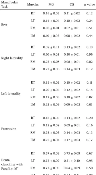
The normalized electromyographic data of the temporal and masseter muscles during rest and mandibular postural tasks for MG and CG are presented in Table 3. Significant statistical differences (p≤0.05), between MG and CG, were observed in the right laterality (right masseter) and left laterality (left masseter). The MG presented higher electromyographic activity under mandibular task, except for right laterality (left temporal) and maximum voluntary contraction with Parafilm (right temporal and left masseter).
Table 4 indicates the average of habitual and non-habitual masticatory efficiency for MG and CG. Significant statistical difference (p≤0.05), between MG and CG, was observed during peanut chewing (right masseter). The MG showed greater electromyographic activity in the dynamic tasks of the mandible.
The average molar bite force values of the right and left sides are shown in Table 5. There was a significant statistical difference (p≤0.05), between MG and CG, in the right bite force, with lower force for MG.
Discussion
The rehabilitation of the breast after removal of cancer can create organic complications that hinder the muscular function, promoting spasms and sensitivity (17).
The stomatognathic system, because it is complex, present’s well-defined physiological functions, requiring mandible requisition, associated with the movements of
Table 1. Comparison of means of the anthropometric measurements [weight (Kg) and height (cm)] and age (years) between mastectomy group (MG) and without the disease group (GC) using t test (p≤0.05)
MG CG p value
Age 56.50±14.50 56.56±14.15 0.99
Weight 65.59±7.53 66.37±9.54 0.79
Height 1.60±0.06 1.62±0.04 0.54
Table 2. Averages means, standard errors (±) and statistical significance (p≤0.05) of the thickness of the right masseter (RM), left masseter (LM), right temporal (RT) and left temporal (LT) during rest and dental clenching in mastectomy group (MG) and without the disease group (CG)
Mandibular task
and muscles MG CG p value
Rest
RT 0.30 ± 0.01 0.33 ± 0.04 0.56
LT 0.29 ± 0.01 0.28 ± 0.01 0.44
RM 0.86 ± 0.04 0.86 ± 0.02 0.92
LM 0.84 ± 0.02 0.83 ± 0.03 0.81
Dental Clenching
RT 0.41 ± 0.03 0.44 ± 0.04 0.50
LT 0.41 ± 0.02 0.41 ± 0.02 0.82
RM 1.05 ± 0.04 1.08 ± 0.03 0.51
Breast cancer and stomatognathic system
structures of the face and neck (18). If a disease occurs in the human body, this system will use adaptive resources to maintain functional efficiency so as not to allow failures to occur, which may be manifested by signs and symptoms of dysfunction (19).
The study of the mechanisms of development and the functions of this complex system can provide parameters useful in the recognition of deviations from normality. Therefore, the objective of this study was to determine if women submitted to unilateral mastectomy would present functional alterations in the stomatognathic system.
We observed that in the mandibular rest, the temporalis and masseter muscles presented electromyographic activity in both groups, however in MG, the activity was greater.
The masticatory, cervical and thoracic musculature present delineated anatomical relationships, with interdependent functions (20) and after invasive treatment in the thoracic region, all postural maintenance may be impaired, promoting muscle imbalance (18), leading to the destabilization of the mandibular positioning, causing functional alteration of the masticatory muscles fibers and consequently muscular hyperactivity (21).
In the protrusion, the activation pattern of the temporalis muscles was greater than that of the masseter in both groups, with a higher normalized electromyographic activity in the MG. These results are in agreement with the pattern described in the literature, where the temporalis muscles are more active than the masseter muscles (22). The mandibular excursion movements are influenced by the position of the head, peripheral impulses and emotional situations (18). After the mastectomy, functional morbidities are observed, highlighting posture changes
Table 3. Means, standard errors (±) and statistical significance (p≤0.05) of the normalized electromyographic data (µV) averages of the right masseter (RM), left masseter (LM), right temporal (RT) and left temporal (LT) for mastectomy group (MG) and without the disease group (CG) in the mandibular task
Mandibular
Task Muscles MG CG p value
Rest
RT 0.16 ± 0.03 0.11 ± 0.02 0.12
LT 0.15 ± 0.04 0.10 ± 0.02 0.24
RM 0.08 ± 0.01 0.07 ± 0.01 0.51
LM 0.10 ± 0.02 0.08 ± 0.02 0.44
Right laterality
RT 0.32 ± 0.11 0.13 ± 0.02 0.10
LT 0.10 ± 0.02 0.10 ± 0.01 0.96
RM 0.27 ± 0.07 0.08 ± 0.01 0.02
LM 0.23 ± 0.05 0.14 ± 0.03 0.12
Left laterality
RT 0.15 ± 0.03 0.10 ± 0.02 0.11
LT 0.20 ± 0.05 0.12 ± 0.02 0.14
RM 0.17 ± 0.03 0.10 ± 0.02 0.07
LM 0.23 ± 0.05 0.09 ± 0.02 0.01
Protrusion
RT 0.18 ± 0.03 0.13 ± 0.02 0.20
LT 0.12 ± 0.02 0.09 ± 0.01 0.16
RM 0.25 ± 0.06 0.14 ± 0.03 0.13
LM 0.25 ± 0.04 0.17 ± 0.04 0.17
Dental clenching with Parafilm M®
RT 0.67 ± 0.09 0.73 ± 0.09 0.67
LT 0.72 ± 0.09 0.71 ± 0.10 0.95
RM 0.73 ± 0.09 0.64 ± 0.09 0.50
LM 0.60 ± 0.05 0.64 ± 0.11 0.77
Table 4. Means, standard errors (±) and statistical significance (p≤0.05) of the normalized electromyographic data (µV) of the right masseter (RM), left masseter (LM), right temporal (RT) and left temporal (LT) for mastectomy group (MG) and without the disease group (CG) in the mandibular task of habitual chewing (peanuts and raisins) and non-habitual chewing (Parafilm M®)
Chewing Muscles MG CG p value
Peanuts
RT 0.79 ± 0.15 0.60 ± 0.16 0.39
LT 0.56 ± 0.07 0.47 ± 0.08 0.42
RM 0.86 ± 0.14 0.52 ± 0.07 0.04
LM 0.80 ± 0.17 0.53 ± 0.08 0.15
Raisins
RT 0.57 ± 0.14 0.53 ± 0.16 0.83
LT 0.48 ± 0.06 0.41 ± 0.07 0.47
RM 0.65 ± 0.14 0.45 ± 0.09 0.26
LM 0.70 ± 0.15 0.35 ± 0.05 0.04
Parafilm M®
RT 0.89 ± 0.16 0.77 ± 0.25 0.69
LT 0.79 ± 0.12 0.61 ± 0.10 0.25
RM 0.74 ± 0.16 0.63 ± 0.11 0.57
LM 0.92 ± 0.18 0.59 ± 0.06 0.09
Table 5. Means, standard errors (±) and statistical significance (p≤0.05) of the right and left molar bite force (N) for mastectomy group (MG) and without the disease group (CG)
MG CG p value
Right molar 139.84 ± 19.22 195.15 ± 14.91 0.03
D.S Esposto et al.
that affect the stomatognathic system, mainly in the mandibular kinematics and promoting alterations of the masticatory function (23).
Our findings showed that in the right and left laterality, the activation of the masseter muscles did not follow the appropriate neuroanatomical pattern, demonstrating that the greater muscle contralateral activity during the function was not found. It was observed that in the mandibular excursion movements, the normalized electromyographic activities of the temporalis and masseter muscles were smaller in the MG when compared to the CG. Women undergoing breast cancer treatment present muscle tension, fatigue, and depression (24). These factors may interfere with the impairment of the masticatory function, causing functional alteration (25), however, variables such as tension, fatigue and depression were not evaluated in this study.
In the maximum voluntary contraction task, the left masseter and right temporalis muscles demonstrated lower electromyographic activity in MG compared to CG. The surgical procedure that reconstructs the breast, after the mastectomy, returning the volume and contour, may not restore the natural function of the breast, altering the stable body balance, destabilizing the movement in the static position (26). In this study, 87.5% of the women submitted to unilateral mastectomy performed breast reconstruction with silicone prosthesis, maintaining the similarity of the contralateral breast (12), which allowed us to verify and include fifteen participants with medium breasts and one participant with large breasts.
In this study, the masticatory performance was evaluated by the use of food tests such as peanuts, representing the consistent food, raisins reproducing the soft food and the Parafilm as inert material. The use of the inert material had the function of demonstrating the joint play movement of the dynamic records, of buccal opening short excursion, requested to reduce effects of the change of muscular length. It was observed that in the habitual mastication (peanuts and raisins) and non-habitual (Parafilm), MG presented higher normalized electromyographic activity when compared to CG. When muscular hyperactivity is observed in dynamic actions of individuals who have suffered functional alterations, it is noticed that the muscular fibers behave in a different way, being more recruitment to maintain the same performance, when in comparison with healthy system (16). This hypothesis may explain the less efficient results of masticatory performance in mastectomized women.
The parameters related to the thickness of the masticatory muscles and molar bite force, in women submitted to mastectomy was not studied until the present moment of this research. This situation justifies
the study of these variables for the scientific community, collaborating in the knowledge of possible morphological changes that will serve for professionals in the medical and dental sciences area (27).
The results these study demonstrated that there were no significant differences between MG and CG, both, in the rest task and in the maximum voluntary contraction. Despite an apparent deficiency in the function of the masticatory muscles of unilateral mastectomized women, there was no change in measures of masticatory muscle thickness when compared to the data collected from healthy women.
Our group comparisons showed that masticatory functional performance in unilateral mastectomized women was lower because there was reduction in maximum molar bite force on both the right and left sides, demonstrating that muscular resistance and dysfunction may occur before and after treatment of a disease (28).
In the literature there are no scientific reports that considered the static and dynamic aspects of mastication muscles in women submitted to unilateral mastectomy, through electromyographic evaluation, masticatory performance, muscular thickness and maximum molar bite force, which makes it difficult to discuss and compare the results of this study.
Although there is no statistical difference in almost all the variables between the groups, the mean values presented by the mastectomy group are higher than the means of the control group.
The lack of muscle balance in mastectomy group can be explained by the posture changes resulting from the surgical procedure (29).
Researches that intends to evaluate this public with the purpose of detailing the morphofunctional changes of the stomatognathic system after the treatment of breast cancer has its scientific and social merit. These, demonstrate the negative impact that the disease and its treatment can promote the functions of the masticatory apparatus, assisting health professionals in oral rehabilitation plans and therapeutic treatments because, based on the results obtained in this study, the masticatory muscular system of these women may be compromised, dysfunctional and fragile.
The present study has some limitations. We do not evaluate the time of mastectomy, posture, masticatory muscular fatigue and depression of participants that could interfere in the muscle activity.
Breast cancer and stomatognathic system
Resumo
Câncer de mama é a neoplasia mais comum que mais acomete mulheres no mundo e proporciona alterações funcionais no corpo humano. Objetivo: avaliar mulheres submetidas à mastectomia unilateral, por meio da atividade eletromiográfica (repouso mandibular, lateralidade direita e esquerda, protrusão, contração voluntária máxima com e sem parafilme, mastigação habitual e não habitual) e espessura (repouso e contração voluntária máxima) do músculo temporal direito (TD), temporal esquerdo (TE), masseter direito (MD) e masseter esquerdo (ME), além da força de mordida molar (direita e esquerda) e comparar os dados com mulheres saudáveis. 32 mulheres foram distribuídas em dois grupos: grupo mastectomia unilateral (GM), média ± desvio padrão 56,50 ± 14,50 anos (n=16) e grupo sem a doença (GC), média ± desvio padrão 56,56 ± 14,15 anos (n=16). Os dados eletromiográficos normalizados, espessura muscular e força de mordida molar máxima foram tabulados e submetidos à análise estatística (SPSS 21.0; teste t de student, p≤0,05). Foram encontradas diferenças estatísticas significantes entre GM e GC na lateralidade direita, para o MD (p=0,02); lateralidade esquerda, para o TE (p=0,01); mastigação com amendoim, para o MD (p=0,04); mastigação com uva-passa, para o ME (p=0,04) e força de mordida molar direita (p=0,03). Não houve diferença estatística significante entre GM e GC para espessura muscular. Os resultados deste estudo sugerem que mulheres submetidas à mastectomia unilateral podem apresentar alterações funcionais do sistema estomatognático, com destaque para hiperatividade muscular, menor eficiência mastigatória e menor força de mordida molar máxima.
Acknowledgements
We gratefully acknowledge the support of Coordenação de Aperfeiçoamento de Pessoal de Nível Superior (CAPES) and National Institute and Technology - Translational Medicine (INCT.TM).
References
1. Athiyaman H, Athiyaman M, Chougule A, Kumar HS. Estimated Risk of Radiation Induced Contra Lateral Breast Cancer Following Chest Wall Irradiation by Conformal Wedge Field and Forward Intensity Modulated Radiotherapy Technique for Post-Mastectomy Breast Cancer Patients. Asian Pac J Cancer Prev 2016;17:6007-6011.
2. Siegel RL, Miller KD, Jemal A. Cancer statistics, 2016. CA Cancer J Clin 2016;66:7-30.
3. Rocha-Brischiliari SC, Oliveira RR, Andrade L, Brischiliari A, Gravena AA, Carvalho MD, Pelloso SM, et al. The Rise in Mortality from Breast Cancer in Young Women: Trend Analysis in Brazil. PLoS One 2017;12:e0168950.
4. Braude L, Laidsaar-Powell R, Gilchrist J, Kirsten L, Juraskova I. An exploration of Australian psychologists’ role in assessing women considering risk-reducing or contralateral prophylactic mastectomy. Breast 2017;32:105-111.
5. Wijayasinghe N, Duriaud HM, Kehlet H, Andersen KG. Ultrasound Guided Intercostobrachial Nerve Blockade in Patients with Persistent Pain after Breast Cancer Surgery: A Pilot Study. Pain Physician 2016;19:E309-E318.
6. Caputo JB, Campos SS, Pereira SM, Castelo PM, Gavião MB, Marques LS. Masticatory performance and taste perception in patients submitted to cancer treatment. J Oral Rehabil 2012;39:905-913.
7. Parsa Yekta Z, Sadeghian F, Taghavi Larijani T, Mehran A. The Comparison of Two Types of Relaxation Techniques on Postoperative State Anxiety in Candidates for The Mastectomy Surgery: A Randomized Controlled Clinical Trial. Int J Community Based Nurs Midwifery 2017;5:61-69. 8. Palma MR, Vanderlei LC, Ribeiro FE, Mantovani AM, Christofaro DG,
Fregonesi CE. The relationship between post-operative time and cardiac autonomic modulation in breast cancer survivors. Int J Cardiol 2016;224:360-365.
9. Shamley D, Lascurain-Aguirrebeña I, Oskrochi R. Clinical anatomy of the shoulder after treatment for breast cancer. Clin Anat 2014;27:467-477.
10. Araújo Neto EA, Alves BCA, Gehrke FS, Azzalis LA, Junqueira VCB, Sousa LVA. Quality of Life of Post-Mastectomy Women Living in a Semi-Arid Region of Brazil. Int J Environ Res Public Health 2017;14:pii: E601. 11. Boudreau SA, Falla D. Chronic neck pain alters muscle activation
patterns to sudden movements. Exp Brain Res 2014;232:2011-2020. 12. Sacchini V, Luini A, Tana S, Lozza L, Galimberti V, Merson M.
Quantitative and qualitative cosmetic evaluation after conservative treatment for breast cancer. Eur J Cancer 1991;27:1395-1400. 13. Taylor R, Jayasinghe UW, Koelmeyer L, Ung O, Boyages J. Reliability and
validity of arm volume measurements for assessment of lymphedema. Phys Ther 2006; 86:205-214.
14. Matsudo VK, Matsudo SM, Araújo TL, Andrade DR, Oliveira LC, Hallal PC. Time trends in physical activity in the state of São Paulo, Brazil: 2002-2008. Med Sci Sports Exerc 2010;42:2231-2236.
15. Siéssere S, Lima NA, Semprini M, de Sousa LG, Issa JPM, Monteiro SAC, Regalo SCH. Masticatory process in individuals with maxillary and mandibular osteoporosis: electromyographic analysis. Osteoporos Int 2009;20:1847-1851.
16. Palinkas M, Cecilio FA, Siéssere S, Borges Tde F, de Carvalho CA, Semprini M. Aging of masticatory efficiency in healthy subjects: electromyographic analysis--Part 2. Acta Odontol Latinoam 2013;26:161-166.
17. Hojan K, Manikowska F, Chen BP, Lin CC. The influence of an external breast prosthesis on the posture of women after mastectomy. J Back Musculoskelet Rehabil 2016;29:337-342.
18. Carvalho-Oliveira M, Salles C, Terse R, D’Oliveira A Júnior ,et al. Association between severe asthma and changes in the stomatognathic system. J Bras Pneumol 2016;42:423-428.
19. Douglas CR, Avoglio JL, de Oliveira H. Stomatognathic adaptive motor syndrome is the correct diagnosis for temporomandibular disorders. Med Hypotheses 2010;74:710-718.
20. Eriksson PO, Häggman-Henrikson B, Zafar H. Jaw-neck dysfunction in whiplash-associated disorders. Arch Oral Biol 2007;52:404-408. 21. Palinkas M, Bataglion C, de Luca Canto G, Machado Camolezi N,
Theodoro GT, Siéssere S. Impact of sleep bruxism on masseter and temporalis muscles and bite force. Cranio 2016;34:309-315. 22. Cecílio FA, Regalo SC, Palinkas M, Issa JP, Siéssere S, Hallak JE. Ageing
and surface EMG activity patterns of masticatory muscles. J Oral Rehabil 2010;37:248-255.
23. Roggia B, Santos VA Filha, Correa B, Rossi ÂG. Posture and body balance of schoolchildren aged 8 to 12 years with and without oral breathing. Codas 2016;28:395-402.
24. Zhang P, Li CZ, Wu CT, Jiao GM, Yan F, Zhu HC. Comparison of immediate breast reconstruction after mastectomy and mastectomy alone for breast cancer: A meta-analysis. Eur J Surg Oncol 2017;43:285-293. 25. Dante AMM, Santos CMD, Palinkas M, Lellis JBM, Siessere S, Semprini
M. Effect of radiation on the performance of the stomatognathic system – a pilot study. J Dent Oral Disord Ther 2014; 2:1-5.
26. Mochizuki H, Tashiro M, Kano M, Sakurada Y, Itoh M, Yanai K. Imaging of central itch modulation in the human brain using positron emission tomography. Pain 2003;105:339-346.
27. Kiliaridis S, Georgiakaki I, Katsaros C. Masseter muscle thickness and maxillary dental arch width. Eur J Orthod 2003;25:259-263. 28. Klassen O, Schmidt ME, Ulrich CM, Schneeweiss A, Potthoff K, Steindorf
K. Muscle strength in breast cancer patients receiving different treatment regimes. J Cachexia Sarcopenia Muscle 2016:1-12. 29. Atanes Mendes Peres AC, Dias de Oliveira Latorre MD, Yugo
Maesaka J, Filassi JR, Chada Baracat E, Alves Gonçalves Ferreira E. Body posture after mastectomy: comparison between immediate breast reconstruction versus mastectomy alone. Physiother Res Int 2017;22:e1642 Epu.