Limites de movimentos mandibulares em crianças****
Mandibular movement range in children
*Fonoaudióloga. Aperfeiçoamento em Interação Ortodontia e Fonoaudiologia promovido pela Faculdade de Odontologia de Ribeirão Preto da Universidade de São Paulo (USP). Monitora do Curso de Aperfeiçoamento em Motricidade Oral do Departamento de Clínica Infantil, Odontologia Preventiva e Social da Faculdade de Odontologia de Ribeirão Preto - USP. Endereço para correspondência: Rua Gonçalves de Magalhães, 388 -Ribeirão Preto - SP - CEP 14030-570 ([email protected]).
**Fonoaudióloga. Pós-Graduada do Departamento de Oftalmologia, Otorrinolaringologia e Cirurgia de Cabeça e Pescoço da Faculdade de Medicina de Ribeirão Preto da USP. Monitora do Curso de Aperfeiçoamento Interação Ortodontia e Fonoaudiologia - Faculdade de Odontologia de Ribeirão Preto - USP.
*** Fonoaudióloga. Doutora em Ciências pela USP. Professora do Departamento de Oftalmologia, Otorrinolaringologia e Cirurgia de Cabeça e Pescoço - Área Fonoaudiologia da Faculdade de Medicina de Ribeirão Preto da USP.
****Trabalho Realizado na Faculdade de Medicina de Ribeirão Preto da USP.
Artigo Original de Pesquisa
Artigo Submetido a Avaliação por Pares
Conflito de Interesse: não
Recebido em 12.12.2008.
Revisado em 26.02.2009; 05.04.2009; 18.05.2009.
Aceito para Publicação em 18.05.2009.
Barbara Cristina Zanandréa Machado*
Ana Paula Magalhães Medeiros**
Cláudia Maria de Felício***
Abstract
Background: identification of the mandibular movement range is an important procedure in the evaluation of the stomatognathic system. However, there are few studies in children that focus on normal parameters or abnormalities. Aim: to determine the average range of mandibular movements in Brazilian children aged 6 to 12 years; to verify the difference between genders, in each age group, and between the different age groups: 6-8 years; 8.1-10 years; and 10.1-12 years. Method: participants of the study were 240 healthy children selected among regular students from local schools of São Paulo State. The maximum mandibular opening, lateral excursion and protrusive movements, and deviation of the medium line, if present, were measured using a digital caliper. Student T test, Analysis of variance and Tukey test were considered significant for p < 0.05. Results: the mean values observed in the studied sample were: 44.51mm for maximum mandibular opening; 7.71mm for lateral excursion to the right; 7.92mm for lateral excursion to the left; 7.45mm for protrusive movements. No statistical difference was observed between genders. There was a gradual increase in the range of mandibular movements, with significant differences mainly between the ages of 6-8 years and 10.1-12 years. Conclusion: during childhood the range of mandibular movements increases. Age should be considered in this analysis for a greater precision in the diagnosis.
Key Words: Stomatognathic System; Range of Motion; Child; Diagnosis.
Resumo
Tema: a determinação dos limites de movimentos mandibulares é um importante procedimento na avaliação do estado funcional do sistema estomatognático, porém poucos são os estudos que focalizam os parâmetros de normalidade ou desvios em crianças. Objetivos: definir as médias dos limites de movimentos mandibulares em crianças brasileiras de 6 a 12 anos de idade; verificar diferenças entre os gêneros, em cada faixa etária, e entre as faixas etária de 6 a 8 anos, 8:1 a 10 anos e 10:1 a 12 anos de idade. Método: participaram 240 crianças, escolares do interior do Estado de São Paulo. Com o auxílio de um paquímetro digital foram mensuradas a máxima abertura mandibular, a protrusão, a excursão lateral direita e esquerda e o desvio da linha média, quando presente. O teste T Student, a Análise de variância e o pós-teste Tukey foram considerados significantes para p < 0,05. Resultados: as médias das medidas da amostra foram: máxima abertura mandibular 44,51 mm, excursão lateral direita 7,71mm, excursão lateral esquerda 7,92 mm e a protrusão 7,45 mm. Não houve diferença estatística entre os gêneros. Houve aumento gradual nos limites dos movimentos mandibulares com o aumento da faixa etária, com diferenças significantes principalmente entre as faixas etárias de 6 - 8 anos e 10:1-12 anos. Conclusão: durante a infância os limites de movimentos mandibulares aumentam e a idade deve ser considerada na análise desses dados para maior precisão no diagnóstico.
Palavras-Chave: Sistema Estomatognático; Amplitude de Movimento Articular; Criança; Diagnóstico.
Referenciar este material como:
Introduction
In the assessment of the functional status of
the stomatognathic system the determination of the
mandibular movement range is an important
procedure (1) since reduced movements have been
considered to be clinical signs in the diagnosis of
problems affecting or related to the orofacial
system, among them temporomandibular disorders
(TMD) (2-6) and neurogenic disorders (7).
There is a great demand for clinical speech
therapy for children aged six to 12 years, but normal
data regarding maximum mandibular opening, lateral
excursion and protrusion in children are scarce both
in the international (3,8-10) and in the Brazilian
(1,6,11) literature.
In addition, there is controversy about the
relationship between mandibular range of
movements and the following variables: gender, age,
body height, facial morphology, gonial angle,
mandibular ramus length, angle of mouth opening,
neuromotor development, and circulatory disorders
(3,9,11-20).
The objectives of the present study were to
define the mean mandibular range of movements
of Brazilian children aged 6 to 12 years, to determine
gender differences in each age range and
differences between the 6-8, 8.1-10 and 10.1-12 year
ranges in order to define normality parameters.
Method
The study was approved by the Human
Research Ethics Committee of the University
Hospital, Faculty of Medicine of Ribeirão Preto,
University of São Paulo (HCFMRP-USP), Process
HCRP nº 89/2007, and all persons responsible for
the children gave written informed consent to
participate in the study.
Subjects
A total of 240 children aged 6 to 12 years from
municipal schools participated in the study. Children
presenting signs and symptoms of TMD, a history of
facial trauma and of head and neck surgery, children
wearing braces, children with neurological disorders
and craniofacial deformities or systemic diseases were
excluded from the study.
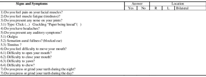
Subjects were selected using the validated
protocol for multiprofessional centers for the
determination of signs and symptoms of TMD
(ProTMDmulti) (21) (enclosed), which permits to
differentiate between asymptomatic and TMD
information provided by the child, the family and
school staff, and with observation during the
evaluation of mandibular movements.
The sample was divided into three groups
according to age range, as follows:
- Group I (6 to 8 years of age) consisting of 89
children, 44 girls and 45 boys;
- Group II (8.1 to 12 years) consisting of 79 children,
38 girls and 41 boys, and
- Group III (10.1 to 12 years of age) consisting of 72
children, 45 girls and 27 boys.
The sample was also subdivided into gender: male
(M) and female (F).
Procedure
A history was taken with the children and the
persons responsible for them in order to obtain the
information needed for the study, such as
identification, age, developmental data, health, and
signs and symptoms of TMD.
During the session for evaluation, the subjects
sat in a chair with a back support, with their feet
resting on the floor. With the aid of a digital Mitutoyo
pachymeter - Series 500 - Absolute Coolant Proof
IP66, with 0.01 repeatability and ± 0.02 mm accuracy,
positioned on point zero at each new measurement,
mandibular movements were measured as follows:
- maximum mandibular opening: the distance
between the incisal ridge of the upper and lower
incisors was considered during maximum mouth
opening up to the painless limit, plus the
measurement of vertical overbite;
- protrusion: with the teeth in occlusion, the
distance from the vestibular surface of the lower
incisors to the incisal ridge of the upper incisors
was measured. The subject was then asked to
protrude his mandible by sliding it along the maxilla
and the horizontal distance from the vestibular
surface of the upper incisors to the incisal ridge of
the lower incisors was measured. The sum resulted
in the measure of mandibular protrusion;
- laterality: the subject was asked to perform
maximum dislocation of the mandible to the right
and the horizontal distance between the line that
passes through the upper central incisors and the
lower central incisors or between the lip frena was
measured. The same procedure was used to
measure left lateral excursion (23).
re-evaluated 20% of the subjects selected at
random. According to the split-half test,
inter-examiner reliability was high (0.91) and correlation
was 0.83.
Data analysis
Data were analyzed by descriptive statistics for
the definition of the means, standard deviations and
confidence limits regarding the mandibular range of
movements. The parametric test one-way ANOVA was
used to analyze the gender and age range variables,
followed by the Tukey post-test to determine
differences between groups. The level of significance
was set at 0.05 in all analyses.
Results
The mean measures for the sample as a whole
were: maximum mandibular opening, 44.51 mm; right
lateral excursion; 7.71 mm, left lateral excursion, 7.92
mm, and protrusion, 7.45 mm.
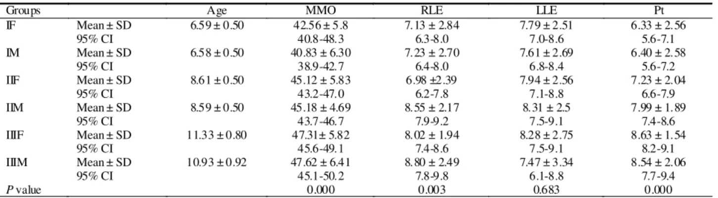
There was no significant difference between
males and females (p> 0.05) for the same age range.
The mean values, standard deviations and
confidence intervals of the measures (in mm)
according to age range and gender are given in
Table 1.
Considering the lack of difference between
genders, the groups were compared according to age
range. There was a significant difference in the
measures of maximum mandibular opening and
protrusion between Groups I, II and III, with the mean
for Group II being higher than the mean for Group I
and lower than the mean for Group III. The measure of
right lateral excursion was significantly lower for Group
I compared to Group III (p < 0.05). The mean values,
standard deviations and confidence intervals of the
measures (in mm) according to age range and to the
results of group comparisons are given in Table 2.
TABLE 2. Means, standard deviations, and confidence intervals according to age and group comparison.
MMO: maximum mandibular opening, RLE: right lateral excursion, LLE: left lateral excursion, Pt: protrusion. ANOVA with one variation factor. Means followed by different superscript letters (a, b, c) differed significantly by the Tukey test (P < 0.05).
Groups Age MMO RLE LLE Pt
I Mean± SD 6.58 ± 0.50 41.69 a ± 6.09 7.18a ± 2.76 7.70 a ± 2.59 6.37 a ± 2.56
95% CI --- 40.4-43.0 6.6-7.8 7.2-8.2 5.8-6.9
II Mean± SD 8.59 ± 0.49 45.15 b ± 5.23 7.79a .b ± 2.39 8.13 a ± 2.52 7.63 b ± 1.99
95% CI --- 44.0-46.3 7.3-8.3 7.6-8.7 7.2-8.1
III Mean± SD 11.18±0.86 47.43 c ± 6.01 8.31b ± 2.18 7.98 a ± 2.98 8.60 c ± 1.74
95% CI --- 46.0-48.8 7.8-8.8 7.3-8.7 8.2-9.0
valor de P 0.0000 0.0000 0.016 0.57 0.0000
TABLE 1. Means, standard deviation and confidence interval according to age and gender.
F: female, M: male, MMO: maximum mandibular opening, RLE: right lateral excursion, LLE: left lateral excursion, Pt: protrusion. ANOVA with one variation factor.
Groups Age MMO RLE LLE Pt
IF Mean ± SD 6.59 ± 0.50 42.56 ± 5.8 7.13 ± 2.84 7.79 ± 2.51 6.33 ± 2.56
95% CI 40.8-48.3 6.3-8.0 7.0-8.6 5.6-7.1
IM Mean ± SD 6.58 ± 0.50 40.83 ± 6.30 7.23 ± 2.70 7.61 ± 2.69 6.40 ± 2.58
95% CI 38.9-42.7 6.4-8.0 6.8-8.4 5.6-7.2
IIF Mean ± SD 8.61 ± 0.50 45.12 ± 5.83 6.98 ±2.39 7.94 ± 2.56 7.23 ± 2.04
95% CI 43.2-47.0 6.2-7.8 7.1-8.8 6.6-7.9
IIM Mean ± SD 8.59 ± 0.50 45.18 ± 4.69 8.55 ± 2.17 8.31 ± 2.5 7.99 ± 1.89
95% CI 43.7-46.7 7.9-9.2 7.5-9.1 7.4-8.6
IIIF Mean ± SD 11.33 ± 0.80 47.31± 5.82 8.02 ± 1.94 8.28 ± 2.75 8.63 ± 1.54
95% CI 45.6-49.1 7.4-8.6 7.5-9.1 8.2-9.1
IIIM Mean ± SD 10.93 ± 0.92 47.62 ± 6.41 8.80 ± 2.49 7.47 ± 3.34 8.54 ± 2.06
95% CI 45.1-50.2 7.8-9.8 6.1-8.8 7.7-9.4
Discussion
The determination of the means and standard
deviations of the mandibular range of movement
according to age and gender is justified by the need
for parameters for the diagnosis of disorders
involving the functionality of the stomatognathic
system. Thus, the objective of the present study
was to determine the measures of maximum
mandibular opening, right and left mandibular
excursion and protrusion in Brazilian children aged
6 to 12 years according to age range and gender, as
well as to compare the measures of the groups
divided into three age ranges.
No significant difference was observed between
genders regarding the measure of excursive
mandibular movements, in agreement with previous
studies (3,6,8,24). The measures studied seem to
follow the same tendency of other physical
characteristics that start to differentiate according
to gender at puberty since gender differences have
been detected in samples of adolescents and adults
(2,10,15,19,20).
Greater amplitude of maximum mandibular
opening, right lateral excursion and protrusion was
observed with increasing age range, corroborating
previous findings (1,3,8-11). Significant differences
in the measures of left lateral excursion and
protrusion were observed in a recent study on
Brazilian children aged 7-10 and 12-14 years (6).
The measures obtained in the present study were
higher than the means obtained for children aged 1
to 1 and a half years (8) and only the lowest age
range presented means close to those of children
aged 3 to 5 years (24).
Compared to the measurements obtained for
children of the same age ranges, the present results
agreed with some studies (6,8), but in general were
inferior to other results (3,11). Characteristics of the
children such as weight an height may perhaps
explain the differences in results, but only
Vanderas(3) considered height, which was positively
correlated with the extension of mandibular
movements. Also, the instrument used in the present
study, which was digital and of high precision, may
have contributed to some differences.
The increased mandibular range of movement
with increasing age ranges (3,6,8,11,24) may be related
to anatomical changes, to the maturation of the
central nervous system, to skeletal growth and to
the maturation of occlusal function and of oral motor
control (25-30).
Clinical evaluation should be based on a very
thorough foundation and therefore normative data
should be investigated in different populations. The
measurement of the mandibular range of movements
provided reliable quantitative data as shown by the
intra- and inter-examiner reliability and also by the
agreement of the present data with previously
published ones.
Thus, these measures may be valid for the
diagnosis and clinical control of patients with
orofacial myofunctional disorders and/or TMD, and
as the measurement of the result of myofunctional
therapy aiming at the liberty, symmetry and control
of mandibular movements.
Conclusion
The present study permitted us to conclude that:
- The mean measures of the range of mandibular
motion in the sample studied were: maximum
mandibular opening, 44.51 mm; right lateral
excursion, 7.71 mm; left lateral excursion, 7.92 mm,
and protrusion 7.45 mm;
- There was no difference between genders;
- The maximum mandibular opening and protrusion
measures gradually increased with increasing age
range. The right laterality measure followed the same
tendency but a difference was demonstrated when
the 6-8 and 10-12 year age ranges were compared.
Anexo
Protocol for multi-professional centers for the determination of signs and symptoms of temporomandibular disorders Name:
PART I. Frequency of signs and symptoms of TMD
PART II. Severity of signs and symptoms of TMD. Instructions: please observe what your symptoms feel like in different situations and point to their severity, scoring from zero to ten. The more severe (strong and frequent) the symptom, the higher the score should be; the less severe, the lower the number. ZERO (0) = no symptom TEN (10) = the worse sensation
In the sequences, the same questions were repeated for the situations WHILE CHEWING, WHILE SPEAKING and WHILE RESTING (when resting, without neither speaking nor chewing). Source: Felício et al (21)
Answer Location
Signs and Symptoms
Yes No R L Bilateral
1) Do you feel pain on your facial muscles? 2) Do you feel muscle fatigue (tiredness)? 3) Do you present any noise on your joints?
3.1) Type: Click (...) Crackling “Paper being knead”( ) 4) Do you have headaches?
5) Do you present any auditory symptoms? 5.1) Otalgia
5.2) Sensation aural fullness? (blocked ear) 5.3) Tinnitus ?
6) Do you feel difficulty to move your mouth? 6.1) Difficulty to open your mouth?
6.2) Difficulty to close your mouth? 6.3) Difficulty to yawn?
6.4) Difficulty to chew?
7) Do you press or grind your teeth during the night? 7) Do you press or grind your teeth during the day?
Table to register answeres – Severity of Signs and Symptoms -
When you wake up
1) Pain on Facial Muscles 0 1 2 3 4 5 6 7 8 9 10
2) Pain on Articulation (TMJ) 0 1 2 3 4 5 6 7 8 9 10
3) Pain on the Neck 0 1 2 3 4 5 6 7 8 9 10
4) Earache 0 1 2 3 4 5 6 7 8 9 10
5) Tinnitus (buzzing) 0 1 2 3 4 5 6 7 8 9 10
6) Blocked Ears 0 1 2 3 4 5 6 7 8 9 10
7) Tooth Sensitiveness 0 1 2 3 4 5 6 7 8 9 10
8) Noise of articulation 0 1 2 3 4 5 6 7 8 9 10
References
1. Leles CR, Moreira Neto JJS, Giro EM, Compagnoni MA. Valores normais da amplitude do movimento mandibular em crianças. Rev Fac Odontol São José dos Campos. 2000;3(2):121-6.
2. Dworkin SF, Huggins KH, Leresche L, Von Korff M, Howard J, Truelove E, et al. Epidemiology of signs and symptoms in temporomandibular disorders: clinical signs in cases and controls. J Am Dent Assoc. 1990;120(3):273-81.
3. Vanderas AP. Mandibular movements and their relationship to age and body height in children with or without clinical signs of craniomandibular dysfunction: Part IV. A comparative study. ASDC J Dent Child. 1992;59(5):338-41.
4. Celic R, Jerolimov V, Knezovic Zlataric D. Relationship of slightly limited mandibular movements to temporomandibular disorders. Braz Dent J. 2004;15(2):151-4.
5. Bianchini EMG, Paiva G, Andrade CRF. Mandibular movements in speech: interference of temporomandibular dysfunction according to pain indexes. Pró Fono. 2007;19(1):7-18.
6. Sousa LM, Nagamine HM, Chaves TC, Grossi DB, Regalo SCH, Oliveira AS. Evaluation of mandibular range of motion in Brazilian children and its correlation to age, height, weight, and gender. Braz Oral Res. 2008;22(1):61-6. 7. Duffy JP. Motor speech disorders: substrates, differential diagnosis e management. St. Louis: Mosby; 1995. 8. Agerberg G. Maximal mandibular movements in children. Acta Odont Scand. 1974;32(3):147-59.
9. Rothenberg LH. An analysis of maximum mandibular movements, craniofacial relationships and temporomandibular joint awareness in children. Angle Orthod. 1991;61(2):103-12.
10. Hirsch C, John MT, Lautenschlager C, List T. Mandibular jaw movement capacity in 10-17-year old children and adolescents: normative values and the influence of gender, age, and temporomandibular disorders. Eur. J. Oral Scie. 2006;114(6):465-70.
11. Hamazaki CM, Kawaura R, Bianchini EMG, Assencio-Ferreira VJ. Verificação da amplitude dos movimentos mandibulares em crianças. Rev. Cefac. 2002;4(1):35-40. 12. Ettala-Ylitalo UM, Lane T. Functional disturbances of the masticatory system in relation to articulatory disorders of speech in a group of 6-8-year-old children. Archs Oral Biol. 1991;36(3):189-94.
13. Laine MT, Pahkala RH, Jaroma SM, Qvarnstrom. Associations among different orofacial dysfunctions in 6-8 year old. Arch. Oral. Biol. 1992;37(11):6-895-9. 14. Dijkstra PU, Hof AL, Stegenga B, Bonti LG. Influence of mandibular length on mouth opening. J. Oral Rehabil. 1999;26(2):117-22.
15. Lewis RP, Buschang PH, Throckmorton GS. Sex differences in mandibular movements during opening and closing. Am. J. Orthod. Dentofacial Orthop. 2001;120(3):294-303.
16. Fukui T, Tsuruta M, Murata K, Wakimoto Y, Tokiwa H, Kuwahara Y. Correlation between facial morphology, mouth opening ability, and condylar movement during opening-closing jaw movements in female adults with normal occlusion. Eur J Orthod. 2002;24(4):327-36. 17. Pahkala RH, Qvarnstrom MJ. Mandibular movements capacity in 19-year-olds with and without articulatory speech disorders. Acta Odontol Scand. 2002;60(6):341-5. 18. Moipolai P, Karic VV, Miller VJ. The effect of the gonial angle, ramus length, age and gender on the temporomandibular opening index. J. Oral Rehabil. 2003;30(12):1195-9.
19. Gallagher C, Gallagher V, Whelton H, Cronin M. The normal range of mouth opening in an Irish population. J. Oral Rehabil. 2004;31(2):110-6.
20. Farella M, Iodice G, Michelotti A, Leonardir R. The relationship between vertical craniofacial morphology and the sagittal path of mandibular movements. J. Oral Rehabil. 2005;32(12):857-62.
21. Felício CM, Mazzetto MO, Rodrigues da Silva MAM, Bataglion C, Hotta THA. Preliminary protocol for multi-professional centers for determination of signs and symptoms of temporomandibular disorders. Crânio. 2006;24(4):258-64.
22. Felício CM, Melchior MO, Rodrigues da Silva MAM. Clinical validity of the protocol for multi-professional centers for the determination of signs and symptoms of temporomandibular disorders. Part II. Crânio. 2009;27(1):62-7.
23. Felício CM. Desordens Temporomandibulares (DTM): diagnóstico fonoaudiológico e terapia. In: Felicio CM. Fonoaudiologia aplicada a casos odontológicos: motricidade oral e audiologia. São Paulo: Pancast; 2001. p. 91-125. 24. Bonjardim LR, Gavião MBD, Pereira LJ, Castelo PM. Mandibular movements in children with and without signs and symptoms of temporomandibular disorders. J. Appl. Oral Sci. 2004;12(1):39-44.
25. Farkas LG, Posnick JC, Hreczko TM. Growth patterns of the face: a morphometric study. Cleft Palate Craniofac. J. 1992;29(4):308-15.
26. Hayasaki H, Yamasaki Y, Nishima N, Naruse K, Nakata M. Characteristics of protrusive and lateral excursions of the mandible in children with the primary dentition. J. Oral Rehabil. 1998;25(4):311-20.
27. Kiliaridis S, Karlsson S, Kjellberg H. Characteristics of masticatory mandibular movements and velocity in growing individuals and young adults. J Dent Res. 1991;70(10):1367-70.
28. Papargyriou G, Kjellberg H, Kiliaridis S. Changes in masticatory mandibular movements in growing individuals: a six-year follow-up. Acta Odontol Scand. 2000;58(3):129-34.
29. Walsh B, Smith A. Articulatory movements in adolescents: Evidence for protracted development of speech motor control processes. J. Speech Lang. Hear Res. 2002;45(6):1119-33.