Printed version ISSN 0001-3765 / Online version ISSN 1678-2690 http://dx.doi.org/10.1590/0001-3765201720160457
www.scielo.br/aabc
Anesthesia of
Epinephelus marginatus
with essential oil of
Aloysia
polystachya
: an approach on blood parameters
CARINE O. FOGLIARINI1
, QUELEN I. GARLET2
, THAYLISE V. PARODI3
, ALEXSSANDRO G. BECKER4
, LUCIANO O. GARCIA5
, BERTA M. HEINZMANN2
, ANA MARIA S. PEREIRA6
and BERNARDO BALDISSEROTTO1
1
Departamento de Fisiologia e Farmacologia, Universidade Federal de Santa Maria, Av. Roraima, 1000, 97105-900 Santa Maria, RS, Brazil 2
Departamento de Farmácia Industrial, Universidade Federal de Santa Maria, Av. Roraima, 1000, 97105-900 Santa Maria, RS, Brazil
3
Universidade Regional Integrada do Alto Uruguai e das Missões, Prédio 9, Av. Batista Bonoto Sobrinho, 733, 97700-000 Santiago, RS, Brazil 4
Centro de Ciências do Mar/ CCMAR, CIMAR – Laboratório Associado, Universidade do Algarve, Campus de Gambelas, 8005-139 Faro, Portugal
5Instituto de Oceanografia, Estação Marinha de Aquicultura, Universidade Federal do Rio Grande/ FURG, Caixa postal 474, 962010-030 Rio Grande, RS, Brazil
6Universidade de Ribeirão Preto, Av. Constabile Romano, 2201, 14100-000 Ribeirão Preto, SP, Brazil
Manuscript received on July 21, 2016; accepted for publication on September 1, 2016
ABSTRACT
This study investigated the anesthetic potential of the essential oil (EO) of Aloysia polystachya in juveniles of dusky grouper (Epinephelusmarginatus). Fish were exposed to different concentrations of EO of A. polystachya to evaluate time of induction and recovery from anesthesia. In the second experiment, fish
were divided into four groups: control, ethanol and 50 or 300 µL L−1 EO of A. polystachya, and each group was submitted to induction for 3.5 min and recovery for 5 or 10 min. The blood gases and glucose levels showed alterations as a function of the recovery times, but Na+ and K+ levels did not show any
alteration. In conclusion, the EO from leaves of A. polystachya is an effective anesthetic for dusky grouper, because anesthesia was reached within the recommended time at EO concentrations of 300 and 400 µL L−1.
However, most evaluated blood parameters showed compensatory responses due to EO exposure.
Key words: anesthetic efficacy, blood gases, glucose, hemoglobin, plasma ion levels.
Correspondence to: Bernardo Baldisserotto E-mail: [email protected]
* Contribution to the centenary of the Brazilian Academy of Sciences.
INTRODUCTION
Several procedures of fish culture such as handling,
blood sampling, transporting and vaccination often
generate a stress response in the animals (Kiessling et al. 2009, Zahl et al. 2012). Firstly, activation of the hypothalamic–pituitary–interrenal axis occurs,
with subsequent release of catecholamines and cortisol. As a consequence, glucose and lactate
increase and osmoregulatory disturbances occur
plants, such as the essential oil (EO) of Lippia alba (Cunha et al. 2010, Azambuja et al. 2011, Becker
et al. 2012, Heldwein et al. 2012, Salbego et al.
2014), Ocimum gratissimum (Silva et al. 2012),
Hesperozygis ringens (Silva et al. 2013, Toni
et al. 2014) and Aloysia triphylla (Gressler et al.
2014, Parodi et al. 2014, Zeppenfeld et al.2014)
showed efficacy and safety for use in aquaculture
procedures.
The EO used in the present study was
obtained from leaves of A. polystachya (Griseb.)
Moldenke (Verbenaceae), an aromatic native plant
widely distributed in subtropical regions of South
America, mainly in Paraguay and North Argentina,
and popularly known as “burrito”, “poleo de
Castilla” or “poleo riojano”. This plant is referred
to as a sedative (Del Vitto and Petenatti 1997) and
is also used against gastrointestinal pain in folk
medicine (Filipoy 1994). Studies with mice and
rats indicated that the hydro-ethanolic extract from
the aerial parts of A. polystachya has anxiolytic
and antidepressant-like effects (Mora et al. 2005,
Hellión-Ibarrola et al. 2006, 2008).
The dusky grouper, Epinephelus marginatus
(Serranidae) has a wide distribution, occurring
along the Mediterranean Sea and in the Indian
Ocean to the southeast of the African continent
(Fennessy 2006). On the west coast of the Atlantic
Ocean, the dusky grouper occurs from Rio de Janeiro to the New Gulf region in Argentinean
Patagonia (Figueiredo and Menezes 1980, Irigoyen
et al. 2005). Since E. marginatus is a target species
for aquaculture (Cunha et al. 2013, Cavalli 2014,
Sanches et al. 2014), the present study investigated
the anesthetic potential of the EO of A. polystachya in dusky grouper juveniles. Some blood parameters
were also analyzed, aiming to evaluate possible
side effects of this EO.
MATERIALS AND METHODS
PLANT MATERIAL AND ESSENTIAL OIL EXTRACTION
Aloysia polystachya (Griseb.) Moldenke
(Verbenaceae) was cultivated in the medicinal plant garden of “Nature’s Pharmacy”, Municipality of
Jardinópolis, SP, Brazil. The leaves were harvested
in September 2012 at 10 am and dried in an oven with forced air circulation at a temperature of 45 °C
for 48 h. The voucher specimen (UPMU No. 1213)
was identified by Dr. Rossi from the Institute of Botany of São Paulo, and a voucher was deposited
in the Herbarium of Medicinal Plants at the
University of Ribeirão Preto, SP, Brazil. The EO
was extracted from dried leaves by hydrodistillation using a Clevenger-type apparatus according to the
European Pharmacopoeia (2007).
ESSENTIAL OIL ANALYSIS
The EO samples were analyzed by GC–MS with an Agilent 6890A gas chromatograph equipped
with a 5973C mass selective detector using a
non-polar HP5-MS fused silica capillary column (5% phenyl, 95% methylsiloxane, 30 m x 0.25 mm i.d.
x 0.25 µm film thickness) and electron ionization
mode at 70 eV. Helium was used as carrier gas
at a flow rate of 1.0 mL min−1; the injector and
detector temperatures were set at 250 and 280 °C, respectively. Oven temperature was kept at 40 °C
for 4 min and then gradually raised to 320 °C at 4 °C min−1. Injections were performed in split inlet
mode (ratio 1:100). Kovats retention indices were
calculated using a homologous series of C7–C31 n-alkanes injected under the same conditions. The
EO constituents were identified by comparison of
the mass spectra and Kovats retention indices with
literature data and with the National Institute of
Library (NIST 2008, Adams 2009). FID analysis
was performed in an equivalent column and using
the same oven parameters as described for GC–MS.
Both injection and detection temperatures were set at 300 °C and the split inlet mode ratio was 1:50.
The percentage of EO compounds was calculated
by under peak area integration.
ANIMALS AND WATER CONDITIONS
Dusky grouper (82.0 ± 2.3 g; 16.7 ± 0.1 cm) juveniles obtained from a fish culture in Rio Grande, southern, Brazil, were maintained for one week in 250 L continuously aerated tanks to acclimate to laboratory conditions. The animals were fed once a day with commercial feed and kept fasted for a period of 24 h prior to the experiments
that were conducted in accordance with the Ethical
Committee and the Animal Welfare Committee of UFSM (process number 074/2014). The water parameters were measured as follows: dissolved oxygen (6.20 ± 0.11 mg L−1) and temperature
(26.29 ± 0.12 °C) with a YSI oxygen meter (model
DO 200A), pH (7.2 ± 0.1) with a pH meter (Hanna Instruments, Woonsocket, RI, USA; model HI 8424), total ammonia nitrogen (0.25 ± 0.06 mg N L−1) measured by the salicylate method (UNESCO 1983), nitrite (0.08 ± 0.03 mg L−1) determined as described by Bendschneider and Robinson (1952) and alkalinity (149.75 ± 0.67 mg CaCO3 L−1) by the method of Baumgarten et al. (1996). In addition, salinity was maintained throughout the experiment at 29 ppt.
EXPERIMENT 1: ANESTHESIA INDUCTION AND RECOVERY
The water conditions for this experiment were similar to those reported for acclimation. Juveniles
were transferred with a net to a 10 L aquarium with the EO from the leaves of A. polystachya at 50, 75, 100, 200, 300 or 400 µL L−1, firstly diluted in ethanol
(1:10). Moreover, the possible anesthetic effect of
ethanol was tested with the highest concentration
used to dilute the EO. The EO concentrations were
chosen based on the study of Parodi et al. (2014) with
the EO of A. triphylla. To evaluate the time required for anesthesia induction, six (n = 6) juveniles were
individually tested using aquaria at the respective concentration. Each animal was used only once and
the anesthesia stages were determined according to Small (2003): Stage 1: sedation – decreased reactivity to external stimuli; Stage 2: partial loss
of equilibrium and erratic swimming; Stage 3: total loss of equilibrium and cessation of locomotion.
The maximum observation time was 30 min. After the induction of anesthesia, juveniles were
transferred to anesthetic-free aquaria to measure
the recovery time. Animals were considered to have recovered when they demonstrated normal swimming and reaction to external stimuli.
EXPERIMENT 2: BLOOD ANALYSIS OF ANESTHETIZED AND RECOVERED FISH
Animals were divided into the following groups (n = 6 per treatment and time of collection): control (without anesthetic), ethanol, 50 or 300 µL L−1 A. polystachya leaf EO. The collection times for each group were: exposure (3.5 min)
and recovery times (5 or 10 min). Each fish was
sampled only once. Recovery was performed in
anesthetic-free aquaria. After exposure or recovery
times, blood was collected from the caudal vein of each fish by heparinized 1 mL syringes and immediately analyzed using an i-STAT portable clinical analyzer with CG8+ cartridge (Abbott Laboratories, Chicago, IL, USA). The parameters measured were: sodium (Na+), potassium (K+),
calcium (Ca2+), bicarbonate (HCO3−), pH, gases (PvO2, PvCO2), glucose, hemoglobin (Hb) and hematocrit (Hct). The clinical analyzer temperature was corrected to the water temperature according to
the manufacturer’s specifications. The efficacy of
fish species (Cooke et al. 2008, Kristensen et al. 2010, Paust et al. 2011).
STATISTICAL ANALYSIS
All data are expressed as mean ± SEM. The
homogeneity of variances between treatments
was calculated with Levene’s test. As the data exhibited homogeneous variances, comparisons
between different groups and times were made
using two-way ANOVA and Tukey’s test. Analyses were performed using Statistica ver. 7.0 software
(StatSoft, Tulsa, OK, USA) with the minimum
significance level set at P < 0.05.
RESULTS
CHEMICAL COMPOSITION
A total of 19 compounds were identified in the EO
obtained from the dried leaves of A. polystachya
(Table I). The main constituents found in this EO
werecarvone (58.76%) and α-limonene (33.68%).
INDUCTION AND RECOVERY TIMES FROM ANESTHESIA
As expected, by increasing EO concentration there was a proportional decrease in the time required
for sedation and anesthesia induction, but not
for recovery. Fish exposed up to 75 µL L−1 A. polystachya EO reached sedation (Stage 1), but no
evidence indicated a possible deep anesthesia (Stage 3) during the evaluation time (maximum 30 min).
Concentrations above 100 µL L−1 EO were able to
induce sedation and anesthesia. Recovery time was
significantly faster at 200 and 300 µL L−1 EO than
at 100 µL L-1, and the highest concentration tested
(400 µL L−1 EO) presented the fastest recovery time (Table II). Mortality was not observed throughout
the anesthesia induction procedure. Ethanol added to the water did not produce any anesthetic effect.
BLOOD PARAMETERS
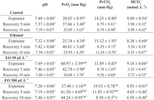
Blood pH was not significantly affected by treatments. The PvO2 and PvCO2 values increased
and decreased, respectively, in fish placed in the
simulated recovery, as well as in those recovering from ethanol exposure for 5 min compared to those
exposed to ethanol. Groupers exposed to both EO
concentrations presented higher PvO2 values than
control fish. The PvO2 values were also higher in
fish exposed to 50 µL L−1 EO than in those exposed
to ethanol. The PvO2 values of fish recovered from 50 µL L−1 EO exposure were significantly lower
than in exposed fish, but in those recovered from
300 µL L−1 EO exposure, these values were higher
than in exposed fish and in the recovered control
and ethanol groups. Groupers exposed to 300 µL L−1 showed significantly higher PvCO2 values than
the control group. In addition, fish recovered for 5 min presented significantly higher PvCO2 values than the control and ethanol groups, but after 10
min recovery these values were significantly lower
than in the ethanol group. The HCO3− concentration decreased in all groups at both recovery times
when compared to exposure, but was not affected
by treatments (Table III).
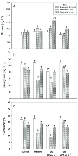
The levels of glucose, hemoglobin and
hematocrit were not significantly different between
exposure times in the control group (Fig. 1).
Glucose levels also did not differ between times
in the ethanol group. The groups treated with 50 or 300 µL L−1 EO showed higher glucose levels after 10 min recovery compared to other times. Fish exposed to 50 µL L−1 EO had increased glucose levels after 10 min recovery compared to the control and ethanol groups, but those exposed to 300 µL L−1 EO had decreased levels after 5 min recovery compared to the ethanol group (Fig. 1a).
hemoglobin and hematocrit levels than the ethanol group and lower hematocrit than the control group.
After 10 min recovery, fish that were exposed to
300 µL L−1 EO presented lower hematocrit and
hemoglobin levels than exposed fish. Fish exposed
to 300 µL L−1 EO showed higher hemoglobin and hematocrit levels after 5 min recovery compared to
the fish that recovered from ethanol exposure for
the same amount of time (Fig. 1b, c). Na+ and K+
levels did not differ between groups or exposure
times (Fig. 2a, b). The lowest Ca2+ levels in the control group were observed after 10 min recovery.
Additionally, lower Ca2+ levels were observed in
animals anesthetized with 50 µL L−1 EO after 10 min recovery compared to the ethanol group at the
same time (Fig. 2c).
DISCUSSION
The main constituents found in the EO obtained from
leaves of A. polystachya were the monoterpenes
carvone (58.76%) and α-limonene (33.68%).
Other studies also demonstrated the presence of
carvone (González et al. 2010) and limonene in
TABLE I
Chemical composition of the essential oil from dried leaves of Aloysia polystachya.
Peak RT Compound RI
experimental RI literature Source %
1 10.23 α-Pinene 931 937 N 0.75
2 11.94 β-Pinene 974 975 N 0.45
3 12.65 β-Myrcene 991 990 N 1.57
4 14.05 α-Limonene 1027 1028 N 33.68
5 14.52 β-E-Ocimene 1039 1038 N 0.40
6 14.91 β-Z-Ocimene 1049 1051 N 0.21
7 16.93 β-Linalool 1100 1098 N 0.93
8 17.69
E-p-Mentha-2,8-dienol 1121 1118 N 0.19
9 18.16 Limonene epoxide 1134 1136 N 0.27
10 20.28 α-Terpineol 1191 1190 N 0.40
11 20.43 1,6-Dihydrocarveol 1195 1195 N 0.29
12 20.50 Dihydrocarvone 1197 1199 N 0.29
13 21.70 E-Carveol 1231 1233 N 0.24
14 22.16 Carvone 1244 1242 N 58.76
15 23.22 Perillal 1275 1274 N/A 0.17
16 23.86 Thymol 1293 1292 N 0.16
17 27.99 β-Caryophyllene 1420 1419 N 0.80
18 29.05 α-Caryophyllene 1454 1455 N 0.15
19 30.74 β-Bisabolene 1509 1509 N 0.29
Total identified: 100.00
TABLE II
Time (in seconds) required for induction and recovery from anesthesia using the essential oil of Aloysia polystachya leaves (EO) in Epinephelus marginatus.
EO concentration
(µL L
−1)
Sedation
Anesthesia
Recovery
50
429.3 ± 12.2
–
–
75
285.8 ± 15.3
–
–
100
210.0 ± 3.5
1555.7 ± 46.7
565.0 ± 9.3
a200
96.3 ± 3.4
313.4 ± 16.8
421.4 ± 17.3
b300
50.7 ± 0.7
163.7 ± 16.5
480.8 ± 8.7
b400
46.5 ± 1.6
90.2 ± 3.2
330.5 ± 13.0
cEquations
y = 684.4 e
(−0.01x)r
2= 0.9529
y = 6424.5 e
(−0.01x)r
2= 0.975
9–
Values are represented as mean ± SEM (n = 6). Different letters indicate significant differences between essential oil concentrations in the recovery stage (P < 0.05). Stages of anesthesia induction are according to Small (2003), where sedation corresponds to Stage 1 and anesthesia corresponds to Stage 3. The equations fitted above represent a relationship between the times of anesthesia and concentrations of EO, where y = time to reach the stage and x = concentration of EO of A.polystachya (in µL L−1).
TABLE III
Blood parameters of Epinephelus marginatus evaluated after exposure (3.5 min) and recovery times from anesthesia induction by essential oil of Aloysia polystachya.
pH PvO2 (mm Hg) PvCO2
(mm Hg)
HCO3− (mmoL L−1) Control
Exposure
7.40 ± 0.06
a20.03 ± 0.95ª
14.25 ± 0.80ª
9.89 ± 0.54
aRecovery 5 min
7.37 ± 0.04
a37.66 ± 3.40
b 8.79 ± 0.81b 5.98 ± 0.32bRecovery 10 min 7.39 ± 0.07a 35.05 ± 3.63b 8.79 ± 0.90b 5.98 ± 0.42b
Ethanol
Exposure
7.32 ± 0.08
a25.74 ± 4.34ª
15.12 ± 1.93
a8.38 ± 0.60ª
Recovery 5 min
7.42 ± 0.06
a40.81 ± 3.68
b 8.29 ± 0.72b5.36 ± 0.24c
Recovery 10 min 7.38 ± 0.03a 25.05 ± 4.28ª 11.24 ± 0.78a 6.55 ± 0.07b
EO 50 µL L−1
Exposure
7.49 ± 0.03
a60.97 ± 3.39ª*
#11.89 ± 0.83ª
9.18 ± 0.66ª
Recovery 5 min
7.46 ± 0.05
a42.79 ± 5.90
b 8.74 ± 1.05ª 5.25 ± 0.43bRecovery 10 min 7.40 ± 0.05a 36.08 ± 3.70b 9.20 ± 0.68ª 5.72 ± 0.32b
EO 300 µL L−1
Exposure
7.28 ± 0.06
a37.48 ± 3.16ª*
19.53 ± 0.78
a*
8.93 ± 0.81ª
Recovery 5 min
7.19 ± 0.05
a61.20 ± 4.69
b*
#11.85 ± 0.93
b*
#4.65 ± 0.46
cRecovery 10 min 7.46 ± 0.07
a44.24 ± 0.95
c*
#8.30 ± 0.3
6c#6.58 ± 0.48
bValues are represented as mean ± SEM (n = 6). Different letters indicate significant difference within the same treatment (P < 0.05). * indicates significant difference from the control group at the same time and # indicates significant difference from the
Figure 1 - Glucose (a), hemoglobin (b) and hematocrit (c) levels in Epinephelus marginatus after exposure to essential oil (EO) of Aloysia polystachya added to the water (n = 6). Values are represented as mean ± SEM. Different letters indicate significant differences between times in the same treatment (P < 0.05). * indicates significant difference from the control group at the same time and # indicates significant difference from the ethanol group at the same time (P < 0.05).
Figure 2 - Plasma Na+ (a), K+
(b) and Ca2+
(c) levels in
Epinephelus marginatus after exposure to essential oil (EO)
this EO (Cabanillas et al. 2003). High contents of α-thujone and β-thujone (which were not found in
the present study) were previously detected in this
EO (Cabanillas et al. 2003, Duschatzky et al. 2004).
The present study demonstrated that A. polystachya EO has an anesthetic effect on
grouper juveniles. This EO induced sedation
at all concentrations tested and anesthetized animals within 3 and 1.5 min (300 and 400 µL L−1, respectively). Recovery time for both concentrations was about 8 min and 5 min, respectively, and no mortality was observed as a
result of anesthesia induction. These findings are
in accordance with literature criteria (Marking and Meyer 1985, Gilderhus and Marking 1987, Keene et al. 1998, Park et al. 2009). Parodi et al. (2014)
tested the anesthetic effect of the EO obtained from
another species of Aloysia, namely A. triphylla, in concentrations ranging between 20 and 800 µL L−1
on two strains (albino and gray) of silver catfish
(Rhamdia quelen), and 200 µL L−1 EO was the best concentration to induce anesthesia in the albino strain, while for the gray strain it was 400 µL L−1
EO. An emulsified mixture composed of Mentha spicata EO and methyl salicylate oil (containing 28.4% L-carvone) anesthetized common carp (Cyprinus carpio) within the recommended time at 395 µL L−1 (Roohi and Imanpoor 2014) and Atlantic salmon (Salmo salar) at 257 µL L−1 (Danner et al. 2011), but M. spicata EO alone induced anesthesia in less than 3 min only at 5000 µL L−1 (Roohi
and Imanpoor 2015). Interestingly, increased EO
concentration promoted recovery time decrease.
Other EOs produced higher recovery times as the EO concentration increased (Cunha et al. 2010,
Heldwein et al. 2012, Silva et al. 2012, 2013, Parodi
et al. 2014). Since EOs are complex mixtures of
compounds, a particular biological activity such as induction and recovery times from anesthesia
depends on the specific chemical characteristics of each EO, including the qualitative composition and
the proportions of each component in the oil (Raut and Karuppayil 2014).
The sedative and anesthetic activity of the
EO of A. polystachya can be explained mainly by the combined action of its major components, the monoterpenoid-derived compounds carvone and limonene, which account for 92.44% of its total chemical composition. Carvone has a central
nervous system (CNS) depressant effect detected in different pre-clinical studies in mice (Sousa et
al. 2007), while limonene acts as an agonist for
adenosine A2A receptors, and consequently can induce sedative effects (Park et al. 2011). Limonene
also inhibited stimulant-induced behavioral changes in mice and rats, by regulating dopamine levels and
5-HT receptor function (Yun 2014). However, it is
likely that a contribution to the activity detected in groupers is made by the minor components, such as limonene epoxide, for which an anxiolytic-like
effect was detected in mice. The CNS effects of this compound were reversed by flumazenil, indicating
a GABAergic mechanism of action (Almeida et al. 2012).
The blood pH values of the groupers, regardless of exposure time or group, were similar to or slightly lower than those reported for red pacu (Piaractus brachypomus) exposed to MS-222 and eugenol at 50, 100 and 200 mg L−1 (Sladky et al. 2001), yellow perch (Perca flavescens), walleye pike (Sander vitreus) and common carp anesthetized
with buffered MS-222 (150 mg L−1) (Hanley et al.
2010); silver catfish transported for 4 h in plastic
bags with eugenol (1.5 or 3.0 µL L−1) and L. alba
EO (10 or 20 µL L−1) added to the water (Becker et
al. 2012). The pH values observed in the blood of
groupers were not affected by A. polystachya EO
exposure, in accordance with the lack of effect of
eugenol and L. alba EO on this parameter in silver
catfish (Becker et al. 2012).
Becker et al. 2012). The handling (simulated recovery) of control groupers increased PvO2 and decreased PvCO2 and HCO3− values, probably
due to hyperventilation. Exposure to 50 µL L−1
A. polystachya EO (and 300 µL L−1 compared to
control fish) increased PvO2, as was also observed by Hanley et al. (2010) in perch, walleye and common carp anesthetized with MS-222. PvCO2 only increased in groupers exposed to 50 µL L−1 A. polystachya EO, as observed by Sladky et al. (2001) in red pacu exposed to MS-222 and eugenol. However, as in groupers exposed to 50 µL L−1 A. polystachya EO, Hanley et al. (2010) reported that PvCO2 did not change in perch, walleye or common carp anesthetized with MS-222.
In the present study, the handling of simulated
exposure did not induce a significant increase in
blood glucose levels, but groupers anesthetized with 50 µL L−1 A. polystachya EO after 10 min recovery showed increased levels compared to those after exposure. Repetitive blood sampling increased blood glucose levels in rainbow trout (Oncorhynchus mykiss) and anesthesia with clove oil or MS-222 did not change this pattern (Wagner et al. 2003).
Hematocrit values found in this study (26–
31%) in fish exposed to control conditions were
similar to those reported by Becker et al. (2012) (26–33%) and Carneiro et al. (2009) (27–30%) in
silver catfish. The lower hematocrit and hemoglobin
values in recovered ethanol and 50 µL L−1 EO (5 min) and 300 µL L−1 EO (10 min) groups compared
to exposed fish indicate possible hemodilution or
decreased red cell number. Contrary to our results, anesthesia with clove oil or MS-222 reduced hematocrit decrease produced by repetitive sampling in rainbow trout (Wagner et al. 2003).
Ionoregulatory homeostasis is important to ensure proper cell function (Hwang et al.
2011). Moreover, stress due to usual aquaculture procedures is known to affect fish ionoregulation
(Ashley et al. 2007). Addition of some sedatives
to water can reduce ion loss in fish (Becker et al. 2012). Ionoregulation in groupers was not affected
by anesthesia with A. polystachya EO, because the only change was lower Ca2+ in fish after 10 min recovery from 50 µL L−1 EO exposure compared to those recovered from ethanol exposure. Anesthesia with H. ringens and L. alba EOs (150, 300 or 450 µL L−1) and recovery did not considerably affect plasma Na+ and Cl− levels either (Toni et al. 2014). Blood Na+ also did not change, but K+ increased in red pacu anesthetized with MS-222 and eugenol (Sladky et al. 2001).
CONCLUSIONS
The EO from the leaves of A. polystachya was an
effective sedative and anesthetic for dusky grouper
juveniles in concentrations similar to those found
for other EOs in other species. Some of the tested
blood parameters showed compensatory responses
due to EO exposure, but most changes returned to
control values after 10 min of recovery.
ACKNOWLEDGMENTS
This study was supported by Fundação de Amparo à Pesquisa do Estado do Rio Grande do Sul/Programa de Apoio a Núcleos de Excelência (FAPERGS/ PRONEX, process 10/0016-8), Conselho Nacional
de Desenvolvimento Científico e Tecnológico
(CNPq, process 470964/2009-0) and Coordenação de Aperfeiçoamento de Pessoal de Nível Superior, Brazil (CAPES) for research fellowships and financial support.
REFERENCES
ADAMS RP. 2009. Identification of essential oil components by gas chromatography/mass spectrometry, 4th ed., Illinois: Allured Publishing Corporation, 804 p.
ASHLEY PJ. 2007. Fish welfare: current issues in aquaculture. App Anim Behav Sci 104: 199-235.
AZAMBUJA CR, MATTIAZZI J, RIFFEL APK, FINAMOR IA, GARCIA LO, HELDWEIN CG, HEINZMANN BM, BALDISSEROTTO B, PAVANATO MA AND LLESUY SF. 2011. Effect of the essential oil of Lippia alba on oxidative stress parameters in silver catfish (Rhamdia quelen) subjected to transport. Aquaculture 319: 156-161.
BAUMGARTEN MGZ, ROCHA JMB AND NIENCHESKY LF. 1996. Manual de análises em oceanografia química, Rio Grande: FURG, 172 p.
BECKER AG, PARODI TV, HELDWEIN CG, ZEPPENFELD CC, HEINZMANN BM AND BALDISSEROTTO B. 2012. Transportation of silver catfish, Rhamdia quelen, in water with eugenol and the essential oil of Lippia alba. Fish Physiol Biochem 38: 789-796.
BENDSCHNEIDER K AND ROBINSON RJ. 1952. A new spectrophometric method for the determination of nitrite in sea water. J Mar Res 11: 87-96.
CABANILLAS CM, LOPEZ ML, DANIELE G AND ZYGADLO JA. 2003. Essential oil composition of Aloysia polystachya (Griseb) Moldenke under rust disease. Flavour Frag J 18: 446-448.
CARNEIRO PCF, KAISELER PHS, SWAROFSKY EAC AND BALDISSEROTTO B. 2009. Transport of jundiá
Rhamdia quelen juveniles at different loading densities: water quality and blood parameters. Neotrop Ichthyol 7: 283-288.
CAVALLI RO. 2014. Maricultura. In: Castello JP. Introdução às ciências do mar, Pelotas: Editora Textos, p. 418-455. COOKE SJ ET AL. 2008. Effects of different capture
techniques on the physiological condition of bonefish
Albula vulpes evaluated using field diagnostic tools. J Fish Biol 73: 1351-1375.
CUNHA MA, BARROS FMC, GARCIA LO, VEECK APL, HEINZMANN BM, LORO VL, EMANUELLI T AND BALDISSEROTTO B. 2010. Essential oil of Lippia alba: a new anesthetic for silver catfish, Rhamdia quelen. Aquaculture 306: 403-406.
CUNHA ME, RÉ P, QUENTAL-FERREIRA H, GAVAIA PJ AND POUSÃO-FERREIRA P. 2013. Larval and juvenile development of dusky grouper Epinephelus marginatus
reared in mesocosms. J Fish Biol 83: 448-465.
DANNER GR, MUTO KW, ZIEBA AM, STILLMAN CM, SEGGIO JA AND AHMAD ST. 2011. Spearmint (l-carvone) oil and wintergreen (methyl salicylate) oil emulsion is an effective immersion anesthetic of fishes. J Fish Wildl Manage 2: 146-155.
DEL VITTO LA AND PETENATTI EM. 1997. Recursos herbolarios de San Luis (República Argentina) Primeira parte: plantas nativas. Multequina 6: 49-66.
DUSCHATZKY CB, MARTINEZ AN, ALMEIDA NV AND BONIVARDO SL. 2004. Nematicidal activity of the
essential oils of several Argentina plants against the root-knot nematode. J Essent Oil Res 16: 626-628.
EUROPEAN PHARMACOPOEIA. 2007. European directorate for the quality of medicines, 6th
ed., Strassbourg. FENNESSY YST. 2006. Reproductive biology and growth
of the yellowbelly rockcod Epinephelus marginatus
(Serranidae) from South-East Africa. Afr J Mar Sci 28: 1-11.
FILIPOY A. 1994. Medicinal plants of the Pilaga of Central Chaco. J Ethnopharmacol 44: 181-193.
FIGUEIREDO JL AND MENEZES NA. 1980. Manual de peixes marinhos do sudeste do Brasil III Teleostei (2) São Paulo, Museu de Zoologia, Universidade de São Paulo, 90 p.
GILDERHUS PA AND MARKING LL. 1987. Comparative efficacy of 16 anesthetic chemicals on rainbow trout. N Am J Fish Manage 7: 288-292.
GONZÁLEZ JOW, GUTIERREZ MM, MURRAY AP AND FERRERO AA. 2010. Biological activity of essential oils from Aloysia polystachya and Aloysia citriodora
(Verbenaceae) against the soybean pest Nezara viridula
(Hemiptera: Pentatomidae). Nat Prod Commun 5: 301-306.
GRESSLER LT ET AL. 2014. Silver catfish Rhamdia quelen
immersion anesthesia with essential oil of Aloysia triphylla
(L’Hérit) Britton or tricaine methanesulfonate: effect on stress response and antioxidant status. Aquac Res 45: 1061-1072.
HANLEY CS, CLYDE VL, WALLACE RS, PAUL-MURPHY J, PATTERSON TA, KEULER NS AND SLADKY KK. 2010. Effects of anesthesia and surgery on serial blood gas values and lactate concentrations in yellow perch (Perca flavescens), walleye pike (Sander vitreus), and koi (Cyprinus carpio). JAVMA - J Am Vet Med A 236: 1104-1108.
HELDWEIN CG, SILVA LL, RECKZIEGEL P, BARROS FM, BALDISSEROTTO B, MALLMANN CA, SCHMIDT D, CARON BO AND HEINZMANN BM. 2012. Participation of the GABAergic system in the anesthetic effect of Lippia alba (Mill) N E Brown essential oil. Braz J Med Biol Res
45: 436-443.
HELLIÓN-IBARROLA MC ET AL. 2008. The antidepressant-like effects of Aloysia polystachya (Griseb) Moldenke (Verbenaceae) in mice Phytomedicine, 15: 478-483 HELLIÓN-IBARROLA MC ET AL. 2006. The
anxiolytic-like effects of Aloysia polystachya (Griseb) Moldenke (Verbenaceae) in mice. J Ethnopharmacol 105: 400-408. HWANG PP, LEE TH AND LIN LY. 2011. Ion regulation in
fish gills: recent progress in the cellular and molecular mechanisms. Am J Physiol-Reg I 301: R28-R47.
(Lowe, 1834) in gulfs of northern Patagonia, Argentina. J Fish Biol 67: 1741‐1745.
KEENE JL, NOAKES DLG, MOCCIA RD AND SOTO CG. 1998. The efficacy of clove oil as an anaesthetic for rainbow trout, Oncorhynchus mykiss (Walbaum). Aquac
Res 29: 89-101.
KIESSLING A, JOHANSSON D, ZAH LIH AND SAMUELSEN OB. 2009. Pharmacokinetics, plasma cortisol and effectiveness of benzocaine, MS-222 and isoeugenol measured in individual dorsal aorta-cannulated Atlantic salmon (Salmo salar) following bath administration. Aquaculture 86: 301-308.
KRISTENSEN T, ROSSELAND BO KIESSLING A, DJORDEVIC B AND MASSABAU JC. 2010. Lack of arterial PO2 downregulation in Atlantic salmon (Salmo salar L.) during long-term normoxia and hyperoxia. Fish Physiol Biochem 36: 1087-1095.
MARKING LL AND MEYER FP. 1985. Are better fish anesthetics needed in fisheries? Fisheries 10: 2-5.
MORA S, DÍAZ-VÉLIZ G, MILLÁN R, LUNGENSTRASS H, QUIRÓS S, COTO-MORALES T AND HELLIÓN-IBARROLA MC. 2005. Anxiolytic and antidepressant-like effects of the hydroalcoholic extract from Aloysia polystachya in rats. Pharmacol Biochem Behav 82: 373-378.
NIST. 2008. Databank NIST/EPA/NIH mass spectral library and search/analysis programs Hoboken: J Wiley & Sons. PARK HM, LEE JH, YAOYAO J, JUN HJ AND LEE SJ.
2011. Limonene, a natural cyclic terpene, is an agonistic ligand for adenosine A2A receptors. Biochem Biophys Res Commun 404: 345-348.
PARK IS, PARK MO, HUR JW, KIM DS, CHANG YJ, KIM YJ, PARK JY AND JOHNSON SC. 2009. Anesthetic effects of lidocaine-hydrochloride on water parameters in simulated transport experiment of juvenile winter flounder,
Pleuronectes americanus. Aquaculture 294: 76-79.
PARODI TV, CUNHA MA, BECKER AG, ZEPPENFELD CC, MARTINS DI, KOAKOSKI G, BARCELLOS LJG, HEINZMANN BM AND BALDISSEROTTO B. 2014. Anesthetic activity of the essential oil of Aloysia triphylla
and effectiveness in reducing stress during transport of albino and gray strains of silver catfish, Rhamdia quelen. Fish Physiol Biochem 40: 323-334.
PAUST LO, FOSS A AND IMSLAND AK. 2011. Effects of chronic and periodic exposure to ammonia on growth, food conversion efficiency and blood physiology in juvenile Atlantic halibut (Hippoglossus hippoglossus L.). Aquaculture 315: 400-406.
RAUT JS AND KARUPPAYIL SM. 2014. A status review on the medicinal properties of essential oils. Ind Crop Prod 62: 250-264.
ROOHI Z AND IMANPOOR MR. 2014. Effects of spearmint (/-carvon) oil and methyl salicylate oil emulsion on
anesthesia of common carp (Cyprinus carpio L., 1758). Aquac Res Dev 5: 1-5.
ROOHI Z AND IMANPOOR MR. 2015. The efficacy of the oils of spearmint and methyl salicylate as new anesthetics and their effect on glucose levels in common carp (Cyprinus carpio L., 1758) juveniles. Aquaculture 437:
327-332.
SALBEGO J ET AL. 2014. The essential oil from Lippia alba
induces biochemical stress in the silver catfish (Rhamdia quelen) after transportation. Neotrop Ichthyol 12: 811-818. SANCHES ES, SILVA FC, LEITE JR, SILVA PKA, KERBER
CE AND SANTOS PA. 2014. A incorporação de óleo de peixe na dieta pode melhorar o desempenho da garoupa-verdadeiraEpinephelus marginatus? Bol Inst Pesca 40: 147-155.
SILVA LL, PARODI TV, RECKZIEGEL P, GARCIA VO, BURGER ME, BALDISSEROTTO B, MALLMANN CA, PEREIRA AMS AND HEINZMANN BM. 2012. Essential oil of Ocimum gratissimum L. anesthetic effects, mechanism of action and tolerance in silver catfish,
Rhamdia quelen. Aquaculture 350: 91-97.
SILVA LL, SILVA DT, GARLET QI, CUNHA MA, MALLMANN CA, BALDISSEROTTO B, LONGHI SJ, PEREIRA AMS AND HEINZMANN BM. 2013. Anesthetic activity of Brazilian native plants in silver catfish (Rhamdia quelen). Neotrop Ichthyol 11: 443-451. SLADKY KK, SWANSON CR, STOSKOPF MK, LOOMIS
MR AND LEWBART GA. 2001. Comparative efficacy of tricaine methanesulfonate and clove oil for use as anesthetics in red pacu (Piaractus brachypomus). Am J Vet Res 62: 337-342.
SMALL BC. 2003. Anesthetic efficacy of metomidate and comparison of plasma cortisol responses to tricaine methanesulfonate, quinaldine and clove oil anesthetized channel catfish Ictalurus punctatus. Aquaculture 218:
177-185.
SOUSA DP, NÓBREGA FFF AND ALMEIDA RN. 2007. Influence of the chirality of (R)-(-)- and (S)-(+)-carvone in the central nervous system: a comparative study. Chirality 19: 264-268.
SOUZA RHD, SONCINI R, GLASS ML, SANCHES JR AND RANTIN FT. 2001. Ventilation, gill perfusion and blood gases in dourado, Salminus maxillosus Valenciennes (Teleostei, Characidae), exposed to graded hypoxia. J Comp Physiol B 171: 483-489.
UNESCO. 1983. Chemical methods for use in marine environmental monitoring. Manual and guides 12, Paris: Intergovernamental Oceanographic Commission.
WAGNER GN, SINGER TD AND MCKINLEY RS. 2003. The ability of clove oil and MS-222 to minimize handling stress in rainbow trout (Oncorhynchus mykiss W). Aquac
Res 34: 1139-1146.
YUN J. 2014. Limonene inhibits methamphetamine-induced locomotor activity via regulation of 5-HT neuronal
function and dopamine release. Phytomedicine 21: 883-887.
ZAHL IH, SAMUELSEN O AND KIESSLING A. 2012. Anaesthesia of farmed fish: implications for welfare. Fish Physiol Biochem 38: 201-218.