julho de 2014
Escola de Engenharia
Juliana de Castro Carvalho
Mechanistic aspects of extracellular
silver nanoparticles synthesis by
filamentous fungi
UMinho|20 14 Juliana de Castr o Car valho Mechanis tic aspects of e xtracellular sil ver nanopar ticles synt hesis b y filamentous fungiDissertação de Mestrado
Mestrado Integrado em Engenharia Biomédica
– Ramo Engenharia Clínica
Trabalho efetuado sob a orientação do
Professor Doutor Nelson Manuel Viana da Silva Lima
e da
Doutora Nicolina Marques Dias
julho de 2014
Escola de Engenharia
Juliana de Castro Carvalho
Mechanistic aspects of extracellular
silver nanoparticles synthesis by
filamentous fungi
iii
Agradecimentos
O espaço limitado desta secção de agradecimentos, seguramente, não me permite agradecer, como devia, a todas as pessoas que, ao longo do meu Mestrado Integrado em Engenharia Biomédica me ajudaram, directa ou indirectamente, a cumprir os meus objectivos e a realizar esta etapa da minha formação académica.
Desta forma, agradeço à Micoteca da Universidade do Minho (MUM) pela oportunidade de realizar a dissertação no seu laboratório. Estou extremamente grata aos meus orientadores Doutora Nicolina Dias e ao Professor Doutor Nelson Lima pelo conhecimento, disponibilidade, estímulo, apoio constante e paciência que sempre dispensaram. Aos investigadores da MUM, Doutor André Antunes, Doutora Célia Soares, Doutora Lúcia Simões e Doutora Marta Simões um muito obrigado pela disponibilidade, companheirismo e atenção prestada ao longo do projecto. Foi um enorme privilégio ter feito parte deste grupo.
Agradeço aos meus amigos que me acompanharam desde sempre, Ana, Catarina, Carina, Liliana, Regina e Patrícia pela amizade, pelos bons momentos, no qual foram e sempre serão muito importantes para mim. À Liliana e à Raquel pela boa disposição e bons momentos compartilhados ao longo destes dois anos.
À minha Família, aos meus pais, Manuel e Armandina, ao meu irmão, José Pedro, à minha tia Eva, aos meus padrinhos, Joaquim e Eduarda, e aos meus primos, Tiago, Diogo e Maria Carolina, um enorme obrigada por acreditarem sempre em mim, pela boa disposição, compreensão e por todos os ensinamentos de vida. A eles dedico todo este trabalho.
O meu profundo e sentido agradecimento a todas as pessoas que contribuíram, de uma forma directa ou indirecta, para a concretização desta dissertação, estimulando-me intelectualmente e emocionalmente.
iv
Resumo
A produção biológica de nanopartículas metálicas foi estudada por muitos investigadores, devido a obtenção de pequenas partículas estabilizadas por proteínas. No entanto, o mecanismo envolvido nessa produção, ainda não foi esclarecido, embora existam diversos mecanismos hipotéticos propostos na literatura.
As estipes dos fungos Aspergillus ibericus MUM 03.49, MUM 03.50 e MUM 03.51; Penicillium chrysogenum MUM 03.18, MUM 97.43 e MUM 92.11; e Neurospora crassa MUM 11.01 e MUM 92.08 foram testados como produtores de nanopartículas de prata (AgNPs) biogénicas. Através da análise das características físico-químicas de AgNPs produzidos por cada um dos filtrados de células fúngicas, a melhor estirpe produtora de cada espécie fúngica foi seleccionada. Deste modo, as estipes seleccionadas P. chrysogenum MUM 92.11 e N. crassa MUM 92.08 foram usadas para estudos posteriores da mutagénese e da modulação de mecanismos bioquímicos da produção de AgNPs.
Os mutantes defeituosos no gene estrutural do nitrato reductase niaD- foram facilmente
recuperados a partir do fungo filamentoso Penicillium chrysogenum MUM 92.11 a partir da sua resistência ao clorato. Apesar das várias tentativas realizadas para a estirpe N. crassa MUM 92.08, a mutagénese não foi bem-sucedida e consequentemente não houve nenhum crescimento de mutantes. O filtrado do tipo selvagem e o mutante foram ambos capazes de produzir nanopartículas de prata após a adição de nitrato de prata no filtrado. Por fim, para verificar se cada composto do filtrado identificado como influência fundamental na produção de nanopartículas foi desenvolvido um ensaio in-vitro. A síntese de AgNPs não ocorreu nos ensaios sem filtrado celular, no entanto, ocorreu nos ensaios com filtrado celular, mesmo após a desnaturação térmica das proteínas, indicando que os aminoácidos podem estar envolvidos na produção de nanopartículas de prata. Outras biomoléculas presentes no filtrado podem também desempenhar um papel-chave no mecanismo de biorredução dos iões de prata, em filtrados fúngicos, que exemplifica o entendimento do mecanismo de produção de AgNPs biogénicas fúngicas.
vi
Abstract
Biological production of metal nanoparticles has been studied by many researchers due to the convenience of the method that produces small particles stabilized by protein. However, the mechanism involved in this production has not yet been elucidated although hypothetical mechanisms have been proposed in the literature.
The fungal strains Aspergillus ibericus MUM 03.49, MUM 03.50 and MUM 03.51; Penicillium chrysogenum MUM 03.18, MUM 97.43 and MUM 92.11; and Neurospora crassa MUM 11.01 and MUM 92.08 were tested as producers of biogenic silver nanoparticles (AgNPs). Through the characterisation of the physiochemical features of the AgNPs produced by each fungal cell filtrate the best producer strain from each fungal species was selected. Therefore, selected strains P. chrysogenum MUM 92.11 and the N. crassa MUM 92.08 were used to further studies of mutagenesis and modulation of biochemical mechanisms for the production of AgNPs.
Defective mutants on the nitrate reductase structural gene niaD- were recovered easily
from the filamentous fungi Penicillium chrysogenum MUM 92.11 by their resistance to chlorate medium. Although several attempts performed for the strain N. crassa MUM 92.08 the mutagenesis was not successful and none of the mutants grew. Wildtype and the mutant filtrate were both able to produce silver nanoparticles after the addition of silver nitrate in the filtrate. Finally, to verify if each filtrate compound identified as key-compound influence the production of nanoparticles an in-vitro assay was developed. The synthesis of AgNPs did not occur without cell filtrate, however it did occur in cell filtrate even after the heat denaturation of proteins, indicating that amino acids could be involved in the production of silver nanoparticles. Other biomolecules present in the filtrate are also believed to play a key-role in the bioreductive mechanism of silver ions in fungal filtrates, which exemplify understanding the production mechanism of fungal biogenic AgNPs.
viii
Abbreviations
Ag+ Ag0 AgNP ATP BSA ddH2O Da EtOH FAD MUM NADPH NR SDS TEMED Tris Tween 80 UV WT Silver ion Reduced silver Silver nanoparticles Adenosine triphosphate Bovine serum albumin Double-distilled water DaltonEthanol
Flavin adenine dinucleotide
Micoteca da Universidade do Minho
Nicotinamide adenine dinucleotide phosphate Nitrate reductase
Sodium dodecyl sulfate
N,N,N',N'-Tetramethyl ethylenediamine Tris(hidroximetil)aminometane
Polyoxyethylene (80) sorbitan monooleate Ultraviolet radiations
x
Index
General introduction, aims and outline ... 1
Chapter I: Fungal screening for optimization of extracellular Bio-AgNPs synthesis. ... 5
1. Introduction ... 7
1.1. Nanoparticles ... 7
1.2. Silver nanoparticles ... 8
1.3. Non-biological vs. biological methods to synthesize AgNPs ... 9
1.3.1. Biosynthesis of silver nanoparticles ... 10
1.4. Fungal-mediated biosynthesis of AgNPs ... 14
1.5. Mechanisms and metabolism of nanoparticles biosynthesis ... 16
2. Materials and methods ... 20
2.1. Fungi ... 20
2.2. Culture and storage conditions ... 20
2.3. Biosynthesis of AgNPs ... 20
2.4. Physicochemical characterisation of the Bio-AgNPs... 21
2.4.1. Ultraviolet-visible spectroscopy ... 21
2.4.2. Dynamic Light Scattering (DLS) ... 21
2.4.3. Inductively Coupled Plasma Optical Emission Spectrometry (ICP-OES) ... 21
2.4.4. Zeta-Potential analysis ... 22
2.4.5. Scanning Electron Microscopy (SEM) ... 22
2.4.6. Energy-dispersive X-ray spectroscopy (EDS) ... 22
3. Results ... 23
3.1. Biosynthesize of AgNPs ... 23
4. Discussion and conclusion ... 32
Chapter II: Mechanism approach of biosynthesis of AgNPs. ... 35
1.1. Nitrogen requirement in filamentous fungi ... 37
1.2. Nitrate assimilation mechanism and NR regulation ... 38
1.3. Mutagenesis ... 40
xi
2.1. Mechanism approach of biosynthesis of AgNPs ... 43
2.1.1. Mutagenesis ... 43
2.1.1.1. Selection of niaD- spontaneous mutants of Penicillium chrysogenum MUM 92.11……….43
2.1.2. Biosynthesis of AgNPs produced by Penicillium chrysogenum MUM 92.11 WT and mutants……….. ... 44
2.1.2.1. Physicochemical characterisation of the Bio-AgNPs produced by Penicillium chrysogenum MUM 92.11 WT and mutants ... 45
2.1.3. Enzymatic assay ... 45
2.1.3.1. Determination of total protein concentration ... 45
2.1.3.2. Electrophoresis with SDS-Polyacrylamide Gel (SDS-PAGE) ... 45
2.1.3.3. Determination of nitrites concentration ... 47
2.1.3.4. Determination of NR specific enzymatic activity ... 48
2.2. In-vitro assay... 49
3. Results ... 50
3.1. Mutagenesis and isolation of niaD- mutants ... 50
3.2. Mechanism approach of biosynthesis of AgNPs ... 53
3.3. Analysis of the NR enzyme ... 57
3.3.1. Electrophoresis with SDS-Polyacrylamide Gel (SDS-PAGE) ... 57
3.3.2. Determination of nitrite concentration ... 58
3.3.3. Determination of NR specific enzymatic activity... 59
3.4. In-vitro assay... 59
4. Discussion and conclusion ... 62
General conclusions and futures perspectives ... 65
xii
Figures list
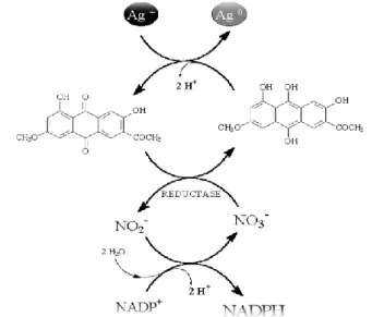
Figure 1 – Hypothetical diagram of the synthesis of silver nanoparticles by the electron shuttle enzymatic metal reduction process, through the NR dependent of NADPH (Adaptation of Rai and Durán 2011)…...18 Figure 2 – Cellular filtrate of Aspergillus ibericus MUM 03.49 (A), MUM 03.50 (B) and MUM 03.51 (C), treated with 1 mM of AgNO3 after 96 h of incubation in the dark. Corresponding
negative controls are shown on the left of each image……….……….23 Figure 3 – Cellular filtrate of Penicillium chrysogenum MUM 97.43 (A), MUM 92.11 (B) and MUM 03.18 (C), treated with 1 mM of AgNO3 after 96 h of incubation in the dark. Corresponding
negative controls are shown on the left of each image………..……24 Figure 4 – Cellular filtrate of Neurospora crassa MUM 11.01 (A) and MUM 92.08 (B), treated with 1 mM of AgNO3 after 96 h of incubation in the dark. Corresponding negative controls are
shown on the left of each image………..24 Figure 5 – UV-visible spectrum of Aspergillus ibericus filtrate with1 mM of AgNO3 aqueous
solution with and its respective strains after 96 h of incubation……….………25 Figure 6 – UV-visible spectrum of Penicillium chrysogenum filtrate with1 mM of AgNO3 aqueous
solution with and its respective strains after 96 h of incubation……….………25 Figure 7 – UV-visible spectrum Neurospora crassa filtrate with1 mM of AgNO3 aqueous solution
with and its respective strains after 96 h of incubation……….……….26 Figure 8 – UV-visible spectrum Aspergillus ibericus MUM 03.49, Penicillium chrysogenum MUM 92.11 and Neurospora crassa MUM 92.08filtrates with1 mM of AgNO3 aqueous solution with and
its respective strains after 96 h of incubation………….………26 Figure 9 – AgNPs sizes distribution by intensity of Aspergillus ibericus MUM 03.49………..27 Figure 10 – AgNPs sizes distribution by intensity of Aspergillus ibericus MUM 03.50………..27 Figure 11 – AgNPs sizes distribution by intensity of Aspergillus ibericus MUM 03.51………..27 Figure 12 – AgNPs sizes distribution by intensity of Penicillium chrysogenum MUM 92.11………..28 Figure 13 – AgNPs sizes distribution by intensity of Penicillium chrysogenum MUM 97.43………..28 Figure 14 – AgNPs sizes distribution by intensity of Penicillium chrysogenum MUM 03.18…...28
xiii
Figure 15 – AgNPs sizes distribution by intensity of Neurospora crassa MUM 11.01………..28 Figure 16 – AgNPs sizes distribution by intensity of Neurospora crassa MUM 92.08………..29 Figure 17 – SEM images of AgNPs synthetized by: Aspergillus ibericus MUM 03.49 (A), Penicillium chrysogenum MUM 92.11(B) and Neurospora crassa MUM 92.08 (C)………..31 Figure 18 – EDS spectrum of the fungi filtrate after 96 h of incubation: (A) A. ibericus MUM 03.49, (B) P. chrysogenum MUM 92.11and (C) N. crassa MUM 92.08………...31 Figure 19 - Stepwise reduction of nitrate ion into ammonia and respectively oxidation states (adapted from Msore-Landecker 1990)…….………..38 Figure 20 – Nitrate assimilation on filamentous fungi (adapted from Msore-Landecker 1990)………..38 Figure 21 - Illustrative model of a possible mechanism of nitrate and nitrite production controls in Aspergillus nidulans (adapted from Moore-Landecker 1990)……….39 Figure 22 - Inoculation grid………44 Figure 23 – Macroscopic characterisation based on the assimilation of P. chrysogenum MUM 92.11 wildtype (WT) and mutants on different nitrogen source plates with 3 days of growth……51 Figure 24 - Cellular filtrate of Penicillium chrysogenum wildtype (A) and mutant (B), treated with 1 mM of AgNO3 after 96 h of incubation in the dark. Corresponding negative controls are shown
on the left of each image……….………..54 Figure 25 – UV-Visible spectrum of P. chrysogenum MUM 92.11 (WT) with its respective mutant (M) with 1 mM of AgNO3 aqueous solution, after 96 h of incubation and their negative controls,
WT and CM, respectively………..………..54 Figure 26 – AgNPs sizes distribution by intensity of Penicillium chrysogenum MUM 03.50 mutant………55 Figure 27 – SEM and STEM images of AgNPs synthetized by P. chrysogenum MUM 92.11: WT (A) and C)) and mutant (B) and D))………56 Figure 28 - EDS spectrum of the P. chrysogenum MUM 92.11 filtrate after 96 h of incubation: (A) wildtype and (B) mutant……….57 Figure 29 – SDS-PAGE of the filtrates WT and mutant (M) of the fungus P. chrysogenum MUM 92.11………..58 Figure 30 – The obtained results of UV-Vis. spectra ranging 250 to 800 nm for each condition of the II assay after 96 h of incubation. The III assay was used as control…….………61
xiv
Tables list
Table 1 – List of fungi that have been used in the production of extracellular AgNPs and their respective size (Adaptation of Rai and Durán 2011)………...……….15 Table 2 – Fugal strains used to study the biosynthesis of AgNPs……….19 Table 3 – Average of size (nm) and polydispersity of each fungus and respective strains analyse by DLS……….……..29 Table 4 – Concentration (103 mg/mL) of silver of each fungus and respective strains filtrate
analyse by ICP-OES………..30 Table 5 – Solution charge (mV) of previously selected strains of each fungus analyse by Zeta-Potential……….30 Table 6 – Mutants growth tests resistant’s to chlorate with different sources of nitrogen (Adaptation of Unkles et al. 1989)..………42 Table 7 - Czapeck-Dock minimal medium (agar-CD or agar-NO3-) composition supplemented with
different source of nitrogen (nitrite, ammonium, glutamate and hypoxanthine) as required………....43 Table 8 – Composition of the separating gel………..…46 Table 9 – Composition of staking gel……….46 Table 10 - UV-Vis. spectra ranging 250 to 800 nm were carried out for each condition of the different assays after 96 h of incubation. In assay I (A-E) different reaction solutions were prepared in microtubes adding the following compounds: β-NADPH, FAD, NR and anthraquinone in phosphate buffer pH = 8. In assay II the same procedure was performed with the WT (A, D and E) and mutant (B and C) filtrate with AgNO3. In assay II E the WT filtrate the proteins were denatured ate 100 ºC for 5 min. In assay III and IV same conditions of assay I were used except the addition of the sodium azide as NADPH inhibitor (A-D) and egg-albumin protein (A-D) respectively in the microtubes………..49 Table 11 – Phenotypic properties of chlorate-resistant mutants of P. chrysogenum MUM 92.11………..52 Table 12 - Penicillium Chrysogenum MUM 92.11 metabolic pathway of nitrate assimilation………..53 Table 13 - Average of size (nm) and polydispersity of the fungus P. chrysogenum MUM 92.11 wildtype and its respective mutant analyse by DLS………...55 Table 14 - Solution charge (mV) of P. chrysogenum MUM 92.11 WT and mutant fungus analyse by Zeta-Potential……….55
xv
Table 15 – Nitrites concentration………58 Table 16 – Determination of NR enzymatic activity……….59 Table 17 – The obtained results of UV-Vis. spectra ranging 250 to 800 nm for each condition of the different assays after 96 h of incubation. ………60
3
1. General introduction, aims and outline
1.1. General introduction
The synthesis of nanoparticles is currently an area of intense scientific interest, because of its physicochemical characteristics that are not found in conventional materials (Krolikowska et al. 2003; Albrecht et al. 2006). Besides the various physical and chemical methods employed for the synthesis of metal nanoparticles, biological methods have better advantageous (Shankar et al. 2004; Panacek et al. 2006).
Many reviews have been published related to the biological synthesis of silver nanoparticles by filamentous fungi and their applications in several areas (Mandal et al. 2006; Chen & Schluesener 2008; Marcato & Durán 2008; Durán et al. 2008, 2009, 2010). However, the mechanism involved in this production has not yet been elucidated although hypothetical mechanisms have been proposed by several authors. (Durán et al. 2005; Rai et al. 2009).
The hypothetic model proposed by Durán et al. (2005) suggests that the silver nanoparticles are synthetized with the presence of the NR enzyme which is present in the nitrogen cycle (Kumar et al. 2007; Kalimuth et al. 2008). Besides the presence of NR enzyme, the fungus secrete the cofactor NADH and NADH-dependent enzymes that might be responsible for the bioreduction of Ag+ to Ag0 and mediates the extracellular reaction process
(Kalimuth et al. 2008; Li et al. 2011).
Filamentous fungi nitrogen metabolism is a process controlled by highly complex regulatory proteins, which ensures greater efficiency in the utilization of available nitrogen sources. For a better comprehension of the nitrate assimilation in filamentous fungi it is essential to understand the nitrogen metabolism, as well as the mechanisms of the control of metabolic regulation (Msore-Landecker 1990).
1.2. Aims
The aims of this dissertation were to optimize the biosynthesis process and characterisation of biogenic AgNPs produced by selected filamentous fungi following with the modulation of the biochemical mechanism of silver reduction for biosynthesis of silver nanoparticles by NR enzyme.
4
1.3. Outline
The theme and relevance of the dissertation are initially described. On the chapter I is selected the best fungal strain from non-pathogenic and non-producers of mycotoxins filamentous fungi and which have the ability to synthesize specific metallic nanoparticles. The chapter II defines the role of NR on the reduction mechanism of biosynthesis of silver nanoparticles and confirmed by the use of defective mutants on the NR structural gene niaD-. The chapter I and II
are divided in introduction, methods, results, discussion and conclusions. Finally, general conclusion and futures perspectives of the dissertation are described.
Chapter I:
Fungal screening for optimization of
extracellular Bio-AgNPs synthesis.
7
1. Introduction
1.1. Nanoparticles
Particles are small size objects classified according to their diameter. Particles with a size between 1 and 100 nm are known as nanoparticles. They have been found to have a diverse range of applications and are currently used in a variety of fields such as nanotechnology, biology, chemistry, physics, botanic, zoology, chemistry, earth sciences, medicine, pharmacology, mycology, microbiology, pathology and biotechnology (Bhushan 2010).
One of the first references to nanotechnology was made by Richard Feynman (Feynman 1959), where he affirmed that one day it would be possible to manipulate objects with atomic dimensions and make structures with a greatly reduced size. In the late 1960s, the first nanoparticles for drug delivery purposes and for vaccines were developed.
In 2010, the ISO/TS 80004 defined the term of nanotechnology as: “The definition of scientific knowledge to control and utilize matter at the nanoscale, where size-related properties and phenomena can emerge” (Luther and Zwech 2013).It is important to refer that the ISO/TS 80004, from the International Organization for Standardization, provides terms, definitions and applications in the field of nanotechnologies. These series of standards were originally elaborated in 1985 by Eric Dexter and are still a current reference for health, safety and environment concerns. In 2011, nanoscale was defined as a size range with approximately 1 to 100 nm (Hatto 2011).
The characteristics of the nanomaterials are dependent upon a number of properties, including particles size and size distribution, solubility and state of aggregation, elemental composition, mass and concentration, shape and crystal structure, surface area, charge, chemistry and the presence of impurities (Castro-Longoria et al. 2011; Tiede et al. 2009).
Among nanomaterials, metallic nanoparticles have been attracting attention mainly due to their unique physicochemical properties, like: conductivity, reactivity, resistance, and durability. Therefore these are particularly interesting for their many potential applications in innumerable scientific fields, namely on the development of nanocomputers, biomolecular detection, catalysis, optical devices, and biomedicine (Ghorbani et al. 2011). In this field, metallic nanoparticles are mostly promising due to their antibacterial properties improved by their large surface area to
8
volume ratio, which is of the interest to researchers because of the increasing microbial resistance to antibiotics, and the development of multiresistant strains.
1.2. Silver nanoparticles
Silver is a well-known antimicrobial agent widely used on infectious diseases (Marshall and Schneider 1977; Drake and Hazelwood 2005). The use of silver and copper ions has also been recommended as superior disinfectants for wastewater generated from hospitals containing infectious microorganisms (Lin et al. 1996). On the other hand, silver ions have the disadvantage of forming complexes and when are used to treated ions may adversely affect human health (Lee et al. 2009).
Among metallic nanoparticles, silver nanoparticles (AgNPs) are recognized potent and broad-spectrum antimicrobial agents being one of the most widely used engineered nanoparticles in consumer products (Shrivastava et al. 2007; Wijnhoven et al. 2009). The AgNPs have been widely investigated for their ability to produced composites further used as disinfecting filter and coating materials. Beyond that, they are used as antimicrobial agents in most of the public places such as elevators and railway stations; in surgically implanted catheters in order to reduce the infections, burn wounds, dental work. Antifungal, inflammatory, angiogenic and anti-permeability activities have been claimed by several authors (Wiederrech 2010).
Silver nanoparticles lead to a rise of number of particles per unit area and, consequently, a maximized antibacterial effect was described (Yeo et al. 2003; Morones et al. 2005).
The volume, V, of spherical nanoparticles is described by: ⁄
And, the area (A) is defined by:
The r is the particle radius.
[1.1]
9
Therefore, the ratio between the area and the volume is given by the follow equation: ( ⁄ ) ⁄
From the equation [1.3] it is possible to extrapolate that the ratio between the area and the volume is inverse to the particle radius. In this way, to improve the proprieties of the nanomaterial and consequently its antimicrobial activity, the size of the particles has to decrease as the area of activated spots increase. In others words, small particles exhibit higher antimicrobial activity than big particles (Panacek et al. 2006; Durán et al. 2010). This outcome can be explained by the fact that high penetration of particle is due to the reduced particle size (White 2001; Durán et al. 2010). Recent publications suggest that silver nanoparticles are shape-dependent on the interaction with the bacterial cells. Pal et al. (2007) and Sharma et al. (2009) confirmed that the triangular form of the silver ions displayed the strongest biocidal action against E. coli when compared with spherical and rod-shaped nanoparticles. Moreover, the high affinity of silver to sulfur or phosphorus is the key element for the antibacterial effect (Yeo et al. 2003; Morones et al. 2005).
1.3. Non-biological vs. biological methods to synthesize AgNPs
The AgNPs can be synthesized by physical/chemical (non-biological) and biological methods. Non-biological methods include irradiation, electrolysis, hydrothermal method, pyrolysis, physical vapor condensation (PVC), arc-discharge methods, chemical reduction and precipitation, and chemical vapor deposition that involve the use of toxic organic solvents and strong reducing chemical agents (for example: N,N-dimethylformamide and sodium borohydride) (Ghorbani et al. 2011). In general, nanoparticles synthesis mediated by non-biological methods requires high temperature and high pressure, consuming energy and producing toxic wastes that are environmentally hazardous, so research efforts have been undertaken for greener production alternatives. Biological methods use microorganisms such as: bacteria (Nair and Pradeep 2002; Lengke and Southam 2006; Husseiny et al. 2007; Shahverdi et al. 2007), filamentous fungi (Ahmad, Mukherjee, et al. 2003; Parikh et al. 2008; Durán et al. 2005; Durán et al. 2010; Anilkumar et al. 2007; Gajbhiye et al. 2009; Govender et al. 2009), actinomycetes (Ahmad et al. 2003; Ahmad et al. 2003) and yeasts (Kowshik et al. 2003); plant extracts (Shankar et al. 2003; Huang et al. 2007; Song & Kim 2009; Bar et al. 2009; Jha et al. 2009) and also peptides (Naik
10
et al. 2002; Tomczak et al. 2007) to synthetize nanoparticles by biological pathways. Many studies have reported that the biological methods represent an inexpensive, less labor-intensive and eco-friendly method, that is causing an emerging focus on the nanotechnology and biotechnology fields (Kowshik et al. 2003; Kalishwaralal et al. 2008). Notwithstanding the production of biosynthesized silver nanomaterial, biological method allows for the production of different types of other metal nanoparticles such as: copper (Ito et al. 2007), zinc (Bai et al. 2006), titanium (Bansal et al. 2005; Prasad et al. 2007), cadmium (Kowshik et al. 2002), magnesium (Gu et al. 2003), zirconia (Bansal et al. 2004), silver and gold (Nair and Pradeep 2002), platinum (Lengke et al. 2006), and palladium (Yong et al. 2002).
Biological methods allow manipulating the properties of metallic nanoparticles by achieving control over the physicochemical parameters that determines their size and shape. Silver nanoparticles can be induced to have different forms such as: spherical, cubic, wire, and triangular shapes (Chen et al. 2010). Rai and Durán (2011) described that form depends on the cultural conditions: culture medium, quantity of biomass, filtrate volume, salt concentration; and physical conditions: pH, temperature and light intensity that affect the maximum yield, presence of light, rate of synthesis and size of nanoparticles.
Furthermore the microbial cultures are easy to handle and also the downstream processing of biomass is much easier as compared to the non-biological methods (Ingle et al. 2008).
1.3.1. Biosynthesis of silver nanoparticles
Biosynthesis of AgNPs mediated by microorganisms occurs within their cell walls (Ahmad et al. 2002) resulting in the production of extracellular or intracellular nanoparticles (Ahmad et al. 2003; Mukherjee et al. 2008; Shaligram et al. 2009; Ingle et al. 2008; Rai et al. 2008). Despite being observed both in prokaryotes and eukaryotes not all organisms are competent for the synthesis of silver nanoparticles.
Besides that, the interaction of AgNPs with biomolecules released by microorganism metabolism, namely proteins will influence surface chemistry of nanoparticles and modify their electronic charge and agglomeration state leading to the improvement of their biological activity
11
(Brett 2006; Liau et al. 1997; Gade et al. 2008; Bhainsa and D’Souza 2006a). Also, the presence of proteins in the AgNPs solution has the function of stabilizing the nanoparticles preventing aggregation (Murdock et al. 2008; Greulich et al. 2009). The aggregation of the metal particles resulting in the geometric shapes observed can be originated by the location of the cysteine/histidine residues.
Some of the organisms used in the biosynthesis of AgNPs are summarized in the next paragraph.
Bacteria
Bacterial-mediated biosynthesis has received the most attention in the area of metal nanoparticle biosynthesis (Das and Marsili 2007). First evidence of synthesis of silver nanoparticles was with the microorganism Pseudomonas stutzeri AG259, obtained in 1984 (Nair and Pradeep 2002; Bhainsa and D’Souza 2006).
It is known that many bacteria are unaffected by silver while most metals are toxic for the majority of microorganisms. Such bacteria have the advantage of being resistant and, frequently accumulate nanoparticles in intracellular spaces. This was observed by e.g. Parikh et al. (2008) in Morganella sp., which reduced metals as a mechanism to decrease toxicity (Hennebel et al. 2009).
The use of bacteria for the synthesis of nanoparticles regularly involves the intracellular synthesis method. In this process, the bacterial cell filtrate is treated with metal salt solution and retained in a shaker in dark at ambient temperature and pressure conditions (Nair and Pradeep 2002; Ahmad et al. 2003).
Nair and Pradeep (2002) demonstrated that the initial step of synthesis of nanoparticles in Lactobacillus sp. consists in the nucleation of clusters of metal ions, and consequent electrostatic interaction between these clusters and the bacterial cell. Parikh et al. (2008) used Morganella sp. and Saifuddin et al. (2009) used Bacillus subtilis to demonstrate that in the presence of silver ions, the supernatant solution produced extracellular silver nanoparticles.
12
Parikh et al. (2008) demonstrated that the reductase together with electron shuttling compounds (Newman and Kolter 2000) and other peptides/proteins are responsible for the reduction of silver ions and the subsequent formation of silver nanoparticles in a similar way as in fungi (Durán et al. 2005).
The advantages of using bacteria for the production of silver nanoparticles include their ease of handling and their capability to be manipulated and enhanced by genetic techniques (Parikh et al. 2008).
Fungi
In Fungi, the reduction of silver ion to silver nanoparticles is associated with a NADH-dependent reductase enzyme produced as secondary metabolite. This was determined in a preliminary protein assay of silver nanoparticle formation by Fusarium oxysporum, Ahmad et al. (2003), and further confirmed by Durán et al. (2005) and Ingle et al. (2008).
Another study using Fusarium moniliforme showed no production of intra- or extracellular silver nanoparticles, suggesting that the reductase enzyme is not present in all fungi. However, a later study with this species detected nitrate reductase (NR) but pointed to the absence of naphthoquinone. These findings are evidence that not only the reductase enzyme is necessary but also the electron shuttle is needed for the ion metal reduction (Durán et al. 2005). Another study conducted by Kumar et al. (2007) confirmed the participation of all these molecules in the formation mechanism of metal nanoparticles, i.e. in the absence of one of these compounds: enzyme, quinone, naphthoquinones or NADPH, silver nanoparticles were not produced (Kumar et al. 2007; Durán et al. 2011). More details about fungal-mediated biosynthesis of AgNPs will be described in section 1.4.
Yeasts
Of all the eukaryotes, yeast species are unquestionably the most studied and applied in bioprocesses, which qualify them as attractive microorganisms for the synthesis of nanoparticles. Nanoparticle biosynthesis by yeasts includes two steps: the synthesis of nanoparticles, where the metal salt solution is added to the yeast culture and incubated in the dark for 24 h;
13
and the recovery of the synthesized nanoparticles, where the cells are separated from the medium by centrifugation and the cell-free extract is used for recovery (Kowshik et al. 2003).
The production of nanoparticles by yeasts is usually intracellular, but a few exceptions are present. Kowshik et al. (2003) reported that the silver tolerant yeast strain MKY3 reduces silver ions to metallic silver resulting in extracellular nanoparticles. This study suggested that the result from the excretion of intracellular nanoparticles as a response to silver stress.
Plants
Plants have been extensively researched regarding the biosynthesis of nanoparticles. The exact mechanism for the plant-mediated synthesis of nanoparticles is still unclear, but several possible mechanisms have been proposed (Ingle et al. 2008; Rai et al. 2008; Thakkar et al. 2010).
In 2007, Li et al. proposed recognition-reduction-limited nucleation besides a growth model to explain the silver nanoparticles production. In this hypothetical mechanism, the recognition phase of the silver ions was trapped on the surface of proteins present in the Capsicum annum extract through electrostatic interaction. They hypothetically said that the proteins probably reduce the silver ions, resulting in the nucleation of silver. Then, the proteins and biomolecules present in the reaction mixture lead to isotropic growth of silver nuclei stabilizing the silver nanoparticles.
Furthermore, in 2008, another method of production was employed where the plant product from Cinnamomum camphora leaf was used to synthesize silver nanoparticles in a continuous flow tubular microreactor (Huang et al. 2007).
It is known that for the extracellular synthesis of silver using plants, the biomolecules act as reducing agents and the heterocyclic compounds act as capping agents for the nanoparticles. The literature describes that the reduction of silver ions and stabilization of the nanoparticles were respectively made by polyol components and the water-soluble heterocyclic components. This study concluded that hydroxyls in the terpenoids present in the leaf extract (citronellol and geraniol) are oxidized to carbonyl groups and hence act as a reducing agent for silver ions (Shankar et al. 2003; Safaepour et al. 2009). This supported the study made by Shankar (2003)
14
through the directly used geraniol extract for the reduction of silver ions and found that geraniol possesses the ability to synthesize silver nanoparticles by reducing silver ions.
Peptides
In 2002, Naik et al. demonstrated the biosynthesis of silver nanoparticles through the use of peptides with silver-binding capability. The authors proposed that the addition of the peptide to a silver ion solution and interaction with preformed nanoclusters or nuclei of silver metal. This interaction generates a chemically reducing environment around the cluster resulting in the reduction of silver ions at the peptide-metal interface. Arginine, cysteine, lysine, and methionine are the amino acid residues that can be used for this production. Advantages of this method include the easy separation from water, and the possibility to produce Au core–Ag (when using tyrosine) (Naik et al. 2002; Durán et al. 2005).
We can conclude that the mutual intermediates in the mechanisms of bacteria, fungi, yeast and plants are NR and an electron shuttle (quinones or naphthoquinones). Peptides also appear to have a reductase-like activity due to their conformation.
When comparing fungi with bacteria, the fungi have been known to secrete much higher amounts of bioactive substances, which make them more suitable for large-scale production and the extracellular biosynthesis by fungi could make downstream processing much easier (Sagar and Ashok 2012).
1.4. Fungal-mediated biosynthesis of AgNPs
The biosynthesis of silver nanoparticles by fungi is a relatively recent research area. Filamentous fungi have emerged up as very good candidates for environmental and friendly synthesis of metal nanoparticles. The use of fungi is potentially interesting since they exhibit metal ions resistance, bioaccumulation (Mandal et al. 2006) and have the ability to secrete large amount of proteins which are desirable features shared by those microorganisms (Vahabi et al. 2011). Moreover, because their biomass is easy to handle, they need simple nutrient possess high wall-binding capacity (Dias et al. 2002; Sanghi and Verma 2009) which made the scale-up synthesis of nanoparticles to a larger scale easier (Durán et al. 2011)
15
Fungi have the capacity to synthetize biogenic, geometric metal particles, in the nanometer range through a bioreductive process, when exposed to metal chloride or nitrate solutions.
The biosynthesis of AgNPs can be performed by intra or extracellular method according to the location where NPs are formed. Nevertheless the formation of NPs in the solution by reduction of metal ions outside the fungal biomass is advantageous from the practical point of view. Screening studies has been done using different species of fungi for extracellular biosynthesis of metal NPs (Table 1). Nevertheless some of those studies used pathogenic strains without regard on the potential presence of mycotoxins in the AgNPs solution (Mandal et al. 2006; Li et al. 2011).
According to Ahmad et al. (2003) the first report on the extracellular synthesis of silver nanoparticles with eukaryotic fungi used the species Fusarium oxysporum. In this study they point out that even though silver nanoparticles have been synthesized using prokaryotes such as bacteria, and eukaryotes such as fungi the nanoparticles grow intracellularly. They also showed that one of the proteins was an NADH dependent reductase that has the responsibility for the reduction of Ag+ ions and the subsequent formation of silver nanoparticles.
Also, Mukherjee et al. (2001) demonstrated the bioreduction of aqueous Ag+ ions with
the Verticillium sp. and verified that the reduction of the metal ions occurs on the surface of the mycelia leading to the formation of silver nanoparticles.
Furthermore, the preliminary study of Bhainsa and D’Souza (2006), used the filamentous fungi Aspergillus fumigatus to reduce silver ions extracellularly proving that this fungus is a good candidate for rapid biosynthesis of silver nanoparticles.
16
Table 1 – List of fungi that have been used in the production of extracellular AgNPs and their respective size (Adaptation of Rai and Durán 2011).
Organism Size range (nm) Author (publication year) Aspergillus clavatus 10–25 Verma et al. (2010)
Aspergillus flavus 8.92 ± 1.61 Vigneshwaran et al. (2007) Aspergillus fumigatus 5–25 Bhainsa & D’Souza (2006)
Aspergillus sp. 20 Gade et al. (2008) Cladosporium cladosporioides 10–100 Balaji et al. (2009) Fusarium acuminatum 5–40 Ingle et al. (2008) Fusarium oxysporum 5–50 Ahmad et al. (2003) Fusarium semitectum 10–60 Basavaraja et al. (2008)
Fusarium solani 5–35 Gade et al. (2009) Penicillium brevicompactum WA 23–105 Shaligram et al. (2009)
Penicillium fellutanum 1–100 Kathiresan et al. (2009) Phanerochaete chrysosporium 100 Vigneshwaran et al. (2006)
Trichoderma asperellum 13–18 Mukherjee et al. (2008) Trichoderma viride 5–40 Fayaz et al. (2010)
Verticillium sp. 25 ± 12 Mukherjee et al. (2001)
1.5. Mechanisms and metabolism of nanoparticles biosynthesis
The synthesis of metal nanoparticles by different microbial species has been reported, but the exact mechanism of nanoparticle biosynthesis is still not well understood. The analysis and identification of active species in the nucleation and growth of metal nanoparticles is complex, mainly due to the interaction process along with metabolic complexity of microorganisms (Das and Marsili 2007).
17
It is not clear why not all organisms are capable to synthetize silver nanoparticles. Recently, Parikh et al. (2011) described the improvement in the biological synthesis and showed that the shape of silver nanoparticles can be altered from nanospheres to nanoprisms by controlling the growth kinetics of a silver resistant bacteria Morganella psychrotolerans.
The mechanism of nanoparticles by intra- and extracellular synthesis is different in numerous biological agents. Extracellular production of nanoparticles is stabilized by proteins and they have the ability to reduce agents secreted by the fungus itself (Durán et al. 2005). Relatively to the intracellular synthesis of nanoparticles, it is known that the cell wall of microorganisms has a major role on their synthesis and also needs a special ion transportation system into the microbial cell (Mann 2001). Also, the fact of the cell wall being negatively charged can interact electrostatically with the positively charged metal ions. Inside of the cell wall, the enzymes reduce the metal ions to nanoparticles and make the smaller sized nanoparticles diffuse through the cell wall.
The metabolic activity of microorganisms can lead to an extracellular precipitation of nanoparticles where the fungi are considered to be extremely good candidates for this processes. The extracellular synthesis of silver nanoparticles by fungi was described in Colletotrichum sp. (Mandal et al. 2006) and Aspergillus fumigatus (Bhanska et al. 2006 cited in Sadowski et al. 2008). Mukherjee et al. (2001) proposed that the synthesis of silver nanoparticles using Vericillum has two-steps. The first involves trapping silver ions at the surface of the fungal cells; and the second, occurs through the reduction of silver ions by the enzymes present in the cell (Mukherjee et al. 2001).
The NR-mediated synthesis is normally related to biosynthesis of nanoparticles using microbes with the extracellular mechanism. The bioreduction of metal ions and synthesis of nanoparticles is carried out by the NR produced by the fungi (Durán et al. 2005; Kumar et al. 2007; He et al. 2007; Gade et al. 2008; Ingle et al. 2008).
The reduction of Ag+ with combinations of biomolecules such as enzymes/proteins,
amino acids, polysaccharides and vitamins is environmentally benign, yet chemically complex. The biosorption and bioreduction process for the fabrication of nanoparticles depends on the
18
source extract that contains proteins, in the microorganisms (Sanghi and Verma 2009) or carboxylic groups, amino groups, proteins and carbohydrates in the plants (Huang et al. 2007).
The role of the biomolecules in the biosynthesis of silver nanoparticles is not yet very clear. It is known that the nanoparticles formed on the surface of the mycelia. The silver ions are first trapped on the surface of the fungal cells via electrostatic interaction among the ions and negatively charged cell wall from the carboxylate groups in the enzymes. The enzymes reduced the metal ions to form silver nuclei, which subsequently grow through further reduction and accumulation (Bansal et al. 2004; Sneha et al. 2010). In 2005, Durán and his work group have proposed a hypothetic model that supports the experimental data for the bioreduction mechanism of the synthesis of extracellular silver and gold nanoparticles formation by the fungus Fusarium oxysporum. This hypothetic model proposes that the silver nanoparticles are synthetized with the presence of the “NR” enzyme which is present in the nitrogen cycle (Kumar et al. 2007; Kalimuth et al. 2008).
Also, Durán et al. (2005) have described the reduction of silver ions with gold nanoparticles as a possible mechanism under the name of electron shuttle enzymatic metal reduction process (Figure 1).
Figure 1 – Hypothetical diagram of the synthesis of silver nanoparticles by the electron shuttle enzymatic metal reduction process, through the NR dependent of NADPH. (Adaptation of Durán et al. 2005)
In this mechanism, the fungus secrete the cofactor NADH and NADH-dependent enzymes, especially NR, that might be responsible for the bioreduction of Ag+ to Ag0 and the
19
Kumar et al. (2007) have shown evidences of the used of NR for synthesis of silver nanoparticle by Fusarium oxysporum. The mixture that contained NR enzyme, silver nitrate and NADPH demonstrated that, gradually, the reaction mixture turned brown which proves the presence of silver nanoparticles. This became the first evidence of the involvement of NR in the synthesis of silver nanoparticles because the metal nanoparticles usually show a strong plasmon resonance extinction bands in the visible spectrum as a consequence of the deep colours suggestive of molecular dyes (Fedlheim and Fos 2002). Besides that, Li et al. (2011) demonstrated that Aspergillus terreus at room temperature synthesized spherical silver nanoparticles polydispersed, with a size range from 1 to 20 nm and found that the reduced NADH is an important reducing agent that mediates the extracellular reaction process.
It is well known that enzymes play an important role in metal transformations, but, their role in the nanoparticle formation is a relatively new and still a developing field of study.
20
2. Materials and methods
2.1. Fungi
The current study investigated the biosynthesis of AgNPs by several fungal strains obtained from Micoteca da Universidade do Minho. We focused on two mesophilic species: Aspergillus ibericus and Penicillium chrysogenum; and a thermophilic one: Neurospora crassa (Table 2).
Table 2 – Fugal strains used to study the biosynthesis of AgNPs.
Species Strain Aspergillus ibericus MUM 03.49 MUM 03.50 MUM 03.51 Penicillium chrysogenum MUM 92.11 MUM 03.18 MUM 97.43 Neurospora crassa MUM 11.01 MUM 92.08
2.2. Culture and storage conditions
The strains were maintained in PDA (Oxoid, UK) plates at 25 ºC for mesophilic or 37ºC for thermophilic strains for 5 to 7 days. Then, the fungi were cut in small agar blocks and set in MGYP (0.3 % malt extract (Oxoid, UK); 1.0 % glucose (Fisher Chemical, USA); 0.3 % yeast extract (Oxoid, UK); 0.5 % peptone (Oxoid, UK)) liquid medium for 72 h at 30 ºC (mesophilic strain) or 37
ºC (thermophilic strain).
2.3. Biosynthesis of AgNPs
For the biosynthesis of AgNPs the methodologies presented by Ahmad et al. (2003) and Durán et al. (2005) were taken into consideration. However, some modifications were put in place, as follow. Fungal cells were grown in MGYP liquid medium, and incubated at 30 °C (mesophilic) and 37 ºC (thermophilic) with 150 rpm for 72 h. The fungal biomass was then harvested followed by extensive washing with Milli-Q deionized water. In a glass flask 10 g (wet
21
weight of biomass) were added to 100 mL of Milli-Q deionized water and incubated with shaking for 72 h (30 and 37 °C for mesophilic and thermophilic strains, respectively at 150 rpm). The fungal biomass in the aqueous suspension was then filtered through a Whatman grade 1 filter paper (Whatman, UK) and the fungal filtrate was finally obtained.
Biosynthesis of Bio-AgNPs was achieved by adding 1 mM silver nitrate (AgNO3 –
Sigma-Aldrich, Germany) to 100 mL of the fungal filtrate, which was incubated in the dark at 30 °C and 150 rpm for up to 96 h. Control flask, without the AgNO3, was incubated at same conditions.
Aliquots of the reaction solution were taken at 96 h of incubation for characterisation of Bio-AgNPs by UV-Vis spectroscopy analysis and Dynamic Light Scattering (DLS) and Zeta-potential. A volume of 25 mL was stored at 4 ºC for ICP-OES analysis. The remaining filtrate was freeze-dried and the samples were ready for further analysis by Scanning Electron Microscopy (SEM/EDS).
2.4. Physicochemical characterisation of the Bio-AgNPs
2.4.1. Ultraviolet-visible spectroscopy
The reduction of Ag+ ions was analysed by UV-Vis spectrophotometry with a specific
surface plasmon absorption band between 380 to 420 nm after 96 h of incubation. This specific surface plasmon absorption band indicates the presence of spherical nanoparticles. Thereby, the density was measured with disposable cuvettes of low volume with 1 mL of extract. It was prepared three measurements by spectrophotometer V-560 (Jasco, Japan) with a wavelength between 300 and 800 nm.
2.4.2. Dynamic Light Scattering (DLS)
The size and the size distribution of AgNPs dispersed in the filtrate were analysed by the Dynamic Light Scattering (DLS) and measured by Malvern Instruments Zatasizer 1000 (Malvern, UK) equipment at 25 ºC. In this method, it was used disposable cuvettes of low volume with 1 mL of filtrate. It was prepared three measurements after 96 h of incubation.
2.4.3. Inductively Coupled Plasma Optical Emission Spectrometry
(ICP-OES)
To determinate the silver concentration in the filtrate Inductively Coupled Plasma Optical Emission Spectrometry (ICP-OES) equipment was used. One milliliter of filtrate with a Sterile Syringe Filter with 0.2 µm cellulose acetate membrane (VWR, North America) before the
22
analysis to remove any organic material existent. The filtrate was diluted up to 5 mL with 2 % (v/v) HNO3. The used instrument was an ICP-OES OPTIMA 8000 from PERKINELMER, with an
absorbance of 338,289 nm. The analysis was performed in triplicate.
2.4.4. Zeta-Potential analysis
The Zeta-Potential analysis was used to determinate the charge of the solution that cover the silver nanoparticles. This analysis was prepared with 1 mL of filtrate with a clear disposable zeta cell after 96 h of incubation. The measurement was made with the Malvern Instruments Zatasizer 1000 (Malvern, UK) equipment at 25 ºC.
2.4.5. Scanning Electron Microscopy (SEM)
Silver nanoparticles topographic analyses were performed in an ultra-high resolution Field Emission Gun Scanning Electron Microscopy (FEG-SEM), NOVA 200 Nano SEM, FEI Company. The freeze-dried samples were analysed with a Backscattering Electron Detector (BESD) at an acceleration voltage of 10 kV.
2.4.6. Energy-dispersive X-ray spectroscopy (EDS)
EDS is an analytical technique used for the elemental analysis of a sample. It depends on the interaction of some source of excitation and a sample. This involves a qualitative analysis that identified the lines in the spectrum and quantitative analysis, which define the concentrations of the elements present. These quantitative analyses involve measuring the line intensities for each element in the sample and for the same elements in calibration standards of known composition.
Chemical analyses of samples were performed with EDS technique, using an EDAX Si(Li) detector with an acceleration voltage of 15 kV and were analysed with an Ultra-high resolution Field Emission Gun Scanning Electron Microscopy (FEG-SEM), NOVA 200 Nano SEM, FEI Company.
23
3. Results
This chapter is focused on the optimization of biosynthesis process and characterisation of biogenic AgNPs produced by selected filamentous fungi. It has the purpose to determinate the best strain of the best fungus from non-pathogenic and non-producers of mycotoxins filamentous fungi and which have the ability to synthesize specific metallic nanoparticles.
3.1. Biosynthesize of AgNPs
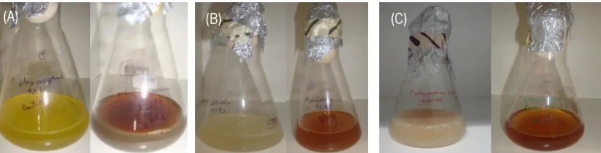
For the study of the biosynthesis of AgNPs the filamentous fungi species of Aspergillus ibericus, Penicillium chrysogenum and Neurospora crassa were used. The production of AgNPs was monitored through the change of colour of the filtrate treated with 1 mM of AgNO3. A
brownish colour of the filtrate was observed after 96 h of incubation for A. ibericus (Figure 2), P. chrysogenum (Figure 3) and for N. crassa strains (Figure 4). A negative control was prepared for each strain without AgNO3.
Different results were found from different species and also within each species. The strain A. ibericus MUM 03.51 produced the most intense brownish filtrate comparing with its negative control and when compared to other A. ibericus strains. All the strains of P. chrysogenum produced brownish filtrate comparing to their negative control. From all the fungi N. crassa MUM 92.08 produced the most brownish filtrate although N. crassa MUM 11.01 did not shown any colour change when compared with the negative control.
Figure 2 – Cellular filtrate of Aspergillus ibericus MUM 03.49 (A), MUM 03.50 (B) and MUM 03.51 (C), treated with 1 mM of AgNO3 after 96 h of incubation in the dark. Corresponding negative controls are shown on the left of each
image.
24
Figure 3 – Cellular filtrate of Penicillium chrysogenum MUM 97.43 (A), MUM 92.11 (B) and MUM 03.18 (C), treated with 1 mM of AgNO3 after 96 h of incubation in the dark. Corresponding negative controls are shown on the left of
each image.
Figure 4 – Cellular filtrate of Neurospora crassa MUM 11.01 (A) and MUM 92.08 (B), treated with 1 mM of AgNO3
after 96 h of incubation in the dark. Corresponding negative controls are shown on the left of each image.
The production of AgNPs was monitored by spectrophotometric analysis and a UV-Vis spectrum was obtained through the excitation of Ago surface plasmon vibrations at wavelengths
ranging from 380 to 420 nm.
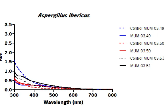
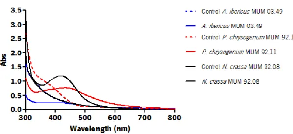
Each species exhibited different absorbance peaks comparing with other species. The strains A. ibericus MUM 03.49, P. chrysogenum MUM 92.11 and N. crassa MUM 92.08 (Figure 8) showed the highest absorbance peak for each species. Differences on peak absorbance were also observed within the same species suggesting intraspecific variability. The strain A. ibericus MUM 03.51 (Figure 5) and the strain N. crassa MUM 11.01 (Figure 7) did not show any absorbance peak on the UV-Vis spectra.
(A)
(A) (B)
25
Figure 5 – UV-visible spectrum of Aspergillus ibericus filtrate with 1 mM of AgNO3 aqueous solution with and its
respective strains after 96 h of incubation.
Figure 6 – UV-visible spectrum of Penicillium chrysogenum filtrate with 1 mM of AgNO3 aqueous solution with and its
26
Figure 7 – UV-visible spectrum Neurospora crassa filtrate with 1 mM of AgNO3 aqueous solution with and its
respective strains after 96 h of incubation.
Figure 8 – UV-visible spectrum Aspergillus ibericus MUM 03.49, Penicillium chrysogenum MUM 92.11 and Neurospora crassa MUM 92.08 filtrates with 1 mM of AgNO3 aqueous solution with and its respective strains after
96 h of incubation.
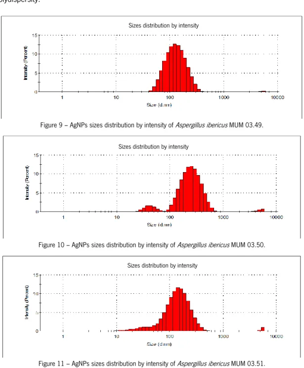
The size distribution of AgNPs produced for each fungal strain (Figures 9 - 16) was determined by Dynamic Light Scattering (DLS). Table 3 represents the average size (nm) and polydispersity of AgNPs. In terms of these physicochemical features different size distribution curves, average size and polydispersity values were obtained for AgNPs produced by each species and within each species. For A. ibericus species the strain A. ibericus MUM 03.49
27
produced AgNPs with a unimodal size distribution, showing the lowest average size, as well as the lowest polydispersity value. The AgNPs produced by P. chrysogenum MUM 97.43 exhibited the lowest average size, however AgNPs produced by P. chrysogenum MUM 92.11 exhibited the best size distribution and the lowest polydispersity values. Overall the strain N. crassa MUM 92.08 produced AgNPs with unimodal size distribution the lowest average size, and the lowest polydispersity.
Figure 9 – AgNPs sizes distribution by intensity of Aspergillus ibericus MUM 03.49.
Figure 10 – AgNPs sizes distribution by intensity of Aspergillus ibericus MUM 03.50.
Figure 11 – AgNPs sizes distribution by intensity of Aspergillus ibericus MUM 03.51.
Sizes distribution by intensity
Sizes distribution by intensity
28
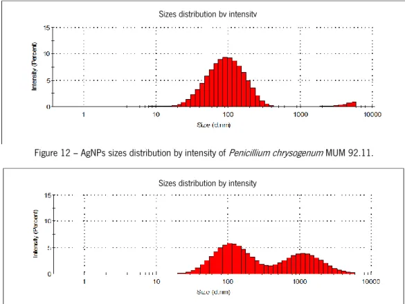
Figure 12 – AgNPs sizes distribution by intensity of Penicillium chrysogenum MUM 92.11.
Figure 13 – AgNPs sizes distribution by intensity of Penicillium chrysogenum MUM 97.43.
Figure 14 – AgNPs sizes distribution by intensity of Penicillium chrysogenum MUM 03.18.
Figure 15 – AgNPs sizes distribution by intensity of Neurospora crassa MUM 11.01.
Sizes distribution by intensity
Sizes distribution by intensity
Sizes distribution by intensity
29
Figure 16 – AgNPs sizes distribution by intensity of Neurospora crassa MUM 92.08. Table 3 – Average of size (nm) and polydispersity of each fungus and respective strains analyse by DLS.
Fungus Average size (nm) Polydispersity
A. ibericus MUM 03.49 122.4 0.179 A. ibericus MUM 03.50 255 0.337 A. ibericus MUM 03.51 141.8 0.390 P. chrysogenum MUM 92.11 141.8 0.242 P. chrysogenum MUM 97.43 105.7 0.672 P. chrysogenum MUM 03.18 164.2 0.435 N. crassa MUM 11.01 105.7 0.325 N. crassa MUM 92.08 24.36 0.269
Inductively coupled plasma optical emission spectrometry (ICP-OES) method was used to determinate the silver concentration. Table 4 represents the silver concentration (103 mg/mL)
in the filtrate after 96 h of incubation with 1 mM AgNO3 of all fungal strains. Theoretical value is
the calculated concentration of silver in 1 mM AgNO3 and real value was obtained by measuring
the concentration of 1 mM AgNO3 in distilled water by ICP-OES. A decrease of silver
concentration in the filtrate was observed for all the strains.
30
Table 4 – Concentration (103 mg/mL) of silver of each fungus and respective strains filtrate analyse by ICP-OES.
Fungus Concentration (103 mg/mL) A. ibericus MUM 03.49 65.578 A. ibericus MUM 03.50 57.225 A. ibericus MUM 03.51 31.335 P. chrysogenum MUM 92.11 43.496 P. chrysogenum MUM 97.43 65.732 P. chrysogenum MUM 03.18 64.079 N. crassa MUM 11.01 22.142 N. crassa MUM 92.08 51.203 Real Value 107.870 Theoretical Value 111. 590
From the previous results the strains A. ibericus MUM 03.49, P. chrysogenum MUM 92.11, N. crassa MUM 92.08 were found the best candidates to producer of AgNPs from each species. Further analyses as zeta-potential, Energy-dispersive X-ray spectroscopy (EDS) and Scanning Electron Microscopy (SEM) were conducted on these strains.
The Zeta-potential analysis (Table 5) clearly shown that the surface charge of the produced AgNPs for P. chrysogenum MUM 92.11 and N. crassa MUM 92.08 was highly negative indicating that the AgNPs surface has an anionic charge.
Table 5 – Solution charge (mV) of previously selected strains of each fungus analyse by Zeta-Potential.
Fungus Charge (mV)
A. ibericus MUM 03.49 - 9.7 P. chrysogenum MUM 92.11 - 25.1
N. crassa MUM 92.08 - 22.2
Real charge (mV) of ddH2O with AgNO3 = -1.1
The morphology and size range and the elemental composition of AgNPs in the freeze-dried filtrate of each selected fungi were analysed by SEM and EDS, respectively. A representative micrograph of the AgNPs was obtained by the SEM microscopy (Figure 17). It was found that the AgNPs exhibited variable shapes but most of them were spherical. Besides a size range of 40.9 to 204.0 nm for AgNPs produced by A. ibericus MUM 03.49 (Figure 17 (A)); a size range of 40.9
31
to 52.5 nm for AgNPs produced by P. chrysogenum MUM 92.11(Figure 17 (B)) and a size range of 43.7 to 72.9 nm for AgNPs produced by N. crassa MUM 92.08 (Figure 17 (C)) was observed. Most of the AgNPs were found as aggregates.
Figure 17 – SEM images of AgNPs synthetized by: Aspergillus ibericus MUM 03.49 (A), Penicillium chrysogenum MUM 92.11 (B) and Neurospora crassa MUM 92.08 (C).
Elemental composition of the AgNPs in the freeze-dried filtrate of selected fungal strain was determined by EDS (Figure 18). It can be observed that the P. chrysogenum MUM 92.11 shown the highest signal on the silver region (proximally at 3 KeV) comparing with A. ibericus MUM 03.49 and N. crassa MUM 92.08. Other elements such as C, N, O, P, S, Cl and K were also found in the freeze-dried filtrate of all the selected strains. In addition the A. ibericus MUM 03.49 and the N. crassa MUM 92.08 filtrate showed the presence of Na and P. chrysogenum MUM 92.11 filtrate shown the presence of Mg.
Figure 18 – EDS spectrum of the fungi filtrate after 96 h of incubation: (A) A. ibericus MUM 03.49, (B) P. chrysogenum MUM 92.11 and (C) N. crassa MUM 92.08.
(A) (B) (C)
32
4. Discussion and conclusion
The potential to manipulate fungus to synthetize nanoparticles involves several key parameters (temperature, agitation, light, control growth and other cellular activities) to achieve an optimized production of nanoparticles. The aim of this study consisted in selecting the best producer of stable biosynthesised AgNPs through the analysis of physicochemical features nanoparticles of selected fungi species.
Therefore, the reduction of Ag+ ions was monitored by the change of colour of the
filtrate from yellow to brown after 96 h of incubation, and the presence of an absorbance peak on UV-Vis spectroscopy analysis at wavelengths ranging from 380 to 420 nm indicated the production of spherical AgNPs. According to previous published data, the changing in the reaction solution colour is due to the excitation of surface plasmon vibrations in the AgNPs (Mulvaney 1996; Elechiguerra et al. 2005). Furthermore, the absorbance band between 380 and 420 nm indicates the formation of spherical or roughly spherical AgNPs (Hasell et al. 2007; Pal et al. 2007). In our study, the fungal strains A. ibericus MUM 03.49, P. chrysogenum MUM 92.11 and N. crassa MUM 92.08 showed the presence of surface plasmon vibrations between 380 and 420 nm suggesting that these strains are the best candidates to produce spherical AgNPs.
Differences of size in AgNPs when comparing the DLS results with the SEM were found. These differences were above two fold which leads to conclude that AgNPs size is overestimated by DLS. In fact, DLS technique measures the hydrodynamic diameter of the particle. It gives the information of the inorganic core along with any coating material and the solvation layer attached to the particle. It means that nanomaterial sizing determined by techniques that use wet dispersion are commonly overestimated (De Palma et al. 2007; Dhawan and Sharmam 2010). In contrast, SEM analysis uses dehydrated samples, where only the inorganic core and the coating material are take into account. In addition, due to poor contrast the measurement of the coating layer can sometimes be underestimated (Gaumet et al. 2008). In addition, the surface plasmon resonance plasmon at 420 nm is characteristic of AgNPs between 20 and 50 nm (Durán et al. 2005).
Zeta-potential analysis showed that the surface charge of the formed nanoparticles was highly negative. This result suggested that the nanoparticle surface has an anionic charge, as
33
stated by Kumar and Mamidyala (2011). It has long been recognized that the zeta potential is a very good index of the magnitude of the interaction between colloidal particles and its measurements are commonly used to assess the stability of colloidal systems (Malvern 2009). In our study, all selected strains had negative surface charge which has been referred to play a role as a cellular signature to study the surface interaction of AgNPs (Kumar and Mamidyala 2011).
The SEM micrographs show the high density of nanostructures, confirming the presence of AgNPs in the selected fungi. The EDS analysis allows confirms the reduction of Ag+
ion to elemental silver (Ag0). The absorption peak on SEM-EDS was observed at approximately 3
keV, which is characteristic of silver nanocrystals (Kumar and Mamidyala 2011; Jain et al. 2011). The presence of other peaks besides silver products as C, N, O, S, Cl and K in the EDS spectra indicated the heterogeneity of the reaction solution. The peaks found for C, N, O and S indicated the presence of fungal proteins as a capping material on the surface of AgNPs (Jain et al. 2011; Durán et al. 2010). Besides that, thick cap surrounding the nanoparticles could also be observed. This is probably due to the biomolecules released from the fungus acting as stabilizing agents of the nanoparticles (Durán et al. 2005; Gade et al. 2008). In our study, the strain P. chrysogenum MUM 92.11 produced roughly spherical AgNPs with a range size of 40.9 to 52.5 nm but most of them were aggregated. The range of sizes of AgNPs produced by N. crassa MUM 92.08 was 43.7 to 72.9 nm and the nanoparticles were found less aggregated.
From the previous results, the best fungal strains for the production of AgNPs were P. chrysogenum MUM 92.11 and N. crassa MUM 92.08. Both strains were used in furthers studies of mutagenesis and modulation of biochemical mechanisms for the production of AgNPs.
Chapter II:
Mechanism approach of biosynthesis
of AgNPs.
37
1. Introduction
1.1. Nitrogen requirement in filamentous fungi
Nitrogen is a major element found in many of the simple compounds and in nearly all of the complex macromolecules of living cells. It is essential to all organisms, to synthesize amino acids and, consequently, proteins. Fungi can use inorganic nitrogen in the form of nitrates, nitrites, ammonia or organic nitrogen to produce amino acids. It is known that fungi are different in their ability to use nitrogen compounds for growth, and also, that they may have a requirement for nitrogen in a specific form (Msore-Landecker 1990).
The principal process of conversion from inorganic nitrogen into organic compounds is done by nitrate assimilation. This conversion occurs in a diversity of microorganism such as: bacteria (Stouthamer 1967; Piéchaud et al. 1967), yeast, fungi (Lewis & Fincham 1970; Cove 1979), algae (Nichols & Syrett 1978; Huskey et al. 1979) and plants (Birkett & Rowlands 1981; Martins et al. 2009).
Extensive studies of nitrogen metabolism and its control have been carried out in several fungi, such as: Neurospora crassa, Aspergillus nidulans, and Saccharomyces cerevisiae. Although certain compounds, like ammonia, glutamate and glutamine, are preferred over others as nitrogen sources, these fungi are able to use many diverse secondary sources, including nitrate and nitrite, purines, proteins, amino acids, acetamide and acrylamide. These secondary nitrogen sources require the synthesis of catabolic enzymes or an activation of previously remaining enzymes (Msore-Landecker 1990).
Synthesis of nitrogen-regulated enzymes involves two conditions: an excitation of nitrogen catabolic repression in required and, secondly, a specific induction by subtract or in the presence of an intermediate pathway. Several works have been developed to study the metabolic pathway of nitrate assimilation of filamentous fungi. Until now, the Ascomycetes (Strauss et al. 1998) and the Aspergillus (Unkles et al. 1989; Daboussi et al. 1989; Ventura & Ramón 1991) have been the most studied fungi for that purposes.
The regulation of nitrogen metabolism on fungi is derived from genetic analysis of structural genes mutant (Msore-Landecker 1990). However, the numerous fungi that are unable to utilize nitrates require a more reduced form of nitrogen, probably because they are unable to reduce the nitrate ion (Msore-Landecker 1990).