This study aimed to synthetize, characterize and evaluate the antimicrobial properties of silver nanoparticles to be used in the development of a root intracanal formulation. Silver nanoparticles (AgNPs) were obtained by reduction of silver nitrate with sodium borohydride and characterized by UV-Visible spectrophotometry, scanning electron microscopy (SEM) and dynamic light scattering (DLS). The antimicrobial activity of nanoparticle formulation was evaluated by determinations of the minimum inhibitory concentration (MIC) and minimal bactericidal concentration (MBC) against different bacterial species by the microdilution method, according to recommendations of the Clinical and Laboratory Standards Institute (CLSI). Three potential vehicles, hydroxyethylcellulose, Carbomer and polyethylene glycol were tested as carriers for formulations containing AgNPs. The efficiency of the synthesis method chosen to produce AgNPs was demonstrated by four characterization techniques. The nanoparticles showed antibacterial activity against all species tested. Incorporation of AgNPs into all experimental vehicles produced stable formulations but the one in hydroxyethylcellulose presented better physical proprieties. The results indicate that silver nanoparticles are potential antiseptic agents to be used in root canals and incorporation in adequate vehicles may favor a broader application.
Development of Intracanal Formulation
Containing Silver Nanoparticles
João Felipe Bonatto Bruniera1, Yara Teresinha Corrêa Silva-Sousa1, Marilisa Guimarães Lara2, André Pitondo-Silva 2, Andrea Marcia Marcaccini1 and Carlos Eduardo Saraiva Miranda1
1University of Ribeirão Preto, School
of Dentistry, Ribeirão Preto, SP, Brazil
2USP - University of São Paulo, School
of Pharmaceutical Sciences of Ribeirão Preto, Ribeirão Preto, SP, Brazil
Correspondence: Profa. Dra. Yara T. C. Silva-Sousa, Rua Célia de Oliveira Meireles, 350, Jardim Canadá, 14024-070 Ribeirão Preto, SP, Brasil. Tel.: +55-16-3623-6002. e-mail: [email protected]
Key words: Endodontics, dental pulp diseases, nanoparticles, silver, antibacterial agents.
Introduction
Physicochemical and biologic properties of nanomaterials, especially the ones containing silver, attracted investigators in recent decades (1). Furthermore, silver nanoparticles show advantageous properties in biocompatibility and antimicrobial activity when compared to the salt precursors (2).
Small quantities of silver ions adsorbed on the surface of silver nanoparticles (AgNPs) and released in the presence of water and oxygen are able to combine with sulfur, nitrogen or oxygen of bacterial organic compounds causing damage to the cell wall and osmotic unbalance (3). Complex formation between silver nanoparticles and proteins may cause bacterial death by compromising cell metabolism (4). Interaction of the particles with bacterial DNA may result in bacteriostatic action, preventing cell reproduction (3). Antibacterial properties of AgNPs can be shown against a large spectrum of microorganisms including gram-positive and gram-negative bacteria (5), fungi (6) and viruses (7). Endodontic infections are mainly caused by bacterial etiology. Therefore, this study investigated the preparation, characterization and evaluation of the antimicrobial properties of silver nanoparticles aiming to develop root intracanal formulations for endodontic therapy.
Material and Methods
Chemicals and Materials
Analytical grade silver nitrate (AgNO3) and sodium
citrate (Na3C6H5O7) were purchased from LabSynth (Diadema, SP, Brazil) and sodium borohydride from Merck (Darmstadt, Germany).
Standard bacterial strains catalogued by American Type Culture Collection (ATCC) and National Collection of Type Cultures (NCTC) were employed to test antimicrobial activity. They included: Escherichia coli (ATCC 25922), Enterococcus faecalis (NCTC 775), Pseudomonas aeruginosa (ATCC 27853), Staphylococcus aureus (ATCC 25923) and Streptococcus mutans (ATCC 25175).
Synthesis of Silver Nanoparticles
To prepare the suspension of AgNPs, a volume of 10 mL silver nitrate solution with 4.0x10⁻⁴ mol/L was added dropwise from a glass burette to a flat-bottom flask (immersed in an ice bath) containing 10 mL of 1.0x10⁻² mol/L sodium citrate, used as a stabilizer, and 10 mL of 2.5x10⁻² mol/L sodium borohydride. The reaction was carried out under constant stirring and protected from light. After the addition of silver nitrate solution was finished, the mixture was then vigorously stirred during 15 min. Furthermore, a higher concentration of reducing agent, 1×10⁻³ mol/L sodium borohydride and a slower rate of silver nitrate addition were also evaluated.
Characterization
303
Silver nanoparticles in endodontics
obtained in a Cary IE spectrophotometer (Varian, Melbourne, Australia). Zeta potential and particle size were determined by using a dynamic light scattering instrument (Zetasizer Nano ZS, Malvern Instruments Ltd. Worcestershire, England). Size distribution and shape of particles were confirmed by scanning electron microscopy (SEM) in magnifications of 20,000×, 50,000×, 80,000× and 120,000×, after sample drying at room temperature and moisture.
Preparation of Intracanal Formulations
T h r e e d i f f e r e n t e x c i p i e n t s ( p o l y m e r i c hydroxyethylcellulose gel, carbomer polymer gel and polyethylene glycol) were tested to formulate the preparation containing 75% (w/w) silver nanoparticle suspension for root intracanal administration. Polymeric hydroxyethylcellulose gel (Cellosize QP100®) were tested in several concentrations (1.0 to 5.0%), associated or not to polyoxyethylene (20) sorbitan monolaurate (Tween 20). Several concentrations of carbomer polymer gel (Carbopol Ultrez 10®) (0.25 to 1.0%) neutralized with triethanolamine in sufficient quantity to reach a pH of 6.0 to 7.0 were also tested as a vehicle for the silver nanoparticles. A semi-solid formulation based on polyethylene glycol was prepared associating Carbowax® 400, 4000 and 6000 in different proportions to 15% Tween 80. The formulations were evaluated by their macroscopic aspect, considering homogeneity and fluidity.
Since the best results were attained by using the formulation containing silver nanoparticles in 1.5% hydroxyethylcellulose gel, this vehicle was selected to verify the antimicrobial activity of the nanoparticles.
Antimicrobial Activity
The minimum inhibitory concentration (MIC) and minimal bactericidal concentration (MBC) of formulation containing silver nanoparticles in 1.5% hydroxyethylcellulose gel were determined in triplicates by the microdilution broth method in 96-well microplates as recommended by the CLSI (9). Briefly, the formulation was serially diluted with Muller Hinton culture broth to silver concentrations ranging from 7,200 to 14 ng/mL and 100
mL were added to the micro wells. The original suspension was diluted with Muller Hinton medium starting with a 1:1 dilution.
The -80 °C stored standard bacterial strains were cultured in Petri dishes containing Muller Hinton Agar (Difco, Detroit, MI, USA) for E. coli, E. faecalis, P. aeruginosa, and S. aureus, and in Muller Hintonwith 5% sheep blood for S.mutans. After incubation of plates for 24 h at 37 °C, isolated colonies were suspended in saline up to a turbidity equivalent to 0.5 in the McFarland scale determined in a Densimat densitometer (Bio Mèrieux, Marcy l´Etoile,
France). The 0.5 value in the McFarland scale is equivalent to about 1.5×10⁸ colony forming units (CFU)per mL. Bacterial suspensions were further diluted (1:10) with Muller Hinton broth (final concentration, 1.5×10⁷ CFU/mL) and 5 µL aliquots added to microplate wells containing the formulation dilutions, finally leading to about 105 or 104 CFU/well of each tested bacterial strain.
All tests included wells containing only bacterial suspensions, as a control of growth, other ones containing only each culture broth (negative control) to ensure its sterility. As positive control, a 0.12% chlorhexidine gluconate solution (CHD) was used against all studied strains.
After 24 h incubation at 37 °C, plates were analyzed to determine MICs against the different strains by considering turbidity as an indication of growth and lack of it as inhibition of growth. MICs were determined as the lowest concentration of test solutions corresponding to the well without bacterial growth (100% inhibition). To determine the MBC, 10 µL aliquots were taken from selected wells that did not show visible bacterial growth and spread on plates containing Agar Muller Hinton medium and incubated for 24 hours at 37 °C. Visual observation indicated presence or absence of growth and MBC was considered as smallest concentration of test solution showing no growth.
Results
Synthesis of Nanoparticles and Characterization The yellow suspension produced by the redox reaction between silver nitrate and sodium borohydride, stabilized by sodium citrate, is characteristic of silver nanoparticles.
Scanning spectrophotometry in the visible range of 300 to 700 nm showed a peak absorbance around 400 nm suggesting the presence of silver nanoparticles (Fig. 1A).
Characterization by dynamic light scattering indicated nanoparticles varying in size between 1 and 100 nm (Fig. 1B) and the analysis also allowed determining the zeta potential of particles, which was -33mV (Fig. 1C). Silver nanoparticle size depending on the concentration of the reducing agent, sodium borohydride, and on the rate of silver nitrate addition (oxidant agent). A higher concentration of sodium borohydride with 1×10⁻³ mol/L and a slower addition rate of silver nitrate were also evaluated. However, the obtained results were not satisfactory. Thus, the addition rate of the oxidant agent was maintained higher and the concentration of reducing agent was standardized in 2.5×10⁻² mol/L.
SEM showed the presence of round particles and confirmed sizes according with the results obtained by dynamic light spreading (Fig. 2).
Intracanal Formulations
J.F
.B
. Bruniera et al.
vehicles of different hydroxyethylcellulose concentrations, 1 to 5%, did not show visible physical alterations after 24 h. When the vehicle employed was carbomer gel, the finished product did not show physical alterations after 24 h, but it did not have adequate fluidity even when the lowest carbomer concentration was used (0.25%). Unstable formulations were obtained when 75% (w/w) silver nanoparticles suspension was added to polyethylene glycol vehicles due to the deficient homogenization obtained.
As previously stated, the formulation containing silver nanoparticles in 1.5% hydroxyethylcellulose gel was considered the best one due to its good physical properties.
Antimicrobial Activity
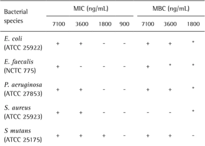
MIC determinations showed that formulation prepared in 1.5% hydroxyethylcellulose gel containing AgNP have bacteriostatic activity against all species tested. MIC values
were very promising ranging from 1,800 to 7,100 ng/mL, depending on the bacterial species. Chlorhexidine gluconate solution (CHD), used as positive control, presented a MIC value < 1,170 ng/mL.
Tests for minimal bactericidal concentration (MBC) indicated that bactericidal activity could only be detected in more concentrated preparations ranging from 3,600 ng/ mL (for E. coli,P. aeruginosa, S. mutans) to 7,100 ng/mL (for E. faecalis). The formulation with silver concentration of 3,600 ng/mL was able to inhibit the growth of S. aureus, however, it was not able to present bactericidal activity against this species. Both MIC and MBC results are shown in Table 1.
Discussion
Silver nanoparticles were synthesized by reduction of
Figure 1. Characterization of silver nanoparticle suspensions. A: Absorption spectrum determined in the region of visible light (300 to 700 nm) showing peak absorbance at 400 nm. B: Particle size distribution determined by dynamic light spreading. C: Zeta potential determined by dynamic light spreading.
Figure 2. SEM micrograph of a suspension of silver nanoparticles showing particle size in nanometers.
Table 1. Minimum inhibitory concentration (MIC) and minimal bactericide activity (MBC) of silver nanoparticle suspensions against different standard bacterial species
Bacterial species
MIC (ng/mL) MBC (ng/mL)
7100 3600 1800 900 7100 3600 1800
E. coli
(ATCC 25922) + + - - + + *
E. faecalis
(NCTC 775) + - - - + * *
P. aeruginosa
(ATCC 27853) + + - - + + *
S. aureus
(ATCC 25923) + + - - - - *
S mutans
(ATCC 25175) + + + - + +
305
Silver nanoparticles in endodontics
silver nitrate by borohydride in sodium citrate medium (8), which was as a stabilizer agent (9) at low temperature. According to Gulrajani et al. (10) smaller nanoparticles are obtained when synthesized at temperatures around 5 °C that favor enucleation and prevent agglomeration of the particles with lower kinetic energy (5).
The presence of nanoparticles in the suspension was confirmed by the characteristic yellow color as suggested by Shukla et al. (5) and by scanning spectrophotometry between 300 and 700 nm that produced an spectrum with a 400 nm absorbance maximum (11,12). According to Pal et al. (13), nanoparticle morphologies are directly related to absorption spectra; round particles produce spectra with a single absorbance peak. Spectra with two peaks indicate disc-shaped particles and triangular ones when three or more peaks are detected (13).
Light dynamic scattering indicated the presence of AgNPs with average sizes between 1 and 100 nm (14) and allowed determination of the zeta potential, a property directly related to particle stability (13,14). Zeta potential values characterize particle surface charges and thus, electrical potential, which is influenced by particle composition and the dispersion medium (15). The particles in this study had a zeta potential of -33 mV, a value considered ideal, according to Mohanraj and Chen (15) and Moraes et al.(16). According to these authors, zeta potentials above +30 or below -30 mV demonstrate nanoparticle stability. The SEM analysis confirmed particle spherical shapes and size distribution between 1 and 100 nm as determined by light dynamic scattering.
Lok et al. (17) suggested that antimicrobial activity of silver nanoparticles is dependent on the ability to cross the microorganism cell walls. Particle sizes ranging from 1 to10 nm in diameter have higher penetration potential and bactericidal power (18). The method described in this report produced particles in this range justifying the results that confirm other observations for S. mutans (6,19), E. coli, P. aeruginosa (20), except S. aureus (21). Against this last species, silver nanoparticles had only bacteriostatic activity. It should be noted that silver particles showed bacteriostatic and bactericidal activity against E. faecalis (22) while, calcium hydroxide a commonly used agent in endodontic therapy only acts as a bacteriostatic agent against this species at pH values higher than 12.5 (23).
To be used for antisepsis in root canal therapy longer than one session, silver nanoparticles must be able to penetrate dental tubules and the lateral channel systems and accessories. In this way, an adequate fluidity is necessary to facilitate penetration and draining of the radicular canal (24). Yamazaki et al. (25) studied dentin permeability and efficiency of Endo-PTC (which is utilized as a lubricant in endodontic instrumentation) in semi-solid pharmaceutical
forms with different excipients as hydroxyethylcellulose or carbomer polymeric gels and associations of polyethylene glycol and surfactants. The results did not show differences between the evaluated preparations in terms of penetration capacity into dentin tubules or efficiency. Thus, similar excipients were tested as vehicles to the silver nanoparticles obtained in this study. The formulation containing silver nanoparticles in 1.5% hydroxyethylcellulose was selected as best due to good physical properties such as homogeneity and fluidity, which certainly it would make easier the gel application into canal.
It may be concluded that synthesis of silver nanoparticles obtained by reduction of silver nitrate, as described, was effective and confirmed by different characterization methods. Antimicrobial properties of the particles were detected against E. coli, E. faecalis, P. aeruginosa, S. aureus and S. mutans. The root intracanal formulation developed with hydroxyethylcellulose polymer gel containing the silver nanoparticles showed adequate homogeneity and fluidity for the use proposed. The study suggests an innovative use of silver nanoparticles as an endodontic antiseptic agent. However, further studies are still necessary to determine in vitro and in vivo cytotoxicity, biocompatibility and pharmacokinetics for viable utilization.
Resumo
O presente estudo teve como objetivo sintetizar, caracterizar e avaliar as propriedades antimicrobianas de nanopartículas de prata visando o desenvolvimento de uma formulação intracanal. As nanopartículas de prata (AGNPS) foram obtidas pela redução de nitrato de prata com borohidreto de sódio e caracterizados por espectrofotometria UV-Visível, microscopia eletrônica de varredura (MEV) e espalhamento de dinâmico de luz (DLS). A atividade antimicrobiana da formulação de nanopartículas foi avaliada por meio das determinações da concentração inibitória mínima (CIM) e a concentração bactericida mínima (CBM) contra diferentes espécies de bactérias pelo método de microdiluição, de acordo com recomendações do Clinical and Laboratory standards Institute (CLSI). Três potenciais veículos, hidroxietilcelulose, carbómero e polietileno glicol foram testados como veículos para as formulações de AGNPS. A eficiência do método de síntese escolhido para produzir AGNPS foi demonstrada por quatro técnicas de caracterização. As nanopartículas apresentaram atividade antibacteriana contra todas as espécies bacterianas testadas. A incorporação de AGNPS em todos os veículos experimentais produziram formulações estáveis, porém, quando utilizado a hidroxietilcelulose foram obtidos melhores propriedades físicas. Os resultados indicam que as nanopartículas de prata são potenciais agentes anti-sépticos para serem usados na terapia endodôntica e a incorporação em veículos adequados pode favorecer uma aplicação mais ampla.
Acknowledgements
J.F
.B
. Bruniera et al.
Professor Elson Longo, PhD, from the Chemistry Department, Federal University of Sao Carlos for the use of scanning electron microscopy equipment at the Interdisciplinary Laboratory of Electrochemistry and Ceramics. The authors are grateful to Coordenação de Aperfeiçoamento de Pessoal do Ensino Superior (CAPES) for granting a CAPES/PROSUP Master Degree scholarship to João Felipe Bonatto Bruniera.
References
1. Cheng L, Zhang K, Melo MA, Weir MD, Zhou X, Xu HH. Anti-Biofilm dentin primer with quaternary ammonium and silver nanoparticles. J Dent Res 2012;91:598-604.
2. Motshekga SC, Ray SS, Onyango MS, Momba MN. Microwave-assisted synthesis, characterization and antibacterial activity of Ag/ZnO nanoparticles supported bentonite clay. J Hazard Mater 2013;6:439-446.
3. Damm C, Münstedt H, Rösch A. The antimicrobial efficacy of polyamide 6/silver-nano- and microcomposits. Mater Chem Phys 2008;108:61-66. 4. Mei L, Lu Z, Zhang W, Wu Z, Zhang X, Wang Y, et al.. Bioconjugated
nanoparticles for attachment and penetration into pathogenic bacteria. Biomaterials 2013;34:10328-10337.
5. Shukla MK, Singh RP, Reddy CR, Jha B. Synthesis and characterization of Agar-based silver nanoparticles and nanocomposit film with antibacterial applications. Bioresou Technol 2011;107:295-300. 6. Nam KY. In Vitro antimicrobial effect of the tissue conditioner
containing silver nanoparticle. J Adv Prosthodont 2011;3:20-24. 7. Duek A, Arkhangelsky E, Krush R, Brenner A, Gitis V. New and
conventional pore size tests in vírus-removing membranes. Water Res 2012;46:2505-2514.
8. Clinical and Laboratory Standards Institute (CLSI). Methods for dilution antimicrobial susceptibility tests for bacteria that grow aerobically. Approved standard (8th Edition, CLSI document M7-A8). NCLLS, Wayne, PA, USA, 2009.
9. Hernández-Sierra JF, Ruíz F, Pena DCC, Martínez-Gutiérres F, Martínez AE, Guillén AJP, et al.. The antimicrobial sensitivity of Streptococcus mutans to nanoparticles of silver, zincoxid, and gold. Nanomedicine 2008;4:237-240.
10. Gulrajani ML, Gupta D, Periyasamy S, Muthu SG. Preparation and application of silver nanoparticles on silk for imparting antimicrobial properties. J Appl Polym Sci 2008;108614-623.
11. Monteiro DR, Gorup LF, Takamiya AS, Camargo ER, Filho ACR, Barbosa DB. Silver distribution and release from an antimicrobial denture base resin containing silver colloidal nanoparticles. J Prosthodont 2012;21:7-15.
12. Lee SJ, Heo DN, Moon JH, Ko WK, Lee JB, Bae MS, et al. Electrospun chitosan nanofibers with controlled levels of silver nanoparticles.
Preparation, characterization and antibacterial activity. Carbohydr Plym 2014;13:530-537.
13. Pal S, Tak YK, Song JM. Does the antibacterial activity of silver nanoparticles depend on the shape of the nanoparticle? a study of the gram-negative bacterium Escherichia coli. Appl Environ Microbiol 2007;73:1712-1720.
14. Mukherjee SG, O´Claonadh N, Casey A, Chamber G. Comparative in vitro cytotoxicity study of silver nanoparticle on two mammalian cell lines. Toxicol in Vitro 2012;26:238-251.
15. Mohanhaj VJ, Chen Y. Nanoparticles - A review. Trop J Pharm Res 2006;5:561-573.
16. Moraes CM, Paula E, Rosa AH, Fraceto LF. Physicochemical stability of poly(lactide-co-glycolide) nanocapsules containing the local anesthetic Bupvacaine. J Braz Chem Soc 2010;21:995-1000.
17. Lok CN, Ho CM, Chen R, He QY, Yu WY, Sun H, et al. Silver nanoparticles: partial oxidation and antibacterial activities. J Biol Inorg Chem 2007;12:527-534.
18. Verran J, Sandoval G, Allen NS, Edge M, Stratton J. Variables affecting the antibacterial properties of nano and pigmentary titania particles in suspension. Dyes Pigments 2007;73:298-304.
19. Bürgers R, Eidt A, Frankenberger R, Rosentritt M, Schweikl H, Handel G, et al. The anti-adherence activity and bactericidal effect of microparticulate silver additives in composite resin materials. Arch Oral Biol 2009;54:595-601.
20. Chen M, Yang Z, Wu H, Pan X, Xie X, Wu C. Antimicrobial activity and the mechanism of silver nanoparticle thermosensitive gel. Int J Nanomedicine 2011;6:2873-2877.
21. Tien DC, Tseng KH, Liao CY, Tsung TT. Colloidal silver fabrication using the spark discharge system and its antimicrobial effect on
Staphyloccocus aureus. Med Eng Phys 2008;30:948-952.
22. Wu D, Fan W, Kishen A, Gutmann JL, Fan B. Evaluation of the antibacterial efficacy of silver nanoparticles against Enterococcus faecalis biofilm. J Endod 2014;40:285-290.
23. Evans M, Davies Jk, Sundqvist G, Figdor D. Mechanisms involved in the resistance of Enterococcus faecalis to calcium hydroxide. Int Endod J 2002;35:221-228.
24. Rossi-Fedele G, Guastalli AR. Osmolarity and root canal antiseptics. Int Endod J 2014;47:314-320.
25. Yamazaki AK, Moura-Netto C, Salgado RJ, Kleine BM, Procopowitsch I. Ex vivo analysis of root canal cleaning using Endo-PTC associated to NaOCl and different irrigant solutions. Braz Oral Res 2010;24:15-20.