DEPARTAMENTO DE CIÊNCIAS BIOMÉDICAS E MEDICINA
Salt marsh plants as source of
bioactive compounds
Maria Isabel Póvoa Rocha
Dissertação de Mestrado em Ciências Biomédicas
Trabalho efetuado sob a orientação de:
Professora Doutora Luísa Barreira
Doutora Luísa Custódio
Declaração de autoria de trabalho
Declaro ser o autor deste trabalho, que é original e inédito. Autores e trabalhos consultados estão devidamente citados no texto e constam da listagem de referências incluída.
Copyright Maria Isabel Rocha
A Universidade do Algarve tem o direito, perpétuo e sem limites geográficos, de arquivar e publicitar este trabalho através de exemplares impressos reproduzidos em papel ou de forma digital, ou por qualquer outro meio conhecido ou que venha a ser inventado, de o divulgar através de repositórios científicos e de admitir a sua cópia e distribuição com objetivos educacionais ou de investigação, não comerciais, desde que seja dado crédito ao autor e editor.
AGRADECIMENTOS
Manifesto aqui o meu sincero reconhecimento à Professora Doutora Luísa Barreira e à Doutora Luísa Custódio, minhas orientadoras científicas, e ao Professor Doutor João Varela por me terem proporcionado a oportunidade de realizar este trabalho. Agradeço o seu esforço, informações, sugestões e disponibilidade que sempre demonstraram no apoio á realização desta dissertação.
Agradeço também aos técnicos de investigação, do grupo MarBiotech pela constante ajuda, apoio, ideias e ensinamentos que me transmitiram, e que permitiram a elaboração de todo este trabalho.
A todos os colegas do grupo MarBiotech, que me acompanharam durante esta jornada, que contribuíram sempre com ideias, e que me ajudaram sempre que necessário. Obrigada pelo bom ambiente de trabalho que criaram, divertido, trabalhador e bem- humorado.
À Maria João Rodrigues, a minha “mini-chefa”, que teve toda a paciência do mundo para me ensinar e instruir novas técnicas, sem nunca desistir e sem nunca desanimar. Que me aturou sempre durante todo este caminho e foi caminhando ao meu lado. À Carolina Bruno de Sousa pela sua infindável boa vontade, generosidade e bom coração que fazem dela um apoio incondicional para a vida. Aos meus colegas de “luta” Ivo Monteiro, Eunice Santos e Tiago Braga pelo constante apoio e animo que sempre me transmitiram, pelo auxílio nos piores momentos e pelos abraços e risadas nos melhores momentos.
A todos os meus colegas de mestrado com quem partilhei os bons e maus momentos desta jornada.
Agradeço à minha afilhada, confidente e amiga Sara Magalhães que sem me aperceber se tornou tão importante na minha vida. És um dos suportes fundamentais na minha vida. Agradeço toda a tua amizade, loucura, partilha de todos os momentos,
melhores e piores, e sobretudo a tua sincera e honesta amizade. Esse suporte e apoio foram importantes para conseguir chegar onde estou hoje.
Agradeço ao Rúben Costa por todas as vezes que me deu na cabeça e que sempre puxou por mim e nunca desistiu, até quando eu já o tinha feito. És daqueles irmãos de coração que ganhamos e que ficam para a vida. Agradeço a tua amizade, companhia, sinceridade, risos, sarcasmos, desabafos e tudo o que tens feito por me ajudar.
A todos os meus demais amigos que sem a sua paciência e apoio não teria sido possível concluir esta tarefa.
Agradeço a todos os amigos “infantinos” pela motivação e compreensão durante esta jornada, obrigada por acreditarem que conseguiria concluir mais esta etapa e por me acompanharem com o vosso espirito e musica.
Agradeço às duas pessoas sem as quais este trabalho não teria sido possível, os meus pais Maria José e Manuel Rocha. Toda a vossa dedicação, compreensão, amor e estímulo foram muito importantes para que conseguisse concluir mais esta etapa. Esta tese é dedicada a vocês que sempre fizeram tudo pelo meu futuro e bem-estar. Toda a vossa persistência, principalmente da mãe, contribuiu em muito para hoje ter a educação que tenho e o conhecimento que alcancei e para ser a pessoa que sou hoje. Muito agradecida! Amo-vos muito.
Finalmente agradeço ao meu namorado Paulo Silva, que apesar de ter aparecido já a meio do trabalho, foi sempre um apoio importante, e que aturou desde os melhores momentos aos piores este feitiozinho nada fácil de aturar. Com muitos “tá bem” e “vamos lá a ver” foste ajudando no que podias e não podias. Deste-me sempre força para continuar, abdicando do “nosso” tempo para que fosse dedicado a este trabalho. Agradeço todo o carinho, amor e compreensão.
Contents Abbreviations ……… vi Abstract ………...….…... viii Resumo ………...………...… ix 1. Introduction ……… 1 1.1 Bioactive compounds ………. 1 1.2 Neurodegenerative diseases ………....… 1 1.2.1 Alzheimer’s disease ………...….. 2 1.2.1.1 Neuropathology of AD ………...…...… 3
1.2.1.2 β-amyloid plaques and Neurofibrillary tangles ………...…….……. 4
1.2.1.3 Cholinesterases ………...………... 5
1.2.2 Parkinson’s disease ………...………... 6
1.2.2.1 Neuropathology of PD ………...…….... 8
1.2.2.2 Tyrosinase ………...………. 10
1.3 Oxidative stress, antioxidants and ND ………...………….. 13
1.4 Neuroinflammation: the role of microglia ………...……….… 20
1.5 Actual treatments for AD and PD ……….…… 21
1.6 Halophytes ………...……….… 22
1.6.1 Carpobrotus edulis ………....… 23
1.7 Aims of the thesis ………...………..… 24
2. Materials and Methods ………...……….... 25
2.1 Plant material ………..………..… 25
2.2 Supplements, chemicals and culture media ………...……...… 25
2.3 Preparation of the extracts ………...………. 26
2.4 Determination of antioxidant activity ………... 26
2.4.1 Radical scavenging activity (RSA) of DPPH ………...……….… 26
2.4.2 Iron (ICA) chelating activity ………...……….. 27
2.4.3 Copper (CCA) chelating activity ………...……… 27
2.4.4 Iron reducing power ………...… 27
2.4.5 Nitric oxide (NO) – scavenging activity ……… 28
2.5.1 Total flavonoid content (TFC) ………...……….... 28
2.5.2 Total condensed tannin content (TCTC) ……… 29
2.5.3 Total phenolic content (TPC) ………. 29
2.6 Enzyme inhibition assays ………...……….. 29
2.6.1 Acetylcholinesterase (AChE) and Butirylcholinesterase (BChE) inhibition………. 29
2.6.2 Tyrosinase (TYRO) inhibition ………... 30
2.7 Neuroprotective activity using cell-based method …………...……….…....30
2.7.1 Protective effect against H2O2 – induced injury in neuronal cells ……….… 30
2.7.1.1 Cell culture and viability determination ………...……... 30
2.7.1.2 Protective effect against H2O2 – induced oxidative stress on SH-SY5Y ………. 31
2.7.2 Anti-inflammatory activity on LPS – stimulated microglia cells ...………... 31
2.7.2.1 Cell culture and viability determination ………...………... 31
2.7.2.2 Quantification of nitric oxide (NO) protection on LPS-stimulated microglia cells ……….. 31
2.8 Statistical analysis ……….…… 32
3. Results ………...……… 33
3.1 Determination of antioxidant activity ………...… 33
3.1.1 Radical scavenging activity (RSA) of DPPH ……….... 33
3.1.2 Iron (ICA) and copper (CCA) chelating activity ………...…… 35
3.1.3 Iron reducing power ………... 36
3.1.4 Nitric oxide (NO)-scavenging activity ………...………... 37
3.2 Total contents of phenolic (TPC), flavonoids (TFC) and condensed tannin (TCTC) ……….. 38
3.3 Enzyme inhibition assays ………...………..… 39
3.3.1 Cholinesterase inhibition ………...……….... 39
3.3.2 Tyrosinase inhibition ………...……….. 40
3.4 Neuroprotective activity using cell-based method ………...…... 41
3.4.1 Protective effect against H2O2-induced injury in neuronal cells ……...…… 41
4. Discussion ………...………..… 47
4.1 Antioxidant activities ………...……….…… 47
4.2 Phytochemical analysis ………. 48
4.3 Enzyme inhibition assays ………...………..… 49
4.3.1 Cholinesterase inhibition ………...……….... 49
4.3.2 Tyrosinase inhibition ………...……….. 49
4.4 Neuroprotective activity ………...……….... 50
4.5 Anti-inflammatory activity on LPS-stimulated microglia cells ……… 51
5. Conclusion and future work ………...……….... 51
vi Abbreviations AA Arachidonic acid ACh Acethylcholine AChE Acethylcholinesterase AD Alzheimer's disease
APP Amyloid precursor protein
Aβ Amyloid-β peptide
BChE Butyrylcholinesterase
BHA Butylated hydroxytoluene
BHT Butylated hydroxytoluene
ChE Cholinesterase
CNS Central nervous system
DCH Docosohexanoic acid
DMEM Dulbecco’s Modified Eagle’s Medium
DMSO Dimethylsulfoxide
DOPA Dihydroxyphenylalanine
DOPA-quinone Dihydroxyphenylalanine DPPH 1,1-diphenyl-2picrylhydrazyl DTNB 5,5-dithio-bis(2-nitrobenzoic) acid EDTA Ethylenediamine tetraacetic acid
IL-1β Interleukin-1β
LB Lewis bodys
LH Unsaturated lipids
LOO˙ Peroxyl radical
LOOH Lipid hydroperoxides
LPS Lipopolysaccharide
MTT 3-(4,5-dimethylthiazol-2-yl)2,5-diphenyl tetrazolium bromide
ND Neurodegenerative diseases
vii
NO Nitric oxide
PBS Phosphate buffer
PD Parkinson's disease
PHFs Paired helical filaments PUFAs Polyunsaturated fatty acids
PV Pyrocatechol violet
RNS Reactive nitrogen species
ROS Reactive oxygen species
RT Room temperature
SNpc Substantia nigra pars compact
SOD Superoxide dismutase
TH Tyrosine hydroxylase
viii Abstract
Neurdegenerative diseases are characterized by the progressive decline of neuronal functions. Oxidative stress is considered the principal initiator of this problem, and contributes to the appearance of neurodegenerative diseases. In this study it was investigated the potential source of biocompounds with neuroprotective features of halophytes species. An initial screening of methanol extract of 26 species was made, using the 1,1-diphenyl-2picrylhydrazyl (DPPH) radical scavenging assay. The species Carpobrotus edulis was selected for the next assays, and used to prepare hexane, dichloromethane, chloroform, ethyl acetate and methanol extracts. The antioxidant activity was evaluated through different and complementary methods, namely the DPPH radical scavenging activity, iron and copper chelating activity, iron reducing power and nitric oxide (NO) scavenging activity, and for the phytochemical analysis, including total flavonoid content (TFC), total condensed tannin content (TCTC) and total phenolic content (TPC). These extracts were also tested for inhibition against enzymes related to neurodegenerative disorders, namely acetylcholinesterase (AChE), butirylcholinesterase (BChE) and tyrosinase (TYRO).
The in vitro anti-inflammatory activity of the extracts was determined through the evaluation of the inhibition of NO production in lipopolysaccharide (LPS)-stimulated N9 microglial cells. The neuroprotective activity was tested through the H2O2- induced injury
in SH-SY5Y cell line, and the cellular viability measured by the MTT method. The methanol extract had the highest activity on the DPPH assay, and also the highest copper chelating activity and NO-scavenging activity. The hexane extract had the highest iron chelating activity, while the ethyl acetate extract had the highest iron reducing power activity. The methanol extract had the highest levels of total flavonoid content (TFC) and total phenolic content (TPC), while the dichloromethane extract had the highest total condensed tannin content (TCTC). All the extracts tested had a protective effect against H2O2- induced injury in SH-SY5Y cells, especially the methanol extract, which also had the
highest anti-inflammatory activity in N9 microglial cells.
Keywords: Halophytes, C. edulis, plant extracts, antioxidant activity, anti-inflammatory activity, enzyme inhibition, nitric oxide, phenolic compounds.
ix Resumo
As doenças neurodegenerativas são caracterizadas pela progressiva degeneração do sistema nervoso. A perda contínua e específica de células neuronais pode conduzir a uma situação de doenças, como a doença de Alzheimer (DA) ou a doença de Parkinson (DP). O stress oxidativo é considerado uma das maiores e mais frequentes causas ou processo iniciador deste tipo de doenças. O envelhecimento conduz a um variado número de problemas para a saúde, incluindo o aumento de espécies reativas de oxigénio (ERO) que podem provocar danos celulares. A DA, a principal causa de demência na população, é caracterizada por distúrbios de comportamento, perda de memória e a diminuição da atividade cognitiva. Na DP os movimentos involuntários, associados a tremores, perda de força muscular e rigidez muscular são sintomas característicos desta doença. Em ambos os casos, a acumulação de neurotoxinas, de algumas proteínas e ERO conduzem a estados de inflamação crónica, perda de capacidade sináptica, levando assim à morte neuronal e ao agravamento dos sintomas. Diversos estudos têm provado a eficácia de compostos antioxidantes na redução da formação de ERO e de fortalecimento dos mecanismos de defesa dos organismos. Torna-se assim fundamental a pesquisa de produtos naturais que possuam propriedades antioxidantes e anti-inflamatórias. As plantas, devido às condições em que habitam, principalmente as halófitas, desenvolveram sistemas antioxidantes eficientes para proteção dos seus sistemas. As halófitas são capazes de sobreviver a ambientes com elevado teor de salinidade. Deste modo, podem desenvolver stress oxidativo, que é regulado por diversos mecanismos que facilitam a retenção e aquisição de água, proteção de cloroplastos, e manutenção da homeostase iónica. Essencialmente, estas ações compreendem a síntese de osmólitos (pequenos glícidos, polióis, aminoácidos e metilaminas), proteínas específicas (ex. enzimas antioxidantes, proteínas transportadoras de iões) e moléculas antioxidantes (ex. compostos fenólicos, carotenóides e vitaminas). Isto pode explicar a utilização de algumas espécies de halófitas em países orientais, na medicina tradicional e na alimentação. Para, além disso, sabe-se que as moléculas presentes em plantas, vulgarmente designadas por fitoquímicos, possuem propriedades antioxidantes e parecem ter uma atividade anti-inflamatória, eventualmente prevenindo o desenvolvimento destas doenças, através da inativação de algumas enzimas, prevenindo o stress oxidativo ou
x ainda evitando a inflamação neuronal. O uso de compostos provenientes de halófitas para a prevenção de doenças neurodegenerativas ainda está pouco estudado. Neste estudo fez-se um screening inicial com 26 espécies de halófitas, fazendo um estudo da atividade antioxidante através do radical 1,1-difenil-2picrylhydrazyl (DPPH). A espécie Carpobrotus edulis foi selecionada para os seguintes ensaios. Foi efetuada uma extração sequencial com os solventes n-hexano, diclorometano, clorofórmio, acetato de etilo e metanol. Assim este estudo teve como objetivo estudar a atividade antioxidante, anti-inflamatória e neuroprotectora de extratos desta espécie. Através dos métodos de redução de radicais livres DPPH, da atividade quelante do cobre e do ferro, da atividade redutora do ferro e potencial de eliminação do óxido nitrico (NO) foi medida a capacidade antioxidante destes extratos. Os teores totais de compostos fenólicos, flavonóides e taninos foram igualmente determinados. Foi também testada a capacidade inibitória destes extratos contra algumas enzimas específicas, como a acetilcolinesterase (AChE), a butirilcolinesterase (BChE) e a tirosinase. Foram determinadas ainda a atividade anti-inflamatória e neuroprotectora em células de neuroblastoma (SH-SY5Y) e células de microglia (linha celular N9). Para o estudo da atividade anti-inflamatória foi utilizado o ensaio de Griess, que é um método simples e rápido para avaliar indiretamente a produção de óxido nitríco em meio aquoso, através da medição de nitritos, que é um produto final estável do óxido nitríco, na presença de oxigénio. Este método permite determinar o potencial inibitório dos extratos sobre a produção de óxido nitríco, na linha celular de células da microglia N9 estimulada com lipopolissacarídeo (LPS). A atividade neuroprotectora foi avaliada através do método de MTT, na linha celular SH-SY5Y estimuladas com peróxido de hidrogénio (H2O2),
previamente tratadas com os extratos, procedendo-se posteriormente à medição da viabilidade celular. Todos os extratos mostraram possuir atividade antioxidante contra o radical DPPH, principalmente o extrato metanólico com uma atividade anti radical de 96.1%, para a concentração de 1 mg/mL. O extrato metanólico permitiu obter ainda a maior atividade quelante do cobre (48.27% a 10 mg/mL), e de no radical NO (41.4% a 10 mg/mL), enquanto na atividade quelante do ferro o extrato de hexano foi o mais ativo (81.32% a 10 mg/mL). A atividade redutora do ferro mais elevada foi obtida com o extrato de acetato de etilo (76.63% a 10 mg/mL). O extrato de metanol apresentou a maior concentração de flavonóides e fenólicos totais, enquanto o extrato de diclorometano
xi apresentou o teor mais elevado de taninos totais. A maior capacidade inibitória da AChE foi obtida com a aplicação do extrato de metanol (43.2% a 10 mg/mL), enquanto o de acetato de etilo foi o mais eficaz a inibir a BChE (60.4% a 1 mg/mL). Os extratos não apresentaram citotoxicidade nas linhas celulares utilizadas nos ensaios de neuroprotecção. Os extratos apresentaram capacidade para proteger a linha celular SH-SY5Y contra o stress oxidativo induzido pela aplicação de água oxigenada, traduzido numa redução da viabilidade para cerca de 50%, e o melhor resultado foi obtido com o extrato de metanol (143.5% de viabilidade celular a 50 μg/mL). A aplicação do extrato de metanol permitiu ainda uma redução significativa da produção de NO por células de microglia. Em conclusão, a espécie halófita C. edulis mostrou ser uma potencial fonte de compostos interessantes para o possível tratamento ou prevenção das doenças neurodegenerativas, em particular o extrato metanólico uma vez que foi o que permitiu obter melhores resultados.
Palavras-chave: Carpobrotus edulis, halófitas, Alzheimer, Parkinson, atividade antioxidante, compostos fenólicos, inibição enzimática, atividade anti-inflamatória, atividade neuroprotectora,
1 1. Introduction
1.1 Bioactive compounds
Bioactive compounds (BioA) can be characterized as crucial and non-crucial constituents that occur in small amounts in plant and animal products, and can influence cellular and physiological activities, contributing to a better health when included in the diet (Kris-Etherton et al., 2002, 2004; Biesalski et al., 2009). Some examples of BioA are flavonoids, isoflavones, phytoestrogens and monotrepenes, which can be found in many vegetables (Kris-Etherthon et al., 2002). These compounds display several biological activities, namely antioxidant, anti-inflammatory and anti-tumoral (Kris-Etherton et al., 2004; Ye et al., 2013). Apart from this activities, the definition of BioA implies that it displays comproved health benefits (Kris-Etherton et al., 2004; Biesalski et al., 2009), and can been taken through the diet or by medication.
Higher variety in fruit and vegetable intake has been associated with a lower risk of several chronic diseases and have been widely used to prevent and treat some disorders (Koppula et al., 2012; Ye et al., 2013). BioA have become popular due to have none or limited side effects, in contrast with synthetic drugs (Koppula et al., 2012). It is increased the search for BioA with a view to discover new antioxidant potential therapeutic compounds to attenuate or treat neurodegenerative diseases (Koppula et al., 2012).
1.2. Neurodegenerative diseases
Neurodegenerative diseases (ND) are often hereditary and sporadic conditions which are characterized by progressive nervous system dysfunction, induced by deposits of abnormal proteins and other factors, which leads to a neuronal degeneration in particular areas of the brain (Ross et al., 2004). Alzheimer's disease (AD) and Parkinson's disease (PD) are examples of ND, and are characterized by the continued loss of specific neuronal cells in association with protein aggregation (Barnham et al., 2004; Ross et al., 2004). The oxidative stress is closely linked to these diseases, and is probably the responsible for the dysfunction of intracellular components, like proteins or neuronal cell death, which contributes to the development of these diseases (Barnham et al., 2004).
2 The increasing age in population is deeply linked to the increase in the prevalence of several forms of ND (Forman et al., 2004). It is expected that by 2025, 75% of the world's population over 60 years of age living in developing countries will suffer of ND (Forman et al., 2004). The search for effective treatments or preventive interventions for ND is extremely important because in the near future the emotional, societal and financial burden of these aging-related disorders will be very high (Forman et al., 2004).
1.2.1 Alzheimer’s disease
Alzheimer's disease (AD) is one of the main causes of dementia in aged population, affecting more than 35 million people worldwide (Prince et al., 2012). It was first identified in 1907 by Alois Alzheimer, in a 51-year-old woman, which exhibited personality changes, progressively worsening memory loss, time disorientation and language disturbances, despite the relatively normal neurological function (Nixon 2002; Forman et al., 2004). After autopsy, her brain revealed evidences of cerebral atrophy (Nixon 2002). There are many factors that contribute to AD, such as hypertension, oxidative stress, inflammation and hypercholesterolemia (Gamba et al., 2011). There are two forms of AD, the sporadic form, which affects 90% of the patients, and the familiar predisposition, that includes the remaining 10% (Nixon 2002). However, the two forms of the disease show the same neuropathology features (Nixon 2002). AD is normally recognized by characteristic symptoms namely behaviour disturbances, memory loss and decreasing of cognitive activity (Chitranshi et al., 2012), induced by the decrease of the forebrain cholinergic neurons, the most affected in this disease (García-Ayllón et al., 2011). With the advances of neuroimaging techniques, it is now possible to detect and measure the metabolism, atrophy and inflammation in brain, and the presence of plaques and neurofibrillary tangles (NFTs), as well the brain activity (Perrin et al., 2009). The research and development of biomarkers in AD is crucial to measure the structural and functional changes in the brain (Perrin et al., 2009). Biomarkers like brain volume, atrophy, level of glucose metabolism in the brain, regional activity changes, amyloid-plaque and NFTs formation, inflammation and oxidative stress are crucial on the preclinical phase of AD to make a diagnosis and prognosis, even before irreversible neurological damage occurs (Perrin et al., 2009; Alzheimer’s & Dementia, 2011).
3 In Europe there are 7.3 million people with AD (Alzheimer-europe.org, 2014). In Portugal it is estimated that there are more than 180 000 cases, and due to the increase of elderly population it is estimated that the number of AD cases will increase (Alzheimer-europe.org, 2014). Every year, 1.4 million Europeans develop dementia meaning that every 24 seconds a new case is diagnosed. It is expected that by 2050 the number of the cases increases to 16.2 million (Forman et al., 2004; Alzheimer-europe.org, 2014).
1.2.1.1. Neuropathology of AD
AD is characterized by extracellular deposits of amyloid-β peptide (Aβ), which form senile plaques and formation of intracellular neurofibrillary tangles (NFTs) composed of paired helical filaments (PHFs) of hyperphosphorylated protein tau (Resende et al., 2008; Fig. 1.1). There are several studies showing that the accumulation of amyloid plaques and NFTs plays an important role in neurodegeneration during and before the clinical phase of the disease (Nixon 2002). These structures are present in brain areas such as those involved in learning and memory, like the cortex and hippocampus (Nixon 2002; Resende et al., 2008). Such areas typically exhibit synaptic and neuronal loss, with cholinergic and glutamatergic neurons being the most affected (Resende et al., 2008). AD is also accompanied by inflammation, and neuronal, axonal and synaptic loss dysfunction (Perrin et al., 2009). These features are also found in cognitively normal elderly individuals and need to reach a certain threshold to be noticed by clinical symptoms (Perrin et al., 2009).
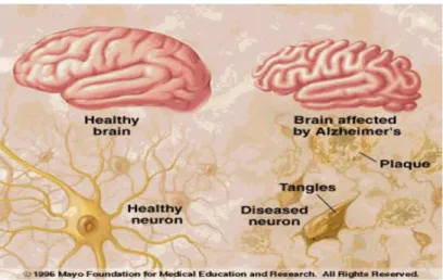
Figure 1.1: Comparison of a healthy brain and a brain affected by AD, showing senile plaques and NFTs tangles leading to cortex atrophy. (Source http://img.docstoccdn.com/thumb/orig/90531693.png).
4 AD has 4 clinical stages, described as “very mild/mild cognitive impairment” (MCI), “mild”, “moderate” and “severe” (Fig. 1.2). In the beginning of the “very mild” stage, the disease is correlated not with plaques or NFTs, but with synaptic and neuronal loss, supporting the idea that plaques and NFTs need to accumulate 10 to 15 years before cognitive decline is noticeable (Perrin et al., 2009).
Figure 1.2: Progressive clinical stages of AD, “very mild/mild cognitive impairment”
(MCI), “mild”, “moderate” and “severe”, correspond to clinical dementia rating (CDR) scores of 0.5, 1, 2 and 3, respectively. These stages are associated with abundant amyloid plaques (red line), the gradual accumulation of NFTs (blue line) and synaptic and neuronal loss in certain brain regions (green line). (Source: Perrin et al., 2009).
1.2.1.2 β-amyloid plaques and neurofibrillary tangles
As stated before, the most common hallmarks of AD is the formation of senile plaques and NFTs (Perrin et al., 2009) (Fig. 1.1).
Senile plaques are composed by extracellular aggregates of Aβ. This peptide is formed from a larger amyloid precursor protein (APP), a transmembrane protein, which, in normal cases, is cleaved first by α-secretase and then by γ-secretase (Goedert et al., 2006; LaFerla et al., 2007; Karran et al., 2011). In AD cases the first cleavage was made by β-secretase, which creates a shorter fragment known as β-amyloid (or Aβ) (LaFerla et al., 2007). When fragments of Aβ aggregate they became toxic, interfering with the normal neuronal function (Butterfield et al., 2001; Perrin et al., 2009). With time these aggregates
5 increase in size and become soluble, originating the senile plaques and thus contributing to cell death (Butterfield et al., 2001; Perrin et al., 2009; Alzheimer’s & Dementia, 2011). The deposition of Aβ plaques is responsible for a chronic inflammatory response involving the activation of microglial cells (Nixon 2002; Dumont and Beal, 2011). Those reactions explains the presence of additional cell types in senile plaques, namely microglia and reactive astrocytes (Butterfield et al., 2001; Nixon 2002). Some of them accelerates the fibrillation of Aβ with the consequent formation of toxic products, which leads to a local development of dystrophic neurites (Butterfield et al., 2001; Nixon 2002). The primary damage to neurites initiates local Aβ overproduction, neurite degeneration-regeneration responses, and secondary inflammatory reactions involved in the removal of cellular debris (Nixon 2002). All these events cause more damage to the neurons through the formation of more toxic products and Aβ (Nixon 2002).
1.2.1.3. Cholinesterases
Neurochemical studies show that the brain of AD patients has a disturbance of acethylcholine (ACh) metabolism, which led to the formulation of the “cholinergic hypothesis” (Carvajal and Inestrosa, 2011). This hypothesis postulates that the accumulation of Aβ and tau protein is connected to the loss of cholinergic neurons (Carvajal and Inestrosa, 2011).
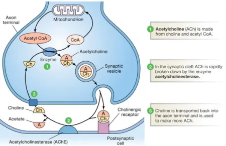
Acetylcholinesterase (AChE) is a selective enzyme responsible for the hydrolysis of the cholinergic neurotransmitter ACh (Fig. 1.3; Ciro et al., 2012; Wang et al., 2012). AChE is present in all cholinergic structures, as well as in a subpopulation of non-cholinergic neurons and in glial cells (Ciro et al., 2012; Wang et al., 2012). Butyrylcholinesterase (BChE) also hydrolyses ACh and is present in a subpopulation of cortical and subcortical neurons and in glial cells (Ciro et al., 2012; Wang et al., 2012). It was demonstrated that intense cholinesterase (ChE) activity occurs in senile plaques and neurofibrillar tangles, which influences the aggregation of Aβ (Ciro et al., 2012). AChE inhibitors have been shown to interfere with the production of Aβ, by decreasing the levels of APP, thus reducing the toxicity effects associated with Aβ production and aggregation (Ciro et al., 2012). Moreover, it is known that the expression of BChE increases as plaques go through the process of maturation, from the diffuse type to the pathologic compact variety (Ciro et
6 al., 2012). Thus, it is suggested that the influence of BChE in the pathology of plaques is greater than that of AChE, being essential in AD plaque maturation (Wang et al., 2012; Ciro et al., 2012). BChE shows to be a great contributor to the loss of ACh in AD brain, and is inhibition can lower the levels of Aβ peptide (Wang et al., 2012).
Figure 1.3: Schematic representation of synthesis of ACh. 1 - Acetylcholine is made from choline and acetyl CoA being afterwards incorporated and stored in presynaptic vesicles. Presynaptic vesicles then move or are emptied into the synaptic cleft, and the diffusion of ACh across the synaptic cleft to the postsynaptic ACh receptors occurs through binding of ACh to postsynaptic ACh receptor. 2 - After signalling, acethylcholine is released from receptors and broken down by acethylcholinesterase to be recycled in a continuous process. 3 – Choline is reuptaked into the presynaptic neuron. (Source: http://peaknootropics.com/wp-content/uploads/2013/08/acetylcholine-metabolism.png).
1.2.2. Parkinson's disease
Parkinson's disease (PD) is a neurodegenerative pathology which affects 2-3% of the population above the age of 65 (Fasano et al., 2006) and 5% above 85 years, and is the second most common ND, after AD (Hasegawa et al., 2008; Bartels et al., 2009; Shulman et al., 2011). In Portugal there are between 13000 and 15000 people affected with this disease (Associação Protuguesa de Doentes de Parkinson, 2012).
7 PD was initially reported by James Parkinson in 1817, and was based on six cases with a disease called then “Shaking Palsy”, which symptoms were involuntary tremulous motion, reduced muscular power, higher propensity to bend the trunk forward, and to pass from walking to a running pace, but where the cognitive abilities have not been injured (Zhang et al., 2000; Bartels et al., 2009). The neuropathological hallmark of PD, the Lewy body (LB), was described around 1912 by Friederichy Lewy (Forman et al., 2004, Bartels et al., 2009; Shulman et al., 2011). Around 1919, Konstantin Tretiakoff associated the loss of pigmented cells in the substantia nigra pars compacta (SNpc; the deep portion of substantia nigra) with the PD progression (Bartels et al., 2009). In the 1950’s Arvid Carlsson found a relationship between the decrease of dopamine and PD (Bartels et al., 2009).
There are two forms of PD: a sporadic form, which affects 95% of all patients, and familial form accounting for about 5% of all cases (Hald and Lotharius, 2005). The familial form is associated with mutations in at least five genes, such as SNCA, PARK, PINK1, DJ1, UCHL1 and LRRK2(Fig. 1.4; Hald and Lotharius, 2005; Tan and Skipper, 2007). The SNCA and PARK1 genes are linked to α-synuclein protein, promoting abnormal protein aggregation like showed in Fig. 1.4 (Tan and Skipper, 2007). Mutations in PARK and UCH-L1 cause a damage in the ability of the cellular machinery to detect and eliminate misfolded proteins (Duaer and Przedbarski., 2003). DJ1 gene have a role in the oxidative stress response, either as a redox sensor protein or as an antioxidant (Tan and Skipper, 2007).
Figure 1.4: Scheme of potential interactions between the encoded proteins of the different PD genes
8 The aetiology of the sporadic form is still unknown (Zhang et al., 2000; Asanuma et al., 2003; Hald and Lotharius, 2005). Currently the most widely accepted hypothesis says that PD is a result of both genetics and environmental factors, leading to the failure of mitochondria, development of oxidative stress and occurrence of dopaminergic cells death (Hasegawa et al., 2008; Bartels et al., 2009; Nakashima et al., 2012). PD manifests through bradykinesia (extreme slowness of movements and reflexes), rest tremor, muscular rigidity, gait abnormalities and postural instability and in some cases dementia (Zhang et al., 2000; Montiel, 2006; Mosley et al., 2006; Zecca et al., 2008; Hauser and Hastings, 2013).
1.2.2.1. Neuropathology of PD
The cell body of dopaminergic neurons project from the SNpc, which is responsible for the body movements, to the caudate nucleus and striatum, where neurons release dopamine (Zhang et al., 2000; Dauer and Przedbarski, 2003; Fasano et al., 2006; Mosley et al., 2006; Hauser and Hastings, 2013). In dopaminergic neurons, which also contain neuromelanin that confers a characteristic pigmentation to the SNpc area, the production of dopamine is affected and decreased, leading to cell degeneration and death (Fasano et al., 2006; Shulman et al., 2011; Hauser and Hastings, 2013). This fact is shown by the depigmentation of SNpc, as shown in Fig 1.5. (Duaer and Przedbarski, 2003). This dopaminergic cell loss in SNpc results in dysfunctional motor movements (Shulman et al., 2011).
9 Figure 1.5: Neuropathology of PD. (A) Schematic representation of the normal dopaminergic neurons whose cell bodies are located in the SNpc (black arrows). These neurons project (thick solid red lines, the nigrostriatal pathway) to the basal ganglia and synapse in the striatum (i.e., putamen and caudate nucleus). (B) Schematic representation showing the degeneration of nigrostriatal pathway in Parkinson’s patients there is a marked loss of dopaminergic neurons that project to the putamen (dashed line) and a much more modest loss of those that project to the caudate (thin red solid line). (C) Immunohistochemical labeling of intraneuronal inclusions, termed Lewy bodies, in a SNC dopaminergic neuron showing the differences between a normal neuron and a neuron from a PD patient. (Source: Duaer and Przedbarski, 2003).
The LB is an intraneuronal proteinaceous cytoplasmic inclusions, mostly composed by α-synuclein, a small acidic presynaptic protein, which is involved in vesicular transport (Fig. 1.5C; Fasano et al., 2006; Shulman et al., 2011; Irwin et al., 2013).
Although the causes of PD are not yet fully understood, data obtained from several studies support the theory that mitochondrial dysfunction and oxidative stress are involved in the progression of the pathogenic disease, which is strongly linked to dopaminergic neuronal loss (Mosley et al., 2006; Hasegawa et al., 2010; Hauser and Hastings, 2013). Besides these two factors, others, such as inflammation, excitotoxic mechanisms and toxic actions of nitric oxide (NO) are involved in PD development (Jenner 2003, Hasegawa et al., 2010; Hauser and Hastings, 2013).
10 The microglia cells are important in surveillance and host defence in brain, aiding in the identification and removal of cells who are programed to death (Jenner 2003; Liu et al., 2003). These cells are particularly sensitive to changes in their environment and turn active in case of injury or infection (Liu et al., 2003; Hald and Lotharius 2005). Several soluble factors are secreted by activated microglia cells, some have neuroprotective function, but the majority are neurotoxic and have proinflammatory activity, such as NO, superoxide anion and fatty acid metabolites (Liu et al., 2003; Hald and Lotharius 2005). Some experiments established a relationship between neuronal degeneration in PD and massive activated microglia, suggesting that activated microglia have a toxic effect in neurons (Liu et al., 2003; Hald and Lotharius 2005).
1.2.2.2. Tyrosinase
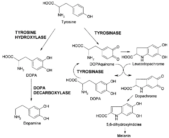
Tyrosinase, also known as polyphenol oxidase, is a copper-containing monooxygenase enzyme widely distributed in organisms (Nerya et al., 2003; Chang 2012). This enzyme catalyses the formation of melanin pigment from tyrosine, oxidation of several phenolic compounds, the synthesis of amino acid based antibiotics among others (Senol et al., 2010; Chang 2012). It is implicated in the hydroxylation of tyrosine to DOPAquinone (dihydroxyphenylalanine-quinone), and oxidation of DOPAquinone to DOPA (dihydroxyphenylalanine), through dopamine-quinone via contributing to neuromelanin (NM) formation (Fig. 1.6; Asanuma et al., 2003; Greggio et al., 2005; Chang 2012).
Dopamine is a potent neuromodulator in the central nervous system (CNS) that can be found in neuronal tissues and body fluids, in the form of large organic cations (Maciejewska et al., 2011). In fact dopamine is linked to PD, but the symptoms are recognized in AD patients too, showing that disturbance of dopamine metabolism may be closely associated also with AD (Silva and Ming, 2007). Catecholamines, dopamine, epinephrine, norepinephrine and DOPA are catechol-containing neurotransmitters, which are involved in cognitive, behavioural, physical, physiological and psychological functions (Silva and Ming, 2007). The oxidation of these molecules can cause severe alterations in brain activity, leading to neuronal death. Metabolic malfunctions of neurotransmitters are known phenomena in the physiology of AD and PD and have also been suggested to be
11 related to the neuropathology of these diseases (Silva and Ming, 2007).
Figure 1.6: Tyrosine metabolism. (Source: Fiore et al., 2004).
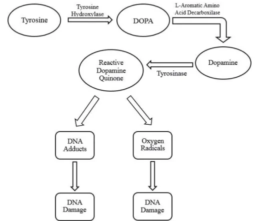
Tyrosine is converted into DOPA through tyrosine hydroxylase (TH), being then carboxylated to form dopamine (Fig. 1.6; Xu et al., 1998; Asanuma et al., 2003). Dopamine, in the presence of tyrosinase, covalently modifies TH, which becomes disabled (Greggio et al., 2005). Tyrosinase can oxidise the catechol ring of dopamine turning it into the high reactive species dopamine-quinone, as showed in Fig. 1.7, which is responsible for the toxicity of the neurotransmitter (Greggio et al., 2005). The oxidation of dopamine inactivates dopamine transporters, the glutamate transporter and the mitochondrial respiration (Xu et al., 1998; Greggio et al., 2005; Hwang 2013). Dopamine-quinones can react with cysteine residues in the cytosol of neurons, and form protein adducts, which lead to irreversible modification or inactivation of functional proteins (Greggio et al., 2005). These quinones can also react with α-synuclein, stabilizing this toxic fibril intermediate, a dominant cause in PD (Greggio et al., 2005).
12 Tyrosinase activity may also result in an increase in toxic effects of hydrogen peroxide (H2O2), being the peroxide a hallmark of oxidative stress and also an inhibitor of
dopamine transporter (Gregiio et al., 2005), increasing the oxidative stress in neurons.
Figure 1.7: Potential pathway for the synthesis of dopamine quinone (DA-quinone) and consequent damages. The DA-quinone is highly reactive and can change DNA or enhanced the formation of reactive oxygen radicals (superoxide, hydroxyl and peroxide). (Source: Stokes et al., 1996).
Neuromelanin is a dark insoluble polymer produced from dopamine oxidation that provide the dark pigmentation to the substantia nigra (Hwang 2013). Tyrosinase can accelerate the induction of catecholamine quinone derivates, due to its oxidase activity leading to dopamine neurotoxicity and neurodegeneration (Khan et al., 2007). Extraneuronal neuromelanin deposition has been observed in the brains of PD patients (Hwang 2013). When this neuromelanin is added to microglia cultures levels of NO increase, causing a strong microglia activation and consequently loss of dopaminergic neurons (Hwang 2013).
13 Since these discoveries, the research of tyrosinase inhibitors as a target for the treatment of PD has become increasingly important. The current treatment relies on the administration of L-dopa, to increase dopamine levels in the brain, though a long-therm therapy may cause adverse reactions (Asanuma et al., 2003).
1.3. Oxidative stress, antioxidants and ND
Oxidative stress has been regarded as a crucial factor in neurodegeneration (Friedman et al., 2011). Reactive oxygen species (ROS) and reactive nitrogen species (RNS), include radical anion superoxide (O2-•), hydroxyl radicals (•OH), hydrogen peroxide
(H2O2), and nitric oxide radical (NO•). ROS and RNS are normal by-products of cellular
metabolism, but react strongly with multiple molecules, leading to neurodegeneration (Behl and Moosmann, 2002; Barnham et al, 2004; Friedman et al, 2011; Hwang 2013; Taylor et al, 2013). Through different pathways, such as direct interaction with redox-active metals and oxygen species via reactions, like Fenton and Haber-Weiss reaction, the radical •OH has produced being the most reactive one of all ROS (Taylor et al., 2013).
The brain is characterized by a high oxygen consumption rate and lipid content, and by a relative shortage of antioxidant enzymes, compared with other tissues, making the CNS especially vulnerable to oxidative stress (Markesbery, 1996; Friedman et al., 2011). The chemical origin of ROS is the reaction between molecular oxygen with redox-active metals iron (Fe3+) and copper (Cu2+) (Barnham et al., 2004). To sustain many functions, the
brain needs an elevated concentration of metal iron, but has a low capacity to manage the oxidative stress and low regeneration ability (Barnham et al., 2004).
Due to high content of polyunsaturated fatty acids (PUFA), like arachidonic (AA), and docosohexanoic acid (DCH), brain are more sensitive to oxidation, being an important mechanism of neurodegeneration (Butterfield and Lauderback, 2002; Floyd and Hensley, 2002). AA and DCH are sensitive to free radical attack and PUFA decreased with the increase of lipid peroxidation (Butterfield and Lauderback, 2002). Lipid peroxidation is a process where molecular oxygen is integrated into unsaturated lipids (LH) and form lipid hydroperoxides (LOOH), like showed in Fig. 1.8 (Minotti and Aust 1992).
14 Figure 1.8: Reactions involved in lipid peroxidation. (Source: Minotti and Aust 1992).
These reactions are promoted by an “initiator” (I˙), which surpass the dissociation energy of an allylic bond and this cause a hydrogen abstraction and formation of a lipid alkyl radical (L˙; Minotti and Aust 1992). This alkyl radical can add oxygen to form lipid peroxyl radicals (LOO˙) which can liberate LOOH via hydrogen abstraction from a neighbour alkyl radical (Minotti and Aust 1992). It is necessary external oxidants, like •OH, to initiate lipid peroxidation, but once started the reaction this will propagate and form new LOOH (Minotti and Aust 1992; Valko et al., 2006). Transition metals, like Fe2+, increase the propagation of lipid peroxidation reaction (Minotti and Aust 1992; Valko et al., 2006). Fe2+ enters into Fenton and Haber-Weiss reaction, producing •OH. The increasing formation of •OH enhances the lipid peroxidation (Fig. 1.8 and 1.9; Minotti and Aust 1992; Behl and Moosmann, 2002; Barnham et al., 2004; Friedman et al., 2011; Hwang 2013; Taylor et al., 2013).
Figure 1.9: Schematic representation of hydroxyl radical production. In the Fenton reaction the
Fe2+ reacts with H
2O2 generating a very reactive and damage OH•. Ferric iron (Fe3+) react with O2-•
through the Haber-Weiss reaction, leading to the regeneration of Fe2+. The production of highly
reactive OH• promote the oxidative stress, lipid peroxidation, mitochondrial dysfunction, increases
in intracellular calcium concentration, and lastly neuronal cell death. (Source:
15 It is known that mitochondria, redox-active metals and inflammation (via activated microglia) are potential causes of oxidative stress in the brain (Fig. 1.10 and Fig. 1.11; Dumont and Beal, 2011). Oxidative stress is therefore considered one of the main causes for the development of different ND, including AD and PD (Halliwell 2001; Behl and Moosmann, 2002; Barnham et al., 2004).
In a brain of an AD patient, the content of Fe2+ and Cu2+ is increased, and can result in the production of free radicals (Jomova et al., 2010). Aβ plaques have been describe as metallic sinks because of the high concentrations of Cu, Fe and zinc (Zn) that have been found in this deposits (Barnham et al., 2004). In vitro studies evidenced that Cu2+ and Fe3+ induced peptide aggregation (Barnham et al., 2004). The homeostasis of Cu, Fe and Zn and their respective proteins, are severely modified in the AD brain (Barnham et al., 2004). Cu2+ and Fe3+ bound to Aβ may induce redox chemical reactions that reduce the oxidation state of both metals leading to the production of H2O2, and promoting the Fenton reaction.
This, in turn, leads to the generation of extremely toxic OH• radicals inducing an inflammatory response by microglial cells (Barnham et al., 2004; Silva and Ming, 2007; Jomova et al., 2010). Aβ cause lipid peroxidation in brain cell membranes, and leads to 4-hydroxy-2-nonenal (HNE) and acrolein formation (Butterfield and Lauderback, 2002). These reactive alkenals can alter the conformation of membrane proteins and are toxic to neurons (Butterfield and Lauderback, 2002). Protein oxidation can lead to diminished specific protein function which can induce cell death (Butterfield and Lauderback, 2002).
16 Figure 1.10: Scheme of the generation and role of free radicals in AD. Several key players,
such as metals and Aβ, can exacerbate free radical production. Once accumulated inside the cell, free radicals can cause lipid, protein, DNA, and RNA damage and exacerbate AD pathogenesis. (Source: Dumont and Beal, 2011).
Dopaminergic neurons are vulnerable to oxidative stress due to the elevated consumption of oxygen, low levels of antioxidant enzymes and the synthesis and storage of dopamine (Taylor et al., 2013; Hwang 2013). The neurotransmitter dopamine is stable when inside synaptic vesicles, however outside the vesicles, as in a damaged neuron, dopamine is metabolised or auto-oxidised by in an oxygen rich environment producing ROS (Taylor et al., 2013; Hwang 2013). The oxidation of dopamine generates H2O2,
reactive quinones and O2-•, leading to a highly oxidative stress conditions (Taylor et al.,
2013). Neuromelanin, present in dopaminergic neurons, is a potent activator of microglia when released from dying cells, thereby increasing the sensitivity of this neurons to oxidative stress-mediated cell death, and promoting neuroinflammation (Hwang 2013; Taylor et al., 2013).
High levels of iron can also be an important factor in the initiation and promotion of neuronal death, due to interaction with neuromelanin and dopamine (Barnham et al., 2004; Taylor et al., 2013; Hwang 2013). Since the SNpc has increased levels of microglia cells, the activation of this cells caused by injuries, is more pronounced in PD (Taylor et al., 2013). Such proneness to pro-inflammatory conditions has been related to elevated oxidative stress produced through the Fenton reaction (Fig. 1.8) (Hwang 2013; Taylor et al.,
17
2013). Dopamine-quinone has the ability to directly modify some proteins, such as α-synuclein, whose dysfunction and accumulation has been linked to PD, being another cause of oxidative stress (Hwang 2013). α-synuclein is a synaptic vesicle protein involved in neurotransmitter release, vesicle turnover, endoplasmic reticulum trafficking and synaptic plasticity, as shown in Fig. 1.11 (Taylor et al., 2013). The covalently modification of α-synuclein, by dopamine-quinone, leads to conversion into a cytotoxic protofibril form (Hwang 2013). The mutation of the α-synuclein gene can increase its expression leading to formation of oligomers and finally insoluble polymers or fibrils (Barnham et al., 2004; Taylor et al., 2013). Both forms have been proved to be neurotoxic by inhibiting the complex I activity of mitochondria thus inducing leakage of dopamine into the cytosol, which in turn promotes dopamine quinone production and consequently increased ROS production and oxidative stress (Fig. 1.11; Jomova et al., 2010; Choi et al., 2012; Hwang 2013; Taylor et al., 2013). Damages caused to the mitochondrial complex I in the electron transport chain leads to a leak of electrons, increasing ROS generation (Hwang 2013).
Figure 1.11: Oxidative stress in PD. The rule of DA and α-synuclein in oxidative stress process. The formation of NM leads to oxidative stress. (Source: Barnham et al., 2004).
18 Oxidative stress occurs when there is an imbalance between ROS production and antioxidant defences leading to oxidation of biomolecules and severe cellular damage (Fig. 1.12; Markesbery, 1996; Halliwell 2001; Orhan, et al 2007; Hauser and Hastings, 2013; Hwang 2013), namely DNA modification, enzymes inactivation, and structural proteins destruction (Behl and Moosmann, 2002; Barnham et al., 2004). Oxidative stress is present in most diseases and contributes to tissue injury. In order to cope with the overproduction of ROS, cells are endowed with several defence systems, being the antioxidants among the most important (Khan 2002; Hwang 2013). Antioxidants are compounds, synthetic or natural, that inhibit or delay the oxidation process by blocking the initiation or propagation of oxidizing chain reactions (Velioglu et al., 1998; Moreno et al., 2006). Natural antioxidants can be phenolic compounds, such as tocopherols, flavonoids and phenolic acids, nitrogen compounds, like alkaloids, chlorophyll derivates, amino acids and amines, and carotenoids as well as ascorbic acid (vitamin C) (Velioglu et al., 1998). Phenolic compounds are secondary metabolites present in a large abundance in plant tissues (Blokhina et al., 2003). Their antioxidants properties arise from their high reactivity as hydrogen or electron donors, from the ability of the polyphenol-derived radical to stabilize the unpaired electron, and from their ability to chelate transition metals ions, stopping the Fenton’s reaction (Blokhina et al., 2003). The ability of flavonoids to alter peroxidation kinetics by modification of the lipid packing order and to decrease fluidity of the membrane, is another mechanism who confers antioxidant properties to these compounds (Blokhina et al., 2003). Synthetic antioxidants like butylated hydroxyanisole (BHA) and butylated hydroxytoluene (BHT) have been used, but it is known now their carcinogenic potential, making the research for natural antioxidant drugs extremely important (Velioglu et al., 1998; Custódio et al., 2011).
19 Moreover the antioxidants can be defined as enzymatic or non-enzymatic (Khan 2002; Hwang 2013). Non-enzymatic antioxidants, like tocopherol (vitamin E) and ascorbic acid (vitamin C), phenolic compounds and other small molecules, and enzymatic antioxidants, such as catalases and peroxidases, which regulates the intracellular level of H2O2, superoxide dismutases (SOD), which catalyses the dismutation of superoxide to
H2O2, and repair enzymes (Khan 2002; Blokhina et al., 2003). All this molecules work
together in order to alleviate the ROS production by directly quenching oxygen radicals before they promote damages to cells (Khan 2002; Blokhina et al., 2003).
Figure 1.12: Schematic representation of how oxidative damage is exerted on brain. The oxidant species noted are produced and interact with cellular targets producing unique oxidation products and they, in some cases in turn, exert oxidative stress upon the tissue also. (Source: Floyd and Hensley 2002).
20 1.4. Neuroinflammation: the role of microglia
Microglia cells are the resident phagocytes and innate immune cells of the brain, and play an important role in inflammatory responses being important in homoeostasis, as well as in various pathologies of the CNS (Liu et al 2012). Reactive microglia are hallmarks of AD, and these brain cells are likely to contribute to the mechanisms of neuronal damage and cognitive loss associated with that disease (Branden et al., 2012). The activation states in microglia are similar to the macrophages and exhibit a functional plasticity during activation states (Branden et al., 2012). In inactive or quiescent state, these cells have a surveillance function, while in an active state they have two functionally distinct states, M1 and M2 with different functions (Branden et al., 2012). Microbial compounds and pro-inflammatory cytokines induce the classical activation of macrophages, the M1 state in microglia cells (Branden et al., 2012. The M2 state, also known as alternative activation state (Liu et al 2012), is responsible for anti-inflammatory activity by blocking the release of pro-inflammatory cytokines, ingesting debris, promoting tissue repair and releasing neurotrophic factors (Branden et al., 2012). The microglia cells respond to exogenous and endogenous stimuli originating a cascade of inflammatory molecules (Liu et al 2012). They can be activated in response to lipopolysaccharide (LPS) stimulation, and in consequence of this activation, cells secrete ROS and pro-inflammatory cytokines, such as interleukin-1β (IL-1β), which cause neuronal injuries (Branden et al., 2012). IL-1β is one of the molecules responsible for neuroinflammation, and has a direct effect on neurons causing glial cells and neurons to release multiple inflammatory mediators leading to self-amplifying neuroinflammation (Zhou Wu et al., 2013).
Microglial cells are activated when the protein β-amyloid form oligomers and amyloid deposits, and migrate towards plaques as they are formed (Fig. 1.13; Perrin et al., 2009). The astrocytes become reactive, and numerous inflammatory mediators, signalling molecules, oxidative processes, complement cascades and modulators of protease activities are released (Perrin et al., 2009). Dendrites and axons become dystrophic, due to this transport process malfunctions, leading to altterations in brain metabolism (Perrin et al., 2009). Apart from the formation of NFTs, neurons suffer many other changes: synapses are lost because they are dysfunctional, and neurons die (Perrin et al., 2009).
21 Figure 1.13: Schematic histological representation of biomarkers in relation to Alzheimer’s disease pathology. Produced by neurons, Aβ42 becomes deposited in plaques, which activate
microglia. Microglia release cytokines (for example IL-1β and TNF-α). Microglia and neurons also produce complement factors that can be activated by amyloid-β aggregates, and cause synapse loss. Tau becomes hyperphosphorylated and aggregates into NFTs in neurons and dystrophic neurites around plaques; its mechanism of release from neurons is uncertain. (Source: Perrin et al., 2009).
1.5. Actual treatments for AD and PD
Some studies show that AChE inhibitors have beneficial effects on cognitive, functional, and behavioural symptoms of AD (Scarpini et al., 2003). There are four cholinesterase inhibitors approved by the US Food and Drug Administration, the tacrine, donepezil, rivastigmine and galantamine (Scarpini et al., 2003). In Europe the availability differs from country to country, and only rivastigmine has been approved centrally by European Agency for the Evaluation of Medical Products (EMEA; Scarpini et al., 2003). Tacrine, the first approved drug for AD, is a potent non-selective inhibitor of both AChE and BChE (Franco et al., 2005). Although this hepatotoxicity and absence of selectivity have reduced is therapeutic use, the search of tacrine analogues is still of interest (Franco et al., 2005). Rivastigmine inhibits both AChE and BChE, while donepezil inhibits only
22 AChE (Bullock et al., 2005). These therapeutic drugs have common adverse events such as nausea, vomiting, diarrhoea, dizziness and weight loss (Hansen et al., 2008).
In PD the widely used and effective treatment is with L-DOPA and has been shown to benefit certain cognitive functions, although detrimental effects can also develop following L-DOPA therapy (Cools 2006). L-DOPA impairs some, but improves other complex cognitive abilities (Cools 2006). A long-term treatment with this medicament can lead to a disabling abnormal involuntary movements, also known as L-DOPA induced Dyskinesia (LID), characterized by uncontrolled and repetitive movement in the axis, arms, legs and oro-facial zone (Bravo et al., 2014). These facts are a serious limitation to the use of L-DOPA, and become important the search for new way of treatments and new therapeutic targets (Bravo et al., 2014).
Because of all these facts presented it becomes even more important the research for natural therapies and also new targets, with the prospect of being more efficient and with fewer side effects.
1.6 Halophytes
Halophytes can live in extreme saline conditions and grow and reproduce in different saline biotypes, such as dunes, rocky coasts, saline depressions, saline inland deserts and in salt marches (Ksouri et al., 2011). In order to survive in these habitats, these plants have developed the ability to resist to toxic ROS, prooduced in response to unfavourable environment conditions: they are endowed with a powerful antioxidant system, including enzymes and secondary metabolites, which can delay the oxidation of molecules through the inhibition of the initiation and propagation of oxidative chain reaction (Ksouri et al., 2011). Due to its unique chemical content, halophytes are a promising source of bioactive compounds, such as vitamins, polyunsaturated fatty acids (PUFAS), and phenolic compounds (Ksouri et al., 2011). The importance of these compounds has been recognized in connection with health promotion, disease risk reduction and decrease in health care costs (Ksouri et al., 2011).
In the last years scientific research has focused in terrestrial and marine natural products with commercial and scientific interests, having found already several thousand
23 novel molecules with biological activities (Haefner 2003). Studies of bioactivities in halophytes from the Coast of Portugal are however limited, despite the great abundance of this plants in this particularly region (Haefner 2003; Custodio et al., 2012)
1.6.1. Carpobrotus edulis
Carpobrotus edulis (Syn. Mesembryanthemum edule; sour fig, Aizoaceae, Fig. 1.14), is a succulent perennial halophyte spread along the coastal areas of Europe, Africa, Australia and California. In the past it was introduced in Portugal to stabilized and fix coastal sand dunes (Conser and Connor 2009). Nowadays it is considered an invasive species which form impenetrable mats that occupy large areas, preventing the development of native vegetation due to the acidification of the soil that they induce (Conser and Connor 2009).
Sour fig is used in traditional medicine in some areas of South Africa, to treat fungal and bacterial infections and some diseases like tuberculosis, throat infections, sinusitis, dysentery, diarrhoea, infantile eczema, mouth ulcers, toothache and oral and vaginal thrust (Martins et al 2011; Ksouri et al., 2011; Custodio et al., 2013). Compounds such as uvaol, β-amyrin, oleanolic acid, catechin, epicatechin and monogalactosyldiacylglycerol have been isolated from C. edulis, and their anticancer activity has been suggested (Martins et al 2011; Custodio et al., 2013). Moreover phenolic compounds from C. edulis, show antioxidant activity, anti-atherosclerotic and anti-inflammatory properties, and protect against congestive heart failure.
Figure 1.14. Carpobrotus edulis. (Source: http://www.onlyfoods.net/wpcontent/uploads/2012/09/ Carpobrotus-Edulis.jpg).
24 1.6. Aims of the thesis
There are several studies on the antioxidant, anti-inflammatory and cytotoxicity activities of different plant species. However, the reports on these bioactivities on halophytes are rare. Halophytes are choose to this work because they are potential source of bioactive compounds, whom normally exhibits interesting activities. In this context, the major goal of this work was to study the in vitro antioxidant, enzyme inhibition, like Acetylcholinesterase, Butirylcholinesterase and Tyrosinase, neuroprotective, anti-inflammatory activities of different extracts of selected halophyte species common in the southern Portugal (Algarve).
These activities were selected in order to:
- Evaluate the neuroprotective potential activity, through the determination of the antioxidant capacity of halophyte extracts;
- Evaluate the anti-inflammatory activity through nitric oxide inhibitory effect of halophyte extracts;
25 2. Materials and Methods
2.1 Plant material
In this work 26 halophyte species were used, namely, Halimione portulacoides, Beta maritima, Carpobrotus edulis, Cotula coronopifolia, Frankenia laevis, Hordeum marinum, Juncus acutus, Limonium algarvense, L. ferulaceum, Limoniastrum monopetalum, Mesembryanthemum crystallinum, Phragmites australis, Plantago coronopus, Polygonum maritimum, Polypogon maritimus, Salicornia ramosissima, Salsola vermiculata, Sarcocornia fruticosa subsp. alpini, S. fruticosa subsp. perennis, Scirpus maritimus, Spartina densiflora, S. maritima, Sporobolus sp., Suaeda vera, Tamarix africana and Lampranthus sp. Biomass was collected in Southern Portugal, namely in Ludo, Praia de Faro and Castro Marim, during the summer of 2010. The aerial parts of the above mentioned species were cleaned from extraneous matter, dried at 40°C for 2-3 days, powdered and stored at -20°C.
2.2 Supplements, chemicals and culture media
Sodium nitrite, sodium acetate, 1,1-diphenyl-2picrylhydrazyl (DPPH), electric eel acethylcolinesterase (AChE) (type-VI-S, EC 3.1.1.7), horse serum butyrylcholinesterase (BChE) (EC 3.1.1.8), galanthamine, acetylthiocholine iodide and butyrylthiocholine chloride, 5,5-dithio-bis(2-nitrobenzoic) acid (DTNB), lipopolysaccharide (LPS) from Escherichia coli, 3-(4,5-dimethylthiazol-2-yl)2,5-diphenyl tetrazolium bromide (MTT), tyrosinase (EC 1.14.1.8.1, 30 U, mushroom tyrosinase), N-(1-Naphthyl) ethylenediamine dihydrochloride (NED) were purchased from Sigma-Aldrich (Germany). Merck (Germany) supplied Folin-Ciocalteau (F-C) phenol reagent, phosphoric acid and ferrospectral. Lonza (Belgium) provided Dulbecco’s Modified Eagle’s Medium (DMEM), fetal bovine serum (FBS), trypsin, L-glutamine and penicillin/streptomycin. Dimethylsulfoxide (DMSO), ethylenediamine tetraacetic acid (EDTA), copper (II) sulphate and additional reagents and solvents were obtained from VWR International (Belgium)
26 2.3. Preparation of the extracts
For the initial screening, dried biomass was powdered and a single overnight methanol (1:40 w/v) extraction was performed, at room temperature with stirring. The extracts were then filtered with filter paper (Whatman no. 4) and concentrated at 40-50ºC under reduced pressure, and dried under a gentle flow of nitrogen. Dried extracts were weighed, and dissolved in DMSO at a concentration of 10 mg/mL.
C. edulis was subjected to sequential extraction with different polarities solvents in order to obtain different compounds with different polarities in each extract. That way we have an arrangement of different compounds with different activities, and can choose the extract(s) who better fit in our study. Dried sample of C.edulis, were extracted in a Soxhlet apparatus for 2 hours with each solvent, in the order: n-hexane, dichloromethane, chloroform, ethyl acetate, and methanol. In each extraction 50 gr of sample and 250 mL of solvent were used. Extracts were filtered with filter paper (Whatman no. 4), and evaporated under reduced pressure. Dried extracts were weighed, dissolved in DMSO to a final concentration of 50 mg/mL and stored at 4ºC.
2.4 Determination of antioxidant activity
2.4.1. Radical scavenging activity (RSA) of DPPH
The DPPH assay was performed according to the method of Brand-Williams et al. (1995), as described by Moreno et al. (2006). Samples (22 μL) were mixed with 200 μL of methanol DPPH solution (120 μM) in 96-wells microplates and incubated for 30 min. at room temperature (RT), in the dark. Hexane, dichloromethane, chloroform and ethyl acetate extracts were tested at the concentrations of 1, 5 and 10 mg/mL. Methanol extract was tested at 0.010, 0.100 and 1 mg/mL. A colour control was made to exclude any absorbance by the crude extract. This control was made by adding 22 μL of extract plus 200 μL of methanol solvent. The DPPH solution was used as a negative control, while butylated hydroxytoluene (BHT, E320) was used as positive control at the concentration of 1 mg/mL. The absorbance was measured at 517 nm using a Biotek Synergy 4 microplate reader and antioxidant activity (%) was calculated according to the following equations: