Vanadium and cadmium in vivo effects in teleost cardiac muscle: Metal
accumulation and oxidative stress markers
S.S. Soares
a,b,⁎
, H. Martins
a, C. Gutiérrez-Merino
c, M. Aureliano
a,ba
Department of Chemistry, Biochemistry and Pharmacy, Faculty of Sciences and Technology, University of Algarve, Campus de Gambelas, 8005-139 Faro, Portugal
b
Centre of Marine Sciences (CCMAR), University of Algarve, Campus de Gambelas, 8005-139 Faro, Portugal
c
Group of Bioenergetics in Neurons and Myocytes, Department of Biochemistry and Molecular Biology, Faculty of Sciences, University of Extremadura, Av. Elvas s/n, 06071 Badajoz, Spain
Received 24 April 2007; received in revised form 9 September 2007; accepted 10 September 2007 Available online 16 September 2007
Abstract
Several biological studies associate vanadium and cadmium with the production of reactive oxygen species (ROS), leading to lipid peroxidation and antioxidant enzymes alterations. The present study aims to analyse and compare the oxidative stress responses induced by an acute intravenous exposure (1 and 7 days) to a sub-lethal concentration (5 mM) of two vanadium solutions, containing different vanadate n-oligomers (n = 1–5 or n=10), and a cadmium solution on the cardiac muscle of the marine teleost Halobatrachus didactylus (Lusitanian toadfish). It was observed that vanadium is mainly accumulated in mitochondria (1.33 ± 0.26 μM), primarily when this element was administrated as decameric vanadate, than when administrated as metavanadate (432 ± 294 nM), while the highest content of cadmium was found in cytosol (365 ± 231 nM). Indeed, decavanadate solution promotes stronger increases in mitochondrial antioxidant enzymes activities (catalase: + 120%; superoxide dismutase: + 140%) than metavanadate solution. On contrary, cadmium increases cytosolic catalase (+ 111%) and glutathione peroxidases (+ 50%) activities. It is also observed that vanadate oligomers induce in vitro prooxidant effects in toadfish heart, with stronger effects induced by metavanadate solution. In summary, vanadate and cadmium are differently accumulated in blood and cardiac subcellular fractions and induced different responses in enzymatic antioxidant defence mechanisms. In the present study, it is described for the first time the effects of equal doses of two different metals intravenously injected in the same fish species and upon the same exposure period allowing to understand the mechanisms of vanadate and cadmium toxicity in fish cardiac muscle.
© 2007 Elsevier Inc. All rights reserved.
Keywords: Vanadium; Decameric vanadate; Cadmium; Oxidative stress; Metal distribution; Heart; Fish
1. Introduction
In recent years, there has been a growing concern over the increase in heavy metals contamination affecting the terrestrial and aquatic environments and ultimately how it would affect human health. The release of pollutants, especially heavy metals, into the aquatic environment is known to cause detrimental effects to the environment and to the living organisms, giving a significant
interest to the study of oxidative stress responses in aquatic organisms induced by toxic metals. Besides the interest and usefulness of piscine models to oxidative stress studies (Kelly et al., 1998), it is becoming apparent that oxidative stress also affects aquatic organisms that are more exposed to environmental pollutants. Fish accumulate heavy metals in higher concentrations in their tissues, mainly through ingestion of contaminated food or by environmental absorption along the gill surface (Kraal et al., 1995), with metals being accumulated mainly in metabolically active tissues (Kock and Hofer, 1998), such as the kidney, liver, gills and digestive tract (Miliou et al., 1998). In fact, several works have reported that some fish species are far more sensitive to heavy metals toxic effects than mammals (Kelly et al., 1998). These studies have associated metals mainly with hepatic and renal Comparative Biochemistry and Physiology, Part C 147 (2008) 168–178
www.elsevier.com/locate/cbpc
⁎ Corresponding author. Department of Chemistry, Biochemistry and Pharmacy, Faculty of Sciences and Technology, University of Algarve, Campus de Gambelas, 8005-139 Faro, Portugal. Tel.: +351 289 800 900x7643; fax: +351 289 819 403.
E-mail address:[email protected](S.S. Soares).
1532-0456/$ - see front matter © 2007 Elsevier Inc. All rights reserved. doi:10.1016/j.cbpc.2007.09.003
toxicity (Palace and Klaverkamp, 1993; Palace et al., 1993; Bagchi et al., 1997; Zikic et al., 1998; Vaglio and Landriscina, 1999). However, recent studies have suggested that heart shows a great vulnerability to metal intoxication (Tort and Madsen, 1991; Sarkar et al., 1995; Correia et al., 1998; Coucelo et al., 1998; Wang et al., 1999; Aureliano et al., 2002; Soares et al., 2006, 2007d). Indeed, one of the physiological responses to metal pollution is the alteration of cardiac function (Tort and Madsen, 1991).
Several biological studies have associated metals, such as lead, cadmium, copper, zinc, mercury, arsenate and vanadium, with the ability to produce reactive oxygen species (ROS), resulting in lipid peroxidation and antioxidant enzymes alterations, leading to oxidative stress (Hu, 2000). Vanadium and cadmium are two metals involved in such effects. In the ocean seawater, the concentrations of vanadium and cadmium range from approxi-mately 1 to 3μg/L and 0.01 to 42 μg/L (Miramand and Fowler,
1998; Hu, 2000). Cadmium are among the most abundant toxic
metals in our environment, with no biological function in superior organisms described so far, which is very toxic even at very low concentrations. The adverse effects of this toxic metal have been studied in many animal species– such as fish – in reproductive, respiratory and haematological systems and in specific target organs, such as the liver and kidney (Verbost et al., 1989; Ricard et al., 1998; Sarkar et al., 1998; Vaglio and Landriscina, 1999; Hu, 2000; Soares et al., 2003; Risso-de Faverney et al., 2004). Besides the toxic effects of this metal has yet to be completely understood, it is well known that cadmium-induced oxidative stress leads to lipid peroxidation and changes in antioxidants enzymes activity (Viarengo, 1989; Correia et al., 1998; Kostic et al., 1993; Shukla
et al., 2000). Although cadmium has been studied most
extensively, toxicity (such as hepatotoxicity) occurs upon exposure to many metals, including vanadium (Dong et al., 1998; Valko et al., 2005). In contrast, vanadium is involved in many physiological systems, although not considered an essential element (Nechay et al., 1986; Harland and Harden-Williams, 1994). Its physiological role is still far from a clear identification. At higher concentrations (N1–10 nM), vanadium becomes toxic to the cells inducing several injury effects at specific target organs, such as liver and kidney, inducing oxidative damage, lipid peroxidation and changes in haematological, reproductive and respiratory systems (Zychlinski et al., 1991; Zaporowska and Wasilewski, 1992; Stohs and Bagchi, 1995; Domingo, 1996;
Byczkowski and Kulkarni, 1998). In order to not generalize the
toxic effects promoted by metallic elements a comparative approach in toxicological studies is always needed to precisely characterize the exact effects promoted by different metals and therefore to define the appropriated stress markers to each metal in study.
Vanadate has a very complex chemistry and different states of protonation and conformations can occur simultaneously in equilibrium in vanadate solutions (Chasteen, 1983; Amado et al.,
1993; Crans, 1994). In biological systems, the most relevant
oxidation states are +4 (vanadyl), which predominates in extra-cellular body fluids, and +5 (vanadate), the most common intracellular form. It is known that these vanadate oligomers interact with several proteins besides affecting numerous biological mechanisms, such as membrane-bound transport systems and
energy transduction (Crans, 1995; Aureliano and Madeira, 1998). Different vanadate-induced effects in biological systems were described to be dependent on the oligomeric species present (Aureliano and Madeira, 1994; Aureliano et al., 2002; Borges et al., 2003; Soares et al., 2003, 2006, 2007a, 2007b, 2007c, submitted for publication, 2007d; Tiago et al., 2004; Aureliano and Gândara, 2005; Gândara et al., 2005; Aureliano et al., 2007). In the present study, besides the comparative study of vanadium and cadmium-induced oxidative stress, we further explored this hypothesis and report the different oxidative stress responses induced by distinct vanadate oligomers on the heart of Halobatrachus didactylus.
Since 1999, our research team has performed investigation on in vivo vanadate-induced oxidative stress responses in fish. In a previously article we reported the major conclusions about subcellular vanadium distribution, lipid peroxidation, antioxidants enzymes activities besides several oxidative stress markers upon in vivo administration of different vanadate oligomers (Soares et al., 2007d). Earlier studies indicated that in vivo exposure to 5 mM vanadate or cadmium solutions intraperitoneally (i.p.) injected affect differently subcellular metal distribution and antioxidant enzymes activities (catalase, CAT; superoxide dismutase, SOD; and glutathione peroxidases, GPx), induce lipid peroxidation, methae-moglobinemia and tissue damage in several organs, namely kidney, liver and heart of the H. didactylus (Correia et al., 1998; Aureliano et al., 2002; Borges et al., 2003; Soares et al., 2003) without, however, establishing a relationship between the promoted effects and/or cellular targets and a specific metal. In this sense, in the present study we intent to further explore the oxidative stress responses induces by these metals in cardiac tissue, trying to answer to several questions that still remain to be addressed: (i) The route of exposure to cadmium influences the acute toxicity effects induced by this metal, as it happens with vanadate oligomers intoxication (Soares et al., 2007d)? (ii) Vanadate oligomers induce similar responses in enzymatic defence mechanisms against oxidative stress than other toxic metals, such as cadmium?
Thus, in the present study we intent to analyse and compare, for the first time, the oxidative stress responses induced by equal doses of two different toxic metals, with distinct metabolisms, intravenous injected on the cardiac muscle of the same marine teleost species (H. didactylus) and upon the same exposure period. Fish were exposed in parallel to both metals in an acute exposure (1 and 7 days) to a sub-lethal concentration (5 mM) of two vanadium, containing different vanadate n-oligomers (n = 1–5 or n=10), and one cadmium solutions. Different patterns of metal accumulation and oxidative stress were observed for vanadium and cadmium. 2. Material and methods
2.1. Metal solutions
Metavanadate solution (50 mM, pH 6.7) was prepared from ammonium metavanadate extra pure (≥98.5%) (NH4VO3,
Riedel-de Haën). Decavanadate solution (50 mM) was obtained by adjusting the pH of the former solution to 4.0 (Aureliano and
Madeira, 1994). However, the pH was always ascertained to 7.0
diluted to the final concentration (5 mM) in 0.9% NaCl (pH 8.0) before in vivo administration. Cadmium solution (5 mM) was prepared from cadmium chloride monohydrate extra pure (≥98%) (CdCl2·H2O, Riedel-de Haën).
2.2. Animals and in vivo treatments
Experiments were made with H. didactylus (Lusitanian toadfish) specimens of both sexes collected from an unpolluted area of the south coast of Portugal (Ria Formosa lagoon). Fish with body mass ranging from 161 to 713 g (358 ± 127 g) and body length ranging from 22 to 35 cm (27 ± 3 cm) were kept in 450 L tanks, in aerated and recirculating seawater, under controlled conditions (20 °C, 35‰ and exposed to natural day/night cycles) for 1 month prior to experimentation. During this acclimatization period, fish were fed three times a week with squid ad libitum and were starved for 2 days, before sacrificed with anaesthetic overdosage of 2-fenoxietanol (bath). For in vivo experiments, 54 individuals were randomly divided in four groups (Placebo, Vn— metavanadate,
V10— decavanadate and Cd — cadmium), each group containing
equal number of males and females and treated with an intravenous (i.v.) injection of 1 mL/kg of 0.9% NaCl, 5 mM vanadium, as metavanadate or decavanadate solutions, or 5 mM cadmium chloride, respectively; sub-groups of six individuals (3 males and 3 females) were sacrificed 1 and 7 days after injection, respectively. Simultaneously, at the beginning of the experiment, a control group (n = 6) was sacrificed to determine basal values (Table 1). All the experiments on animals follow the National Research Council's guide for the care and use of laboratory animals.
2.3. Toxicity test (24 h LC50)
A statistic bioassay (Reed and Muench, 1938) was performed to determine the 24 h-lethal concentrations of vanadate oligomers
(monomeric and decameric species) and cadmium chloride that significantly decrease the number of survival H. didactylus fish by 50% (24 h LC50). Fish were transferred from the stock tank to
the experimental aquaria and, after acclimatization, i.v. injected with nominal vanadate and cadmium chloride concentrations of 0 (control), 20, 40, 60 and 80 mM, respectively. For each concentration level, 6 fish were equally distributed in experimen-tal aquaria with moderate aeration, the same water quality characteristics as the stock tank, a constant temperature (20 °C) and controlled photoperiod (12 h dark: 12 h light). The experimental aquaria were observed frequently and as fish died they were immediately removed. Fish mortality and behaviour were recorded every 2 h.
2.4. Isolation of subcellular fractions
Blood samples were collected through caudal vein puncture using heparin as anticoagulant and were kept at 4 °C. The blood samples were centrifuged at 500 g for 10 min, in order to isolate red blood cells (RBC) from plasma (the leukocyte and upper erythrocyte layers were removed). After blood collection, the heart was immediately removed, weighted and the ventricle excised. The heart and ventricles were weighed in order to calculate the relative cardiac mass– [heart mass (g)/body mass (g)] × 100– and the relative ventricular mass – [ventricle mass (g)/body mass (g)] × 100. Mitochondrial and cytosolic cardiac muscle subcellular fractions were prepared according to the procedure described elsewhere (Aureliano et al., 2002; Soares
et al., 2007d). Protein content was determined by Bradford
method (Bradford, 1976), using the Sigma protein dye reagent and bovine serum albumin (BSA) as protein standard.
2.5. Subcellular metal distribution
Vanadium and cadmium subcellular distribution analysis were performed in dried sub-samples of heart tissue (mito-chondrial and cytosolic fractions) and blood (plasma and RBC) by atomic absorption spectroscopy (AAS), as previous described (Soares et al., 2006, 2007d). Vanadium lamp was operated at 318.2 nm, with slit width of 0.2 nm and the instrument was calibrated against a series of solutions contain-ing 6.25, 12.5, 25, 50 and 100 ppb of vanadium. Cadmium lamp was operated at 228.8 nm, with slit width of 0.5 nm and the instrument was calibrated against a series of solutions contain-ing 0.625, 1.25 and 2.5 ppb of cadmium. The detection and quantification limits of the instrument for these analysis conditions, determined according ISO 8466-1, were 5 ± 1 and 14 ± 3 ppb of vanadium and 0.23 ± 0.08 and 0.51 ± 0.03 ppb of cadmium, respectively. The method was validated using a certified reference material (TORT-1′s vanadium and cadmium content 1.4 ± 0.3 mg/kg and 26.3 ± 2.1 mg/kg, respectively), purchased from National Research Council of Canada. Vanadium and cadmium recovery ranged from 104 to 105% and from 161 to 182% of the certified value, respectively. The precision of the method was also acceptable, ranging from 1 to 5% and from 29 to 38%, as percent relative standard deviation, for vanadium and cadmium analysis, respectively.
Table 1
Vanadium (V) and cadmium (Cd) amounts; catalase (CAT), superoxide dismutase (SOD), total and selenium-dependent glutathione peroxidases (total GPx and Se–GPx, respectively) activities; lipid peroxides (TBARS — thiobarbituric acid-reactive substances); reduced glutathione (GSH); total NADH oxidase activity; reactive oxygen species (ROS) and superoxide anion radical (O2
.-) basal/control values in cardiac tissue of H. didactylus Basal/control values V amount in heart tissue 463 ± 163 nmole/g dry mass Cd amount in heart tissue 71.2 ± 8.9 nmole/g dry mass CAT activity (mitochondria) 4 ± 1μmol/min/mg protein CAT activity (cytosol) 2 ± 0μmol/min/mg protein SOD activity (mitochondria) 44 ± 14 U/mg protein SOD activity (cytosol) 10 ± 2 U/mg protein
Cytosolic total GPx activity 0.193 ± 0.38μmol/min/mg protein Cytosolic Se–GPx activity 0.143 ± 0.033μmol/min/mg protein Lipid peroxidation 0.862 ± 0.100μM MDA/g tissue
29.7 ± 5.3 a.u./min/mg protein Reduced GSH content 2.64 ± 0.68 nmol GSH/mg protein Total NADH oxidase activity 1.83 ± 0.39 nmol NADH/min/mg protein Mitochondrial overall
ROS production
33.2 ± 4.1 a.u./min/mg protein Mitochondrial O.-2production 10.81 ± 0.76 nmol O2.-/min/mg protein
Values are stated as mean and standard deviation (STD) of six independent observations (n = 6).
2.6. Enzymatic antioxidant defence system
Antioxidant responses in cardiac subcellular fractions (mitochondrial and cytosolic) of H. didactylus were determined evaluating changes in antioxidant enzymes activities by UV/ visible spectroscopy. The catalase (CAT) activity was deter-mined in mitochondrial and cytosolic fractions using the direct determination of hydrogen peroxide consumption described
(Clairborne, 1985). The superoxide dismutase (SOD) activity
was evaluated in mitochondrial and cytosolic fractions with the xanthine oxidase–cyrochrome c method modified as described elsewhere (Aureliano et al., 2002; Soares et al., 2007d). The total (total GPx) and the selenium-dependent (Se–GPx) glutathione peroxidases activities were both evaluated in the cytosolic fraction according to the methods previously described (Lawrence and Burk, 1976; Günzler and Flohé, 1985).
2.7. Lipid membrane degradation
The evaluation of free radical-induced damage in lipid membranes of the fish heart was determined by analysis of lipid peroxidation in the total homogenate of cardiac tissue. The evidence of lipid membrane oxidation was monitored using two independent assays: (i) measurement of thiobarbituric acid-reactive substances (TBARS) formation as described elsewhere
(Wills, 1987) and (ii) detection of cis-parinaric acid (AcPn)
degradation following a method previously described (Tyurina et al., 2000; Beg et al., 2001).
2.8. Glutathione content and NADH oxidase activity as oxidative stress markers
Reduced GSH content was determined in fresh cardiac homogenates, from the reduction of 5, 5′-dithiobis (2-nitrobenzoic acid) (DTNB) by sulfhydryl groups (ɛ412= 13.2 mM– 1cm– 1), as described elsewhere (Gândara et al., 2005). Total NADH oxidase activity was measured from the rate of oxidation of NADH (at 340 nm) and, as previously reported, dimethylsulfoxide (used as a solvent for CoQ10) was found to have no effect on this activity
up to 1% (v/v) of the assay medium (Martín-Romero et al., 2002).
2.9. Reactive oxygen species production
The overall rate of in vivo reactive oxygen species (ROS) production in H. didactylus's cardiac tissue upon exposure to vanadate oligomers and cadmium, as well as in vitro ROS generation in mitochondria samples incubated with vanadate and cadmium concentrations ranging from 0 to 1 mM total vanadium, were determined from the kinetics of increasing of fluorescence (λexc= 495 nm, λem= 520 nm) of the probe
dichlorodihydrofluorescein (H2DCFDA) (Zhang et al., 2001;
Rao and Ramasarma, 2000). In vitro superoxide anion radical
(O2.-) production in cardiac mitochondria was measured from the
rate of nitroblue tetrazolium reduction (NBT) reduction, as described elsewhere (Auclair and Voisin, 1985).
2.10. Oxidative DNA damage
DNA isolation from whole blood cells and from heart cells was performed with Qiagen genomic DNA-Kit according to the manufacturer's instructions. DNA digestion and analysis were performed according to Shigenaga et al. (1990) with slight modifications as described elsewhere (Ivancsits et al., 2002). The nucleosides were subjected to high performance liquid chromatography (HPLC) analysis of 8-hydroxy-2′ deoxygua-nosine (8-OHdG), a well-established marker for oxidative stress. Separation was performed at a flow rate of 0.5 mL/min. Nucleosides were measured by UV-detection at 270 nm. The 8-OHdG was detected at 0.6 V. Amounts of 8-8-OHdG and dG were determined using calibration curves of these two nucleosides. The rate of 8-OHdG formation was calculated by relating the amount of 8-OHdG to the amount of 105dG.
2.11. Statistical analysis
All parameters studied are presented as average and standard deviation (STD) of measurements taken from six individuals in each group (n = 6). Statistical analyses of the data were performed using Mann–Whitney non-parametric test. Differ-ences from controls were considered significant at P b 0.05. For all parameters analysed, control and placebo groups showed no-significant differences between them (P N 0.05) and therefore these two groups were considered together (Control group) for results analysis and presentation.
3. Results
3.1. Vanadate and cadmium LC50 determination upon
intravenous administration
In the present study, the standard 24 h mean lethal concentration (24 h LC50) values (at 20 °C) determined for
both vanadate oligomers and cadmium chloride solutions i.v. injected in H. didactylus individuals were approximately 40 mM total vanadium (4.68 g/L) and 80 mM cadmium (14.67 g/L), respectively. During the time course of acute vanadium and cadmium exposure, no mortality occurred in the controls.
However, and despite the LC50 values determined in the
present work, we choose a sub-lethal metal concentration (5 mM) well known to induced toxic effects in other biological systems than the cardiac and/or different fish species, as described in previous studies (Soares et al., 2003, 2006; Gândara et al., 2005).
3.2. Changes in relative cardiac and ventricular masses Decavanadate solution had different effects on cardiac and ventricular masses (Fig. 1). Decameric vanadate oligomers increased relative ventricular mass (0.100 ± 0.026%) 7 days after exposure, relatively to Control group (0.078 ± 0.008%), while metavanadate solution induced a decrease in relative cardiac mass (0.135 ± 0.012%) and relative ventricular mass
(0.068 ± 0.004%) 1 day after exposure, relatively to Control group (0.152 ± 0.014% and 0.078 ± 0.008%, respectively). In contrast, these parameters were not significantly affected 1 or 7 days upon administration of cadmium chloride. The monitoring of these parameters is of great significance once it is known that there is a strict relationship between cardiac tissue morphological integrity and the maintenance of adequate heart activity (Coucelo et al., 1998).
3.3. Metal subcellular distribution following in vivo administration The amount of vanadium and cadmium in H. didactylus heart and blood (Table 1) are within the range of values reported for fish (Aureliano et al., 2002; Miramand and Fowler, 1998; Al-Saleh and Shinwari, 2002). As recently reported for Sparus aurata species, the basal vanadium content in heart is about 10 times lower than the value described for hepatic tissue (Gândara et al., 2005). Fig. 1. Variation of relative (A) cardiac and (B) ventricular masses (%) of H. didactylus (n = 6) intravenously injected with metavanadate, decavanadate and cadmium chloride solutions (5 mM; 1 mg/kg), 1 and 7 days post-exposure (mean ± STD). ⁎Significantly different from Control (P b.05).
Fig. 2. Vanadium and cadmium concentrations (inμmole and nmole per g of dry mass, respectively), on (A and D) blood plasma, red blood cells (RBC) and cardiac (B and E) mitochondria and (C and F) cytosol of H. didactylus individuals (n = 6), 1 and 7 days after intravenous administration of (A, B, and C) vanadate oligomers and (D, E and F) cadmium chloride solutions (mean ± STD). ⁎Significantly different from Control (P b 0.05).
Following i.v. administration, vanadium and cadmium were primarily distributed in blood plasma, being vanadium amount (220 ± 20 and 115 ± 20 μmole/g dry mass upon exposure to metavanadate and decavanadate solutions, respectively) approx-imately from 7 to 14-fold higher than cadmium (16 ± 5μmole/g dry mass) (Fig. 2A and D). In the heart mitochondria, vanadium accumulates from 10 to 30-fold more than cadmium. Vanadium, preferentially when administrated in the form of decameric vanadate, was mainly accumulated in cardiac mitochondria (1.55 ±0.005 and 1.12 ± 0μmole/g dry mass after 1 and 7 days of exposure, respectively) (Fig. 2B). On contrary, the highest cadmium concentrations in cardiac tissue were found in cytosol (329 ± 160 and 391 ± 311 nmole/g dry mass after 1 and 7 days of exposure, respectively) (Fig. 2F). Apparently, and as previously reported for S. aurata (Soares et al., 2006), vanadium distribution
in fish cardiac muscle is dependent on the administration of decameric vanadate (2 to 10-fold higher vanadium accumulation than upon metavanadate solution administration). These results are in agreement with the changes observed in antioxidant enzymes activities induced by vanadate (mitochondrial catalase and superoxide dismutase activities) and cadmium (cytosolic glutathi-one peroxidases activities), described in the next section. 3.4. Antioxidant enzymes activities
It was observed that only decameric vanadate induces variations in mitochondrial antioxidant defence system. In cardiac mitochondria, it is reported a +147% and +100% increase of catalase (CAT) activity, in comparison with Control (4 ± 1μmol/ min/mg protein), 1 and 7 days upon exposure to decameric
Fig. 3. Antioxidant enzymes activity of H. didactylus heart (n = 6): (A) mitochondrial catalase (CAT); (B) cytosolic CAT; (C) mitochondrial superoxide dismutase (SOD); (D) cytosolic SOD; (E) total glutathione peroxidases and (F) selenium-dependent glutathione peroxidases, 1 and 7 days upon intravenous administration of both vanadate and cadmium solutions (mean ± STD). ⁎Significantly different from Control (P b 0.05).
vanadate, respectively (Fig. 3A). Mitochondrial superoxide dismutase (SOD) activity was also increased, by + 164% and +119% relatively to Control (44 ± 14 U/mg protein), after 1 and 7 days of exposure, respectively (Fig. 3C). Furthermore, in cytosolic fraction, SOD activity was affected by vanadate (Fig. 3D): 1 day upon metavanadate administration SOD activity was decreased by −50%, while 7 days post-injection both vanadate solutions induced a decrease of SOD activity (−40%).
In cytosol, the main target of cadmium accumulation, only this metal affects both total and selenium-dependent (Se–GPx) glutathione activities (+60% and + 45%, respectively) (Fig. 3E and F), relatively to Control (19 ± 6 and 13 ± 4 μmol/min/mg protein, respectively). Cadmium also promoted an increase in CAT activity (111%), 7 days after exposure (Fig. 3B). Conversely, a decrease of CAT activity was described in fish exposed to 5μM cadmium (Pruell and Engelhardt, 1980).
3.5. Lipid peroxidation
Lipid peroxidation propagation in fish heart tissue (estimated by TBARS test) remained unchanged (0.862 ± 0.100μM MDA/g tissue) upon vanadate or cadmium exposure. Lipid peroxidation products were quantified, upon exposure to both vanadate and cadmium solutions, through a more sensitive method: the cis-parinaric acid (cPnA) assay. Fluorescence measurements of cPnA confirmed the previous results obtained with TBARS test. In fact, no lipid peroxidation propagation was detected 1 or 7 days after exposure to both decameric and metameric vanadate species or cadmium (29.7 ± 5.3 a.u./min/mg protein). This observation reveals that metal effects on lipid membrane oxidation, besides dependent of metal concentration, mode of administration, fish species and target organ, vary with the time after administration (Soares et al., 2007d). In this sense, membrane lipid peroxidation was additionally recorded in earlier time of exposure (12 and 24 h), however no significant changes in lipid degradation products were observed upon exposure to either vanadate oligomers or cadmium chloride (data not shown).
3.6. Oxidative stress markers
No vanadate or cadmium effects were detected on GSH levels upon 1 or 7 days of exposure to metal solutions. In order to identify potential changes in GSH amount after exposure to either vanadate oligomers or cadmium chloride, GSH measurements in heart muscle were performed in earlier times of exposure (12 and 24 h). It was observed a 135% (P b 0.001) increased in cardiac GSH levels 24 h upon 5 mM (total vanadium) decameric vanadate and an enhancement of 171% and 120% 12 and 24 h after metavanadate injection, respectively (Fig. 4). Upon cadmium exposure, GSH content in H. didactylus cardiac muscle increased by 180% and 172% 12 and 24 h after i.v. administration, respectively (Fig. 4). Moreover, the exposure to both metals does not changed membrane NADH oxidase activity (1.83 ± 0.39 nmol NADH/min/mg protein).
3.7. In vivo and in vitro reactive oxygen species production Overall pro-oxidant activity upon in vivo vanadate and cadmium exposure had been determined by quantitative analysis of ROS production and shows that, in fish heart mitochondria, ROS generation keeps almost constant following in vivo exposure to vanadate oligomers or cadmium chloride (variation b10%). No significant changes were observed on the basal value of in vivo O2.- radical production 10.81 ± 0.76 nmol O2.-/min/mg protein
after exposure to metal solutions. Furthermore, in vitro O2
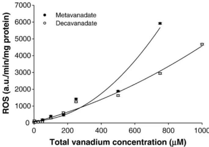
.-production in cardiac mitochondria was increased by both vanadate solutions, although monomeric vanadate showed a strong effect (Fig. 5).
3.8. Oxidative DNA damage
Oxidative DNA damage analysis shows that neither vanadate nor cadmium significantly increased 8-OhdG formation (0.62 ± 0.17 8-OhdG/105dG) in whole blood or cardiac cells, indicating that vanadium and cadmium-induced oxidative stress is not responsible for in vivo related DNA damage.
Fig. 4. Reduced glutathione (GSH) content variation (nmol GSH/mg protein) in the cardiac tissue of H. didactylus (n = 6) following 12 and 24 h in vivo exposure (intravenous administration) to vanadate and cadmium. Variation is calculated based on basal values (Section 3.6.). ⁎Significantly different from Control (P b 0.05).
Fig. 5. In vitro effects of metavanadate and decavanadate solutions on the overall rate of reactive oxygen species (ROS) production (a.u./min/mg protein) in H. didactylus cardiac mitochondria (mean ± STD; n = 6).
4. Discussion
In the present work it are described the effects of a sub-lethal concentration (5 mM) of vanadium (administered as metava-nadate or decavametava-nadate solution) and cadmium on subcellular distribution, antioxidant enzyme activity, mitochondrial ROS production and lipid peroxidation. Vanadium and cadmium intravenous injections induced metal accumulation and higher levels of antioxidant enzymes and GSH, although the degree of response differs between metals. It is also demonstrated that the administration route plays an important role in metal-induced oxidative stress and antioxidant defence responses in fish.
The LC50 values determined in the present study (Section
3.1.) are considerable higher, for both metals, than the values previous reported for other teleost fish species exposed to vanadate and cadmium chloride in the water or even upon in vivo administration: from 3.10 to 60.00 mg V/L (Catostomus latipinnis, flannelmuoth sucker; Ptychocheilus lucius, Colorado squawfish; Xyrauchen texanus, razorback sucker; Gila elegans, bonytail) (Hamilton, 1995; Hamilton and Buhl, 1997) and from 24.66 to 30.40 mg Cd/l (Poecilia reticulata, guppy; Oreo-chromis niloticus, Nile tilapia) (Yilmaz et al., 2004;
Garcia-Santos et al., 2006) or 68 mg Cd/kg fish in Oncorhynchus
mykiss (rainbow trout) (Tort and Madsen, 1991), respectively. In fact, it is well known that fish species vary in their sensitivity to metals (being different LC50values a manifestation of different
individual vulnerabilities), as well as, the route and/or the time period of exposure contribute to the magnitude of the promoted toxic effects. It is also known that the uptake of metals also depends on the route of administration and, in fact, the vanadium concentrations measured in the present study are up to 10-fold higher from those reported in the same species i.p. injected with equal metal concentration (5 mM total vanadium)
(Aureliano et al., 2002). Furthermore, vanadate and cadmium
seem to have different toxicological patterns in the heart of the toadfish, once the different metal solutions induced different patterns of subcellular metal distribution (Section 3.3.) and affected differently the antioxidant enzymes (Section 3.4.) studied. Vanadium was mainly accumulated in mitochondria and decameric vanadate increases mitochondrial antioxidant enzymes activities, while cadmium was mainly accumulated in cytosol and promotes changes only in cytosolic catalase (CAT) and glutathione peroxidases (GPx) activities.
Previous studies report opposite effects from those presented herein on antioxidant enzymes activities upon vanadate or cadmium i.p. administration in H. didactylus (Correia et al., 1998; Aureliano et al., 2002) or in S. aurata i.v. injected with 1 mM total vanadium (Soares et al., 2007d). Therefore, besides dependent on the metal administrated, the antioxidant responses induced by metal toxicity may depend on the total metal concentration administrated, on the way of exposure and/or vary between fish species, as described elsewhere (Soares et al.,
2007d). Therefore, the differences in metal accumulation and
sensitivity that arise either from differences in metabolic rates and/or from evolutionary diversity may be considered in toxicological studies. Presently, we are also interested to study pelagic marine teleosts, like gilthead seabream (Sparus aurata)
(Soares et al., 2006, 2007d), for instance, with higher
metabolism relatively to the sedentary and benthic H. didactylus, in order to correlate metal toxicity with fish metabolism.
Once no membrane lesions due to lipid peroxidation (Section 3.5) are occurring and it were observed antioxidant responses to metal-induced oxidative stress (Section 3.4.), it is suggested that, H. didactylus species has an efficient antioxidant response which confers cellular protection against either vanadate or cadmium cytotoxicity. Therefore, this fish species seems to be a good model to study biological resistance to oxidative stress injury once it has an efficient antioxidant mechanisms against vanadium and cadmium-induced oxidative stress, with no membrane lipid peroxidation detected. Similar lipid peroxida-tion results from those presented herein were report byCorreia
et al. (1998)in H. didactylus i.p. injected with 5 mM cadmium
chloride. However, toxic metal effects on lipid membrane oxidation, besides dependent on metal concentration, mode of administration, fish species and target organ, may vary with the time after administration (Gândara et al., 2005; Soares et al.,
2007d). Therefore, lipid peroxidation is not a good stress
biomarker to monitor in toxicological studies, being subcellular accumulation and antioxidant enzymes activities satisfactory approaches to oxidative stress damages.
Even though that vanadium may participate in Fenton-like reactions (Stohs and Bagchi, 1995), together with the proposed mechanisms of vanadate action that involves its bioreduction and ROS production (Capella et al., 2002; Zhang et al., 2003), previous results point out to a depression in the overall rate of ROS production (Gândara et al., 2005). This observation, and the lack of a prooxidant activity in the present work (Section 3.7.), is in good agreement with previous reports showing that vanadate supplementation diminished oxidative stress in certain experimental conditions, such as in rat-induced hepatocarcino-genesis (Chakraborty et al., 2000) and in rat diabetic tissues (Genet et al., 2002). A similar in vivo study (Gândara et al., 2005), reported an antioxidant action promoted by both vanadate oligomers (5 mM total vanadium) in liver, which was consistent with the observed increase in GSH content. However, in the present study, no vanadate or cadmium effects were detected on GSH levels upon 1 or 7 days of exposure to metal solutions (Section 3.6.).
NADH oxidase activity (Section 3.6.) and the associated O2
.-production (Section 3.7.) kept constant upon in vivo exposure to both vanadate oligomers and cadmium chloride solutions. In order to further explore the contribution of different vanadate oligomers on oxidative stress promotion, it was observed that in vitro mitochondrial O2
.-production was increased by both vanadate solutions, although monomeric vanadate showed a strong effect on cardiac mitochondria (Fig. 5). Therefore, decameric vanadate seems to induce stronger changes in antioxidant defence system in vivo (Section 3.4.), with no promotion of ROS, while in vitro metavanadate solution exhibits a higher prooxidant ability in toadfish heart (Section 3.7.). The results presented above suggested a potent in vivo blockade of ROS production in cardiac tissue by both vanadate and cadmium solutions. Therefore, total NADH oxidase activity
and linked O2.- production in cardiac muscle, upon in vivo
exposure to both vanadate or cadmium, can account for a blockade of the oxidative stress.
Altamirano-Lozano et al. showed that i.p. injection of vanadium pentoxide solutions in mice resulted in organ specific differences of DNA damage, being highest in liver, heart and kidney (Altamirano-Lozano et al., 1996, 1999). Taken together, the absence of genotoxic effects in our study does not exclude possible vanadium induced DNA alterations in other cell types.
Although biochemical data do not support oxidative stress– once there was neither lipid peroxidation nor evidences of ROS promotion – GSH enhancement and the in vitro reported prooxidant activity corroborate the mitochondrial function impairment reported (Soares et al., 2007b). In fact, in a previous in vivo study it was suggested that, besides accumulated in mitochondria, decameric vanadate inhibits mitochondrial oxygen consumption (IC50= 400 nM) and induces membrane
depolariza-tion (IC50= 196 nM) more strongly than monovanadate (IC50of 23
and 55μM, respectively), through the promotion of changes in the redox steady-state of complex III, pointing out that mitochondria is a toxicological target for decameric vanadate and the importance to take into account the contribution of decameric vanadate species to the vanadate toxic effects (Soares et al., 2007b).
In conclusion, both vanadium and cadmium significantly affected cardiac antioxidant enzymes activities in vivo, besides not inducing ROS production. However, they have different intracel-lular targets and affect differently antioxidant defence mechan-isms. Because the results obtained in the present study (i.v. administration) differ from the obtained with i.p. administrations of metal solutions and from those obtain with different fish species, it is suggested that the administration method, as well as, the fish metabolic rate are determinant in oxidative stress responses induced by vanadium and cadmium and it should be taken into account when interpreting and comparing results from in vivo metal exposure. Moreover, the i.v. administration seems to be the more reliable route of intoxication once the drug is injected directly in the blood stream, and not in the body peritoneal cavity, as it happens with the i.p. injection. Furthermore, besides decameric vanadate seems to induce in vivo stronger changes in antioxidant defence response system, in vitro assay pointed that both vanadate oligomers induce ROS production in the toadfish heart, exhibiting monomeric vanadate an higher prooxidant ability than decameric species. Considering the contradictory effects observed in vivo and in vitro, we intent to further explore this issue to clarify the involvement of different vanadate oligomers in oxidative stress induced by vanadium.
Acknowledgements
This work was support by Joint Spanish-Portuguese Grant HP2004-0080 (to M.A.), by POCTI program funded through FEDER for the research project POCTI/38191/QUI/2001 (to M.A.). The authors thank to Joint Portuguese-Spanish CRUP project E-106/05. S.S. Soares was supported by a PhD grant [SFRH/BD/8615/2002] from the Portuguese Foundation for Science and Technology (FCT). The authors gratefully acknowledge to Professor José J. G. Moura the collaboration
in the present work, allowing the NMR studies at the Laboratório de Ressonância Magnética Nuclear, REQUIMTE, Departamento de Química, Universidade Nova de Lisboa. References
Al-Saleh, I., Shinwari, N., 2002. Preliminary report on the levels of elements in four fish species from the Arabian Gulf of Saudi Arabia. Chemosphere 48, 749–755.
Altamirano-Lozano, M., Alvarez-Barrera, L., Basurto-Alcantara, F., Valverde, M., Rojas, E., 1996. Reprotoxic and genotoxic studies of vanadium pentoxide in male mice. Teratog. Carcinog. Mutagen. 16, 7–17.
Altamirano-Lozano, M., Valverde, M., Alvarez-Barrera, L., Molina, B., Rojas, E., 1999. Genotoxic studies of vanadium pentoxide (V(2)O(5)) in male mice. II. Effects in several mouse tissues. Teratog. Carcinog. Mutagen. 19, 243–255. Amado, A., Aureliano, M., Ribeiro-Claro, P.J., Teixeira-Dias, J., 1993. Combined
Raman and 51V NMR spectroscopic study of vanadium (V) oligomerization in aqueous alkaline solutions. J. Raman Spectrosc. 24, 669–703.
Auclair, C., Voisin, E., 1985. Nitroblue tetrazolium reduction. In: Greenwald, R.A. (Ed.), Handbook of methods for oxygen radical research. CRC Press Inc., Boca Raton, Florida, pp. 123–132.
Aureliano, M., Madeira, V.M.C., 1994. Interactions of vanadate oligomers with sarcoplasmic reticulum Ca2+-ATPase. Biochim. Biophys. Acta 1221, 259–271.
Aureliano, M., Madeira, V.M.C., 1998. Energy transduction mechanisms as affected by vanadium(V) species: Ca2+-pumping in sarcoplasmic reticulum.
In: Nriagu, J.O. (Ed.), Vanadium in the environment, Part 1: Chemistry and Biochemistry. John Wiley & Sons, Inc., New York, pp. 333–357. Aureliano, M., Gândara, R.M.C., 2005. Decavanadate effects in biological
systems. J. Inorg. Biochem. 99, 979–985.
Aureliano, M., Joaquim, N., Sousa, A., Martins, H., Coucelo, J.M., 2002. Oxidative stress in toadfish (Halobatrachus didactylus) cardiac muscle: acute exposure to vanadate oligomers. J. Inorg. Biochem. 90, 159–165. Aureliano, M., Soares, S.S., Tiago, T., Ramos, S., Gutiérrez-Merino, C., 2007.
Biological effects of decavanadate: muscle contraction, mitochondrial toxicity and in vivo oxidative stress. ACS Symposium Series 974, 249–263. Bagchi, D., Vuchetich, P., Bagchi, M., Hassoun, E., Tran, M., Tang, L., Stohs, S.J., 1997. Induction of oxidative stress by chronic administration of sodium dichromate [chromium VI] and cadmium chloride [cadmium II] to rats. Free Radic. Biol. Med. 22, 471–478.
Beg, M.U., Al-Muzaini, S., Saeed, T., Jacob, P.G., Beg, K.R., Al-Bahloul, M., Al-Matrouk, K., Al-Obaid, T., Kurian, A., 2001. Chemical contamination and toxicity of sediment from a coastal area receiving industrial effluents in Kuwait. Arch. Environ. Contam. Toxicol. 41, 289–297.
Borges, G., Mendonça, P., Joaquim, N., Aureliano, M., Coucelo, J.M., 2003. Acute effects of vanadate oligomers on heart, kidney, and liver histology in the Lusitanian toadfish (Halobatrachus didactylus). Arch. Environ. Contam. Toxicol. 45, 415–422.
Bradford, M., 1976. A rapid and sensitive method for the quantification of microgram quantities of protein utilizing the principle of protein-dye binding. Anal. Biochem. 72, 248–254.
Byczkowski, J.Z., Kulkarni, A.P., 1998. Oxidative stress and pro-oxidant biological effects of vanadium. In: Nriagu, J.O. (Ed.), Vanadium in the environment, Part 1: Chemistry and Biochemistry. John Wiley & Sons, Inc., New York, pp. 235–263.
Capella, L., Gefé, M., Silva, E., Affonso-Mitidieri, E., Lopes, A., Rumjanek, V., Capella, M., 2002. Mechanisms of vanadate-induced cellular toxicity: role of cellular glutathione and NADPH. Arch. Biochem. Biophys. 406, 65–72. Chakraborty, A., Selvaraj, S., Sudarshan, M., Dutta, R.K., Ghugre, S.S., Chintalapudi, S.N., 2000. Modulatory role of vanadium on trace element profile in diathylnitrosamine-induced rat hepatocarcinogenesis. Nucl. Instrum. Methods Phys. Res., Sect. B 170, 156–162.
Chasteen, N., 1983. The biochemistry of vanadium. Struct. Bond. (Berl.) 53, 105–138.
Clairborne, A., 1985. Catalase activity. In: Greenwald, R.A. (Ed.), Handbook of methods for oxygen radical research. CRC Press, Boca Raton- Florida, pp. 283–284.
Correia, V., Joaquim, N., Coucelo, J.M., Azevedo, J., Coucelo, J.A., 1998. [Cádmio e célula muscular cardíaca– Biomarcadores de stress oxidativo – (Trabalho experimental)] Portuguese. Rev. Port. Cardiol. 17, 911–916. Coucelo, J.M., Joaquim, N., Correia, V., Azevedo, J., Coucelo, J.A., 1998.
[Análise histológica de alterações induzidas experimentalmente por cádmio no tecido muscular cardíaco através de método computorizado de análise de imagem] Portuguese. Rev. Port. Cardiol. 17, 735–740.
Crans, D.C., 1994. Aqueous chemistry of labile oxovanadate: relevance to biological studies. Comments Inorg. Chem. 16, 1–33.
Crans, D.C., 1995. Interaction of vanadates with biogenic ligands. Met. Ions Biol. Syst. 31, 147–209.
Domingo, J.L., 1996. Vanadium: a review of the reproductive and development toxicity. Reprod. Toxicol. 10, 175–182.
Dong, W., Simeonova, P.P., Gallucci, R., Matheson, J., Wang, S., Hubbs, A., Luster, M.I., 1998. Toxic metals stimulate inflammatory cytokines in hepatocytes through oxidative stress mechanisms. Toxicol. Appl. Pharmacol. 151, 359–366.
Gândara, R.M.C., Soares, S.S., Martins, H., Gutiérrez-Merino, C., Aureliano, M., 2005. Vanadate oligomers: In vivo effects in hepatic vanadium accumulation and stress markers. J. Inorg. Biochem. 99, 1238–1244. Garcia-Santos, S., Fontainhas-Fernandes, A., Wilson, J.M., 2006. Cadmium
tolerance in the Nile tilapia (Oreochromis niloticus) following acute exposure: assessment of some ionoregulatory parameters. Environ. Toxicol. 21, 33–46. Genet, S., Kale, R.K., Baquer, N.Z., 2002. Alterations in antioxidant enzymes and oxidative damage in experimental diabetic rat tissues: effect of vanadate and fenugreek (Trigonellafoenum graecum). Mol. Cell. Biochem. 236, 7–12. Günzler, W.A., Flohé, L., 1985. Glutathione peroxidase. In: Greenwald, R.A. (Ed.), Handbook of methods for oxygen research. CRC Press, Boca Raton, FL, pp. 285–290.
Hamilton, S.J., 1995. Hazard assessment of inorganics to three endangered fish in the Green River, Utah. Ecotoxicol. Environ. Saf. 30, 134–142. Hamilton, S.J., Buhl, K.J., 1997. Hazard evaluation of inorganics, singly and in
mixtures, to flannelmouth sucker Catostomus latipinnis in the San Juan River, New Mexico. Ecotoxicol. Environ. Saf. 38, 296–308.
Harland, B., Harden-Williams, B., 1994. Is vanadium of human nutritional importance yet? J. Am. Diet. Assoc. 94, 891–894.
Hu, H., 2000. Exposure to metals. Occup. Environ. Med. 27, 983–996. Ivancsits, S., Pilger, A., Diem, E., Schaffer, A., Rüdiger, H.W., 2002. Vanadate
induces DNA strand breaks in cultured human fibroblasts at doses relevant to occupational exposure. Mutat. Res. 519, 25–35.
Kelly, K., Havrilla, C., Brady, T., Abramo, K., Levin, E., 1998. Oxidative stress in toxicology: established mammalian and emerging piscine models. Environ. Health Perspect. 106, 375–384.
Kock, G., Hofer, R., 1998. Origin of cadmium and lead in clear softwater lakes of high-latitude, and their bioavailability and toxicity to fish. In: Braunbeck, T., Hinton, D.E., Streit, B. (Eds.), Fish ecotoxicology. Birkhause Verlag, Boston, pp. 64–77.
Kostic, M.M., Ognjanovi, B.I., Dimitrijevic, S., Zikic, R.V., Stajn, A.S., Rosic, G.L., Zivkovic, R.V., 1993. Cadmium-induced changes of antioxidant and metabolic status in red blood cells of rats: in vivo effects. Eur. J. Haematol. 51, 86–92.
Kraal, M.H., Kraak, M.H., de Groot, C.J., Davids, C., 1995. Uptake and tissue distribution of dietary and aqueous cadmium by carp (Cyprinus carpio). Ecotoxicol. Environ. Saf. 31, 179–183.
Lawrence, R.A., Burk, R.F., 1976. Glutathione peroxidase activity in selenium-deficient rat liver. Biochem. Biophys. Res. Commun. 71, 952–958. Martín-Romero, F.J., Gutiérrez-Martín, Y., Henao, F., Gutiérrez-Merino, C.,
2002. The NADH oxidase activity of the plasma membrane of synaptosomes is a major source of superoxide anion and is inhibited by peroxynitrite. J. Neurochem. 82, 604–614.
Miliou, H., Zaboukas, N., Moraitou-Apostolopoulou, M., 1998. Biochemical composition, growth, and survival of the guppy, Poecilia reticulata, during chronic sublethal exposure to cadmium. Arch. Environ. Contam. Toxicol. 35, 58–63.
Miramand, P., Fowler, S.W., 1998. Bioaccumulation and transfer of vanadium in marine organisms. In: Nriagu, J.O. (Ed.), Vanadium in the environment. Part 1: Chemistry and Biochemistry. John Wiley & Sons, Inc., New York, pp. 167–197.
Nechay, B., Nanninga, L., Nechay, P., Post, R., Grantham, J., Macara, I., Kubena, L., Philips, T., Nielsen, F., 1986. Role of vanadium in biology. Fed. Proc. 45, 123–132.
Palace, V., Klaverkamp, J., 1993. Variation of hepatic enzymes in three species of freshwater fish from precambrian shield lakes and the effect of cadmium exposure. Comp. Biochem. Physiol. 104C, 147–154.
Palace, V., Majewski, H., Klaverkamp, J., 1993. Interactions among antioxidant defences in liver of rainbow trout (Oncorhynchus mykiss) exposed to cadmium. Can. J. Fish. Aquat. Sci. 50, 156–162.
Pruell, R.J., Engelhardt, F.R., 1980. Liver cadmium uptake, catalase inhibition and cadmium thionein production in the killifish (Fundulus heteroclitus) induced by experimental cadmium exposure. Mar. Environ. Res. 3, 101–111. Rao, A.V., Ramasarma, T., 2000. NADH-dependent decavanadate reductase, an
alternative activity of NADP-specific isocitrate dehydrogenase protein. Biochim. Biophys. Acta 1474, 321–330.
Reed, L.J., Muench, H., 1938. A simple method of estimating fifty per cent end-points. Am. J. Hyg. 27, 493–497.
Ricard, A.C., Daniel, C., Anderson, P., Hontela, A., 1998. Effects of subchronic exposure to cadmium chloride on endocrine and metabolic functions in rainbow trout Oncorhynchus mykiss. Arch. Environ. Contam. Toxicol. 34, 377–381. Risso-de Faverney, C., Orsini, N., de Sousa, G., Rahmani, R., 2004.
Cadmium-induced apoptosis through the mitochondrial pathway in rainbow trout hepatocytes: involvement of oxidative stress. Aquat. Toxicol. 69, 247–258. Sarkar, S., Yadav, P., Bhatnagar, D., 1998. Lipid peroxidative damage on cadmium exposure and alterations in antioxidant system in rat erythrocytes: a study with relation to time. Biometals 11, 153–157.
Sarkar, S., Yadav, P., Trivedi, R., Bansal, A.K., Bhatnagar, D., 1995. Cadmium-induced lipid peroxidation and the status of the antioxidant system in rat tissues. J. Trace Elem. Med. Biol. 9, 144–149.
Shigenaga, M.K., Park, J.W., Cundy, K.C., Gimeno, C.J., Ames, B.N., 1990. In vivo oxidative DNA damage: measurement of 8-hydroxy-2 V-deoxyguano-sine in DNA and urine by high-performance liquid chromatography with electrochemical detection. Methods Enzymol. 186, 521–530.
Shukla, G.S., Chiu, J., Hart, B.A., 2000. Cadmium-induced elevations in the gene expression of the regulatory subunit of gamma-glutamylcysteine synthetase in rat lung and alveolar epithelial cells. Toxicology 151, 45–54. Soares, S.S., Aureliano, M., Joaquim, N., Coucelo, J.M., 2003. Cadmium and vanadate oligomers effects on methaemoglobin reductase activity from Lusitanian toadfish: in vivo and in vitro studies. J. Inorg. Biochem. 94, 285–290. Soares, S.S., Martins, H., Aureliano, M., 2006. Vanadium distribution following decavanadate administration. Arch. Environ. Contam. Toxicol. 50, 60–64. Soares, S.S., Gutiérrez-Merino, C., Aureliano, M., 2007a. Decavanadate induces
mitochondrial membrane depolarization and inhibits oxygen consumption. J. Inorg. Biochem. 101, 189–196.
Soares, S.S., Gutiérrez-Merino, C., Aureliano, M., 2007b. Mitochondria as a target for decavanadate toxicity in Sparus aurata heart. Aquat. Toxicol. 83, 1–9. Soares, S.S., Gutiérrez-Merino, C., Aureliano, M., 2007c. Decavanadate toxicity
effects following in vivo administration. In: Aureliano, M. (Ed.), Vanadium compounds/vanadate oligomers in biological systems: Chemistry, Biochem-istry and Biological effects. Research Signpost, India, pp. 167–197. Soares, S.S., Martins, H., Duarte, R.O., Moura, J.J.G., Coucelo, J.,
Gutiérrez-Merino, C., Aureliano, M., 2007d. Vanadium distribution, lipid peroxidation and oxidative stress markers upon decavanadate in vivo administration. J. Inorg. Biochem. 101, 80–88.
Soares, S.S., Henao, F., Aureliano, M., Gutiérrez-Merino, C., submitted for publication. Vanadate-induced necrotic death in neonatal rat cardiomyocytes through mitochondrial membrane depolarization.
Stohs, S.J., Bagchi, D., 1995. Oxidative mechanisms in the toxicity of metal ions. Free Radic. Biol. Med. 18, 321–336.
Tiago, T., Aureliano, M., Gutiérrez-Merino, C., 2004. Decavanadate binding to a high affinity site near the myosin catalytic centre inhibits F-actin-stimulated myosin ATPase activity. Biochemistry 43, 5551–5561.
Tort, L., Madsen, L., 1991. The effects of heavy metals cadmium and zinc on the contraction of ventricular fibres in fish. Comp. Biochem. Physiol. 99C, 353–356. Tyurina, Y.Y., Shvedova, A.A., Kawai, K., Tyurin, V.A., Kommineni, C., Quinn, P.J., Schor, N.F., Fabisiak, J.P., Kagan, V.E., 2000. Phospholipid signaling in apoptosis: peroxidation and externalization of phosphatidylserine. Toxicology 148, 93–101.
Vaglio, A., Landriscina, C., 1999. Changes in liver enzyme activity in the teleost Sparus aurata in response to cadmium intoxication. Ecotoxicol. Environ. Saf. 43, 111–116.
Valko, M., Morris, H., Cronin, M.T., 2005. Metals, toxicity and oxidative stress. Curr. Med. Chem. 12, 1161–1208.
Verbost, P.M., Flik, G., Pang, P.K., Lock, R.A., Wendelaar Bonga, S.E., 1989. Cadmium inhibition of the erythrocyte Ca2+ pump. A molecular
interpretation. J. Biol. Chem. 264, 5613–5615.
Viarengo, A., 1989. Biochemical effects of trace metals. Mar. Pollut. Bull. 16, 153–158.
Wang, R., Wang, X.T., Wu, L., Mateescu, M.A., 1999. Toxic effects of cadmium and copper on the isolated heart of dogfish shark, Squalus acanthias. J. Toxicol. Environ. Health 57, 507–519.
Wills, D., 1987. Evaluation of lipid peroxidation in lipids and biological membranes. In: Snell, K., Mullock, B. (Eds.), Biochemical toxicology: A pratical approach. IRL Press, Oxford, pp. 127–152.
Yilmaz, M., Gul, A., Karakose, E., 2004. Investigation of acute toxicity and the effect of cadmium chloride (CdCl2d H2O) metal salt on behavior of the
guppy (Poecilia reticulata). Chemosphere 56, 375–380.
Zaporowska, H., Wasilewski, W., 1992. Haematological effects of vanadium on living organisms. Comp. Biochem. Physiol. 102C, 223–231.
Zhang, Z., Huang, C., Li, J., Leonard, S.S., Lanciotti, R., Butterworth, L., Shi, X., 2001. Vanadate-induced cell growth regulation and the role of reactive oxygen species. Arch. Biochem. Biophys. 392, 311–320.
Zhang, Z., Leonard, S., Huang, C., Vallyathan, V., Castranova, V., Shi, X., 2003. Role of reactive oxygen species and MAPKS in vanadate-induced G(2)/M phase arrest. Free Radic. Biol. Med. 34, 1333–1342.
Zikic, R.V., Stajn, A.S., Ognjanovic, B.I., Saicic, Z.S., Kostic, M.M., Pavlovic, S.Z., Petrovic, V.M., 1998. The effect of cadmium and selenium on the antioxidant enzyme activities in rat heart. J. Environ. Pathol. Toxicol. Oncol. 17, 259–264. Zychlinski, L., Byczkowski, J.Z., Kulkarni, A.P., 1991. Toxic effects of
long-term intratracheal administration of vanadium pentoxide in rats. Arch. Environ. Contam. Toxicol. 20, 295–298.