Vol-7, Special Issue-Number5-July, 2016, pp310-317 http://www.bipublication.com
Research Article
A comparison between gray and white mineral trioxide aggregate (MTA) and
calcium enriched mixture (CEM) cytotoxicity in L929 and Saos-2 cell lines
Robab Farhang1, Maryam Ehsani1, Ebrahim Zabihi2,3, Fatemeh Vejdani1, Maryam Raoof4,
Nayyer Seifizadeh5, Asiyeh Khalilpoor6 and Niusha Hajizadeh1*
1
Department of Endodontics, School of Dentistry, Babol University of Medical Sciences, Babol, Iran & Dental Materials Research Center,
3Department of Pharmacology & Physiology, School of Medicine,
Babol University of Medical Sciences, Babol, Iran.
School of Dentistry, Babol University of Medical Sciences, Babol, Iran.
2
Cellular and Molecular Biology Research Center, Babol University of Medical Sciences, Babol, Iran
4Department of Endodontics, School of Dentistry,
Kerman University of Medical Sciences, Kerman, Iran
5Department of Biochemistry, School of Medicine,
Babol University of Medical Sciences, Babol, Iran.
6Department of Hematology, School of Paramedicine,
Babol University of Medical Sciences, Babol, Iran. *Corresponding Author: Niusha Hajizadeh
ABSTRACT
The purpose was to compare the cytotoxic effects of two commonly used mineral trioxide aggregate (MTA) (gray & white) with the recently introduced calcium enriched mixture (CEM) in L929 and Saos-2 cell lines and to determine the oxidative stressinduced by each cement. Sterile discs of each cement were submerged and incubated in complete media for 24, 48 and 72-h. The cell lines were exposed to two fold serial dilutions (1, 0.5, 0.25 X) of each extract. In addition to trypan blue (TB) dye exclusion test, MTT assay was carried out after 24-h. To determine the oxidative stress mediated cell toxicity, malondialdehyde (MDA) concentration and total ferric reduction activity potential (FRAP) were measured in the media.The MTT and TB results with 48-h and 72-h CEM extracts showed that both extracts are cytotoxic in L929 cell line at 1 X and 0.5 X dilutions, while neither white MTA (WMTA) nor gray MTA (GMTA) showed any toxic effects in this cell line. On the other hand all three cements (undiluted 48-h and 72-h extracts) were cytotoxic against Saos-2 cell line. The oxidative stress marker (MDA) was proportionate to the cytotoxic effects of CEM while there was no significant changes in reductive capacity of the media measured by FRAP in both cell lines.The cytotoxic effects of CEM on L929 and Saos-2 cell lines seems to be higher than WMTA and GMTA and it is increased by the time of extraction up to 72-h.. However, CEM, WMTA and GMTA showed similar toxic effect in Saos-2 cell line proportionate to the oxidative stress measured by TBARS. It seems that while all three cements extracts don't change the antioxidant activity of the media, they could significantly increase lipid peroxidation, especially CEM with Saos-2 cell line which elucidate triggering oxidative stress mechanisms without having direct oxidative activity.
Key words: Cytotoxicity, CEM, MTA,L929 cell line, Saos-2 cell line
INTRODUCTION:
The root canal filling materials seal off the root of the teeth and prevent bacterial penetration and
An ideal root-end filling material must be biocompatible, antibacterial, nontoxic and radio-opaque. Moreover, it should be non-soluble and non-absorbable in the oral cavity. It should also be economical, easy to use and compatible with the oral and periradicular tissues (1-2). Original
mineral trioxide aggregate (OMTA) was
introduced by Torabinejad in Loma Linda university as a treatment for root canal perforation treatment (3-4). Some good properties of OMTA are its biocompatibility, low cytotoxicity, low moisture sensitivity and inducing hard tissue formation (5). These characters make MTA as a selective material for root canal filling, direct pulp capping, pulpotomy and root opacification (6). On the other hand, MTA is difficult to manipulate and has low radiopacity. Also it is expensive and it hardens slowly. The search for finding better materials for this aim is ongoing. The CEM is one of newly introduced material that contains calcium compound substances such as calcium phosphate, calcium sulphate, calcium hydroxide, calcium oxide, calcium silicate and calcium carbonate (7-8). CEM has shown some advantages such as
antibacterial property, tissue compatibility,
capability of inducing hard tissue formation, one session treatment, easy to use, non sensitivity to blood and saliva, stable color and low price (7). In this study, we aimed to compare the cytotoxicity induced by WMTA, GMTA and CEM on two cell lines and its correlation to lipid peroxidation and oxidative stress.
MATERIALS AND METHODS Cements extraction
According to the manufacturer's instructions, white and gray MTA-Angelus (WMTA & GMTA) (Angelus, Londrina, Brazil) powders were mixed with distilled water and CEM powder (Asgari, Shahid Beheshti University, Iran) was
99% humidity and 5% CO2 under sterile condition.
Using a 24-well cell culture plate, each disk was submerged in 1ml complete medium and incubated at 3 different time of extraction (24-h., 48-h. or 72-h.). The corresponding supernatant of each well was considered as 1 X dilution (concentration). To prepare other dilutions (0.5
and 0.25 X), either 500 µl or 250 µl of this supernatant was added to 750 µl and 500 µl of
complete medium respectively. Cell culture
The L929 and Saos-2 cell lines (Pasteur institute, Tehran, Iran) were cultured in 75 cm2 flask with Roswell Park Memorial Institute(RPMI)-1640 medium containing L-glutamine (PAA cell culture company, Austria), supplemented with 10% fetal bovine serum (FBS; Gibco BRL,USA) and 1% PenStrep® (100U/ml penicillin, 10 mg/ml Streptomycine) (Sigma-Aldrich, Germany). After the cells were grown to 75-80% of confluence, they were washed with PBS and detached using 0.25% trypsin/EDTA and the cell count was performed. Then, 1ml of Saos-2 cell suspension (1.3×10⁴ cells per ml) or 1 ml of L929 cell suspension (1.2×10⁴ cells per ml) were seeded in 24-well culture plate and incubated. The supernatant was replaced after 24-h with different concentrations of each cements extracts. Complete culture medium without extracts was added to 3 wells of confluent cells as control group. All
plates were incubated in 37˚C, 99% humidity and
5% CO2.
Viability test using MTT assay
The supernatant of each well was collected after 24h and cells were rinsed by PBS. Subsequently,
200 µl of MTT solution (5mg/ml) were added to
each well and incubated for 4-h in 37◦C. Finally,
the purple coloured formazan precipitate was
dissolved in 800 µl of acidic isopropyl alcohol
as ΔOD of each well divided by the mean of ΔOD
of the corresponding control group×100. Viability test using trypan blue assay
Trypan blue (TB) dye exclusion test was carried out only at 1 X dilution of each cements extract. Following 24-h exposure of 75% confluent cells with extracts in a 6 wells plate, the number of viable cells was counted using 0.4% TB.
Determination of malondialdehyde (MDA) concentration
After exposure to different cement extracts, the MDA concentration (as a marker of lipid peroxidation) in both cell lines supernatants was
measured by thiobarbituric acid reactive
substances (TBAR) assay, according to the method of Ohkawa (9).
Determination of ferric reducing potential (FRAP) of medium
To measure the total antioxidant power of the medium, the FRAP assay was performed on cell culture medium according to the method of Benzie and Strain (10)(Benzie & Strain, 1996). The antioxidant power of each medium sample was reported as micromolar equivalent of FeSO4 (mg/dl).
Statistical Analysis
Each dilution of cements' extract has been tested in triplicate and the results are presented as mean ± standard error. Means were compared between the groups using Student’s t-test, one-way analysis of variance (ANOVA) and Tukey’s post hoc tests. Statistical significance was set at p < 0.05.
RESULTS:
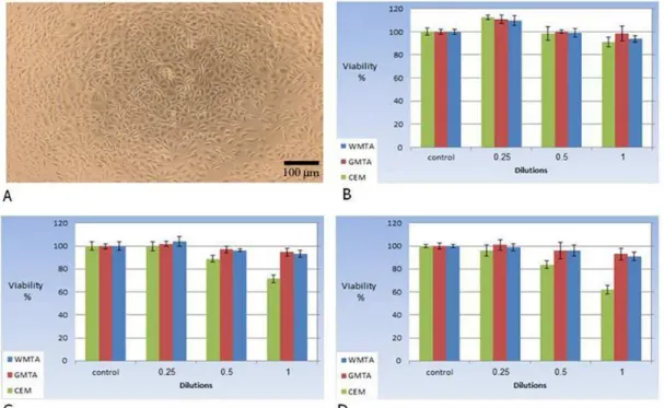
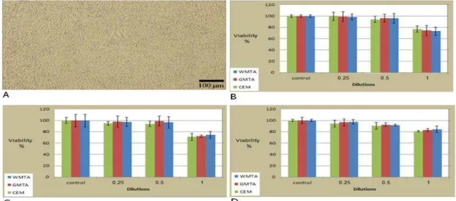
Comparision of L929 and Saos-2viability after exposure to three cements extracts
The MTT results for all three cements extracts (at 3 different extraction times and 3 different dilutions) after exposure to either L929 or Saos-2 cell lines are depicted in Fig. 1 and Fig. 2 respectively. The viability of L929 cells after exposure to 0.25 X dilution of all 3 cements extracts (with 24-h extraction time) had a minor increase compared to the control group. There is no significant difference between the viability of control group and 24-h extracts of the other 3 cements at 0.5 X and 1 X dilutions in L929 cell line (Fig. 1B).
Evaluation of CEM cytotoxicity against L929 and Saos-2 cell lines
There was no significant difference between the control group and 48-h extracts of WMTA and GMTA. However, CEM extract at 0.5 X and 1 X dilution showed significant cytotoxicity compared to the control group (Fig. 1C). Also there was no significant difference between the control group and 72-h extracts of WMTA and GMTA. However, CEM extract at 0.5 X and 1 X dilution showed significant cytotoxicity compared to the control group (Fig. 1D) too.All 3 tested cements extracts at 0.25 X dilution showed less cytotoxicty compared to their 1 X dilution.
Cytotoxicity assay on Saos-2 cell line showed that among all 24-h extracts, only1 X dilution was significantly toxic (Fig. 2B). Same results were observed with 48-h and 72-h extracts (Fig. 2C& 2D) The results of trypan blue assay, performed with undiluted (1 X) cement extracts, showed that by increasing the extraction time from 24 to 72hours, the cytotoxicity of the extracts had increased in both cell lines (Fig. 3A & 3B).
Figure 3: (A)The effect of extraction time on L929 cell viability after exposure to 1 X cements extracts using TB assay. (B) The effect of extraction time on Saos-2 cell viability after exposure to 1 X cements extracts using TB assay.
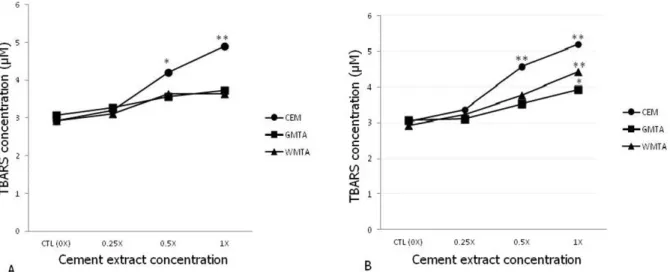
Elevated lipid proxidation in L929 and Saos-2 cell lines by CEM
The result of MDA measurement after exposure to different dilution of the cement extracts in the culture medium of L929 and Saos-2 cell lines are depicted in Figure4. It seems that in both cell lines, CEM extract could significantly increase the level of MDA at lower concentrations compared to the other two cements (Fig. 4A & 4B).
Measure the total antioxidant power of the all cement extract showed,no significant changes in the total antioxidant power of culture medium (between tested cement extracts and the control) that were observed in Figure 5A & 5B.
Figure 5: Culture medium antioxidant power (FRAP assay) (equivalent to µM FeSO4) for L929 (A) and Saos-2 (B) cell lines after exposure to different dilution of 3 cements.
DISCUSSION:
In the current study, in vitro cytotoxicity and oxidative stress induced by two highly used mineral trioxide aggregate cements (WMTA and GMTA) were compared to the recently invented calcium enriched material (CEM) in two different cell lines. The 72-h extract of CEM seems to be able to induce cell toxicity in both L929 and Saos-2 cell lines at much lower concentrations compared to the other two cements (WMTA & GMTA), (Fig. 1D & 2C). This cytotoxic effect seems to be proportionate to CEM capacity to
indirect oxidative stress induced by the cements extracts.
Having low cytotoxicity against periapical tissues is one of the main criteria for a good endodontic material. The release of cytotoxic substances from hardened cements could be a time dependant phenomenon (11). Endodontic materials with higher cytotoxicity have greater chance to lead to degeneration, necrosis or late recovery of the periapical tissues (12-13).
properties compared to MTA (16), including the ability to induce hard tissue formation (17), settable in moist conditions (8) and having the sealing ability comparable to MTA and better than IRM (18).
Although, there is still no consensus on the ideal assay procedure for comparing different dental materials cytotoxicity against apical tissues, the in vitro tests (including the MTT assay on normal and cancerous cell lines) using set materials extracts, have been extensively used (14, 19-23). According to ISO standards, which determine instructions for dental material assessment, endodontic sealer samples must be submerged in culture medium for 1-3 days. Moreover, the ratio of tested material surface area to the extraction medium volume is another important factor affecting the cytotoxicty assessment which should be about 0.5 -0.6 cm2/ml (19). In this study, the surface area of disc shaped set cements were 42 mm2 extracted by 1 ml complete medium for 24, 48 and 72-h. The time and exposure surface area have profound effects on the amount of cytotoxic materials released into the extraction medium (13). In some older studies, the cements cytotoxicities have been assessed using freshly mixed materials (5, 21, 24) however in more recent studies, it is the set form of the cements which are mostly used (11, 14, 25-26). Since the surface-area/extraction-volume ratio for all three cements disks has been the same, the extraction time seems to have more important effects on the observed cytotoxicities. The cytotoxicity of CEM seems to be increased more than two other cements which indicates slower leakage of its cytotoxic components into the medium (Figs 1D& 2D). Although this sustained release of cytotoxic substances might lead to higher cytotoxic effects in vitro but with in vivo interstitial fluid drainage, the CEM cytotoxicity might be alleviated in clinical use. The cytotoxic effects of all 3 cement extracts seems to be highly dose dependent and non of these tested materials showed cytotoxic effects at 2 times dilutions (0.5 X) which might signify the importance of periapical fluid dilution
and its drainage in the assessment of these cements biocompatibility. In fact a mild increase in the mean of L929 cell viability after exposure to low concentration (0.25 X) of 24-h extracts (Fig. 1B) might be of benefit in tissue repair after root canal procedures by these cements especially by CEM.
The two cell lines used in this study, L929 a fibroblast derived cell line and Saos-2 an osteosarcoma derived cell line are the most relevant models in vitro to periapical and dental tissues (26). These two cell lines have shown
somehow similar sensitivity to cytotoxic
substances (27). However, having data of these two cell lines with different origin might be useful for a better comparison of the cements biocompatibility.
The MTT results of both cell lines indicate that despite CEM undiluted extract (1X) shows a bit more cytotoxic effect compared to GMTA and WMTA, at lower concentrations there is no significant difference (Fig. 1 & 2). With the in vivo constant dilution and tissue fluids drainage into the blood, the cytotoxicity of CEM doesn’t seem to be an important issue while its other physicochemical properties might be useful. The results of TB assays show that the difference between these 3 cements is not significant (Fig. 3A & 3B).
Oxidative stress has been considered as an important mechanism for cytotoxicty of xenobitics
including dental materials (28). Cytotoxic
significant oxidative stress induced by high concentration of the cements extracts (1 X) especially with CEM (Fig.4A&4B). It seems that while CEM extract show a bit higher cytotoxicity in both tested cell lines (L929 and Saos-2) compared to WMTA and GMTA, this difference is not significant at lower dilutions and practically CEM extract might have positive proliferative effects on these two cell lines. All three cements induce their cytotoxic effects, at least partly, through indirect oxidative stress and lack direct oxidative capacity.
Conflict of interest statement
The authors have declared no conflict of interest.
ACKNOWLEDGMENT
The authors like to acknowledge Shahid Beheshti Dental Research Center, Cellular and Molecular Biology Research Center of Babol University of Medical Sciences and Dental Materials Research Center of School of Dentistry of Babol.
REFERENCES:
1. Johnson B, Whierspoon D. Periradicular
surgery. Pathways of the pulp. 2006:724-85.
2. Gartner AH, Dorn SO. Advances in endodontic
surgery. Dent Clin North Am. 1992
Apr;36(2):357-78.
3. Lee SJ, Monsef M, Torabinejad M. Sealing
ability of a mineral trioxide aggregate for repair of lateral root perforations. J Endod. 1993 Nov;19(11):541-4.
4. Torabinejad M, Watson TF, Pitt Ford TR.
Sealing ability of a mineral trioxide aggregate when used as a root end filling material. J Endod. 1993 Dec;19(12):591-5.
5. Torabinejad M, Chivian N. Clinical
applications of mineral trioxide aggregate. J Endod. 1999 Mar;25(3):197-205.
7. Asgary S, Eghbal MJ, Parirokh M, Torabzadeh
H. Sealing ability of three commercial mineral trioxide aggregate and an experimental root-end filling material. Iranian Endodontic Journal. 2006;1:101-5.
8. Asgary S, Shahabi S, Jafarzadeh T, Amini S, Kheirieh S. The properties of a new endodontic material. J Endod. 2008 Aug;34(8):990-3.
9. Ohkawa H, Ohishi N, Yagi K. Assay for lipid
peroxides in animal tissues by thiobarbituric
acid reaction. Anal Biochem. 1979
Jun;95(2):351-8.
10.Benzie IF, Strain JJ. The ferric reducing ability of plasma (FRAP) as a measure of "antioxidant power": the FRAP assay. Anal Biochem. 1996 Jul 15;239(1):70-6.
11.Badole GP, Warhadpande MM, Meshram GK,
Bahadure RN, Tawani SG, Tawani G, et al. A comparative evaluation of cytotoxicity of root canal sealers: an in vitro study. Restor Dent Endod. 2013 Nov;38(4):204-9.
12.Ghoddusi J, Tavakkol Afshari J, Donyavi Z,
Brook Z, Disfani R, Esmaeelzadeh M. Cytotoxic effect of a new endodontic cement and mineral trioxide aggregate on L929 line
culture. Iranian Endodontic Journal.
2008;3(2):17-23.
13.Spangberg L, Pascon EA. The importance of
material preparation for the expression of cytotoxicity during in vitro evaluation of biomaterials. J Endod. 1988 May;14(5):247-50.
14.Alanezi AZ, Jiang J, Safavi KE, Spangberg LS,
Zhu Q. Cytotoxicity evaluation of
endosequence root repair material. Oral Surg Oral Med Oral Pathol Oral Radiol Endod. 2010 Mar;109(3):e122-5.
end filling material. J Biomed Mater Res A. 2008 Dec 1;87(3):706-9.
17.Asgary S, Eghbal MJ, Parirokh M, Ghanavati
F, Rahimi H. A comparative study of histologic response to different pulp capping materials and a novel endodontic cement. Oral Surg Oral Med Oral Pathol Oral Radiol Endod. 2008 Oct;106(4):609-14.
18.Asgary S, Eghbal MJ, Parirokh M, Torabzadeh
H. Sealing ability of three commercial mineral trioxide aggregate and an experimental root-end filling material. Iranian Endodontic Journal. 2006;1:101-5.
19.International Organization for Standardization (ISO). Biological evaluation of medical devices. Part 5: Tests for in vitro cytotoxicity.
Geneva: International Organization for
Standardization; 2009. p. 1-34.
20.Kasten FH, Felder SM, Gettleman L,
Alchediak T. A model culture system with human gingival fibroblasts for evaluating the cytotoxicity of dental materials. In Vitro. 1982 Jul;18(7):650-60.
21.Osorio RM, Hefti A, Vertucci FJ, Shawley AL.
Cytotoxicity of endodontic materials. J Endod. 1998 Feb;24(2):91-6.
22.Spangberg L, Langeland K. Biologic effects of
dental materials. 1. Toxicity of root canal filling materials on HeLa cells in vitro. Oral
Surg Oral Med Oral Pathol. 1973
Mar;35(3):402-14.
23.Dahl JE, Frangou-Polyzois MJ, Polyzois GL.
In vitro biocompatibility of denture relining materials. Gerodontology. 2006 Mar;23(1):17-22.
24.Chong BS, Owadally ID, Pitt Ford TR, Wilson
RF. Cytotoxicity of potential retrograde root-filling materials. Endod Dent Traumatol. 1994 Jun;10(3):129-33.
25.Asgary S, Moosavi SH, Yadegari Z, Shahriari S. Cytotoxic effect of MTA and CEM cement in human gingival fibroblast cells. Scanning electronic microscope evaluation. New York State Dental Journal. 2012 Mar;78(2):51-4.
26.Damas BA, Wheater MA, Bringas JS, Hoen
MM. Cytotoxicity comparison of mineral
trioxide aggregates and EndoSequence
bioceramic root repair materials. J Endod. 2011 Mar;37(3):372-5.
27.Ribeiro DA, Marques ME, Salvadori DM.
Lack of genotoxicity of formocresol,
paramonochlorophenol, and calcium hydroxide on mammalian cells by comet assay. J Endod. 2004 Aug;30(8):593-6.
28.Lee DH, Lim BS, Lee YK, Ahn SJ, Yang HC.
Involvement of oxidative stress in
mutagenicity and apoptosis caused by dental resin monomers in cell cultures. Dent Mater. 2006 Dec;22(12):1086-92.
29.Krifka S, Seidenader C, Hiller KA, Schmalz G,
Schweikl H. Oxidative stress and cytotoxicity generated by dental composites in human pulp cells. Clin Oral Investig. 2012 Feb;16(1):215-24.