PROTON BEAM RADIOTHERAPY: THE USE IN ONCOLOGICAL THERAPY
Radioterapia com feixe de prótons: o uso na terapêutica oncológica
Pedro Henrique Alves Soares1
Hugo Gonçalves Dias1 Fernando Ribeiro Amaral1 Marcella Oliveira Rabelo1 Luís Gustavo Biondi Soares1 Eliana Cavacami2
Abstract: Understanding new approaches in the treatment of neoplasias is fundamental to instituting new
indications and therapeutic options for patients with such pathology, especially in the current epidemiological
scenario that shows an increase in the total cancer mortality rate. Proton therapy is an option with less effect
on healthy tissues adjacent to the tumor. The objective of this work was to perform a literature review on
the technique, advantages, limitations and uses of proton therapy in oncological practice. Consultation
of national and international databases was made to achieve this goal. The use of proton therapy has
been increasing worldwide, with conclusive studies showing its beneficial effects on uveal melanoma,
hepatocellular carcinoma, esophageal cancer, among others. Due to the high cancer mortality in the world
and the various adverse effects related to the healthy tissues adjacent to the tumor with conventional
radiotherapy, there is a need for research on the real effects of proton therapy.
Keywords: Proton therapy; Radiotherapy; Neoplasms.
________________
Corresponding author: Pedro Henrique Alves Soares. E-mail: [email protected]
1 Universidade Estadual de Montes Claros. 2 Universidade Federal de Minas Gerais.
REVISTA UNIMONTES CIENTÍFICA
Montes Claros, I Congresso Nacional de Oncologia da Associação Presente - Agosto de 2017.
241
Resumo: Compreender novas abordagens no tratamento das neoplasias é fundamental para instituir novas
indicações e opções terapêuticas aos pacientes portadores de tal patologia, principalmente, no atual cenário
epidemiológico que revela aumento na taxa de mortalidade total por câncer. A protonterapia se apresenta
como uma opção com menor efeito sobre tecidos saudáveis adjacentes ao tumor. O objetivo deste trabalho
foi realizar uma revisão bibliográfica sobre a técnica, as vantagens, as limitações e os usos da protonterapia
na prática oncológica. Consulta a bases de dados nacionais e internacionais foi feita para alcançar tal
objetivo. O uso da protonterapia vem aumentando em todo o mundo, com estudos conclusivos mostrando
seus efeitos benéficos sobre o melanoma uveal, carcinoma hepatocelular, câncer de esôfago, dentre outros.
Devido à alta mortalidade por câncer no mundo e aos diversos efeitos adversos relacionados aos tecidos
saudáveis adjacentes ao tumor com a radioterapia convencional, surge necessidade de investigação sobre
os reais efeitos da protonterapia.
REVISTA UNIMONTES CIENTÍFICA
INTRODUCTION
The rapid industrial growth and
technological progress, and the intense process of
urbanization in recent decades have enabled the
increase in life expectancy of the world population
and control of various diseases. In contrast, the
aging of the population increased the incidence of
chronic and degenerative diseases, such as cancer
and cardiovascular diseases¹
Cancer has therefore become an important
public health problem in Brazil due to increased
population exposure to risk factors; This is
evidenced by the death rate over the years, in
1979, the neoplasias represented 7.91% of the total
deaths in Brazil. Whereas in 2014 this number
reached 16.23% of the total number of deaths in the
country, revealing the importance of proper therapy
to decrease both mortality and morbidity associated
with this disease.
1,2Due to genetic abnormalities, cancer cells
acquire the ability to proliferate more rapidly than
normal cells and irregular, this division becomes
autonomous, regardless of physiological stimulus
for growth, i.e., cells remain growing even after the
end of the stimulus that generated such changes.
The malignant lesion also has the ability to invade
and destroy surrounding tissue and spread to distant
sites, metastasizing.
The early diagnoses are critical to establish
the appropriate therapy according to the stage of
cancer and determine the prognosis and survival of
patients involved. Some cancers rely on strategies
for screening to promote early diagnosis, others will
benefit from various procedures such as endoscopy,
histopathology, cytology, imaging and laboratory
studies with tumor markers.
1,3The treatment has as main goals to cure,
prolongation of life and improvement in the
quality of life of patients. The main modalities of
treatment are surgery and radiotherapy (RT) and
chemotherapy (QT). The combination of methods
is used in the majority of cases, using the QT or
RT prior to surgical practice. The main objective
of treatment is to destroy the cancerous cells with
the preservation of healthy tissues surrounding the
tumor.
4Generally, surgery and RT are appropriate
for local and regional disease, curing some cancers
in early stages. QT can cure some cancers and have
effective action in spread diseases, as in Hodgkin’s
disease and non-Hodgkin lymphomas of high
degree and leukemias, in addition to the palliative
use in other diseases.
The proton therapy or radiation therapy with
protons beam mainly used in eye neoplasms as
uveal melanoma, emerges as a therapeutic option
with the ability to decrease the radiation dose used
in surrounding tissues, keeping the tumor’s dose
high.
5It is presented as a method highly accurate
and with radiation of high energy, using beams
of protons instead of X-rays and electrons for
treatment of cancer. Many specialized centers in
proton therapy are being built around the world,
especially in developed countries, with optimistic
results in the treatment of certain types of cancer.
The present work of review is the product of
bibliographical, qualitative and exploratory research
of data obtained in books and scientific production
indexed in electronic databases of national and
international data such as Bireme, Scielo and
Pubmed, published between January 2000 and
July 2017. For this study, the following keywords
in Portuguese and in English were used: terapia
com prótons (proton therapy), efeitos de radiação
(radiation effects), radioterapia (radiotherapy),
neoplasias (neoplasms).
REVISTA UNIMONTES CIENTÍFICA
Montes Claros, I Congresso Nacional de Oncologia da Associação Presente - Agosto de 2017.
243
The inclusion criteria were those who
responded to the research objectives in the period,
mentioned above. Articles without full availability
of content and duplicated articles were excluded.
LITERATURE REVIEW
PROTON THERAPY
The use of protons for therapy in cancer
was proposed in 1946 by Robert Wilson, but the
first tests were in fact in Uppsala, Sweden, in 1957.
With the development of diagnostic techniques
enhanced in image, such as computed tomography,
positron emission tomography and nuclear
magnetic resonance, the proton therapy gained great
visibility, allowing the more accurate location of
tumor.
4According to data from the Particle Therapy
Co-Operative Group (PTCOG) of November 2016,
there are 54 international facilities in operation,
making a total of 131240 treated by protons.
6TECHNIQUE, ADVANTAGES AND
LIMITATIONS
The radiation exerts its effect to destroy the
ability of cells to divide and grow in both cancerous
cells and normal cells, because it is not selective for
a particular cell type.
Because of its technique and physical and
radiobiological properties, proton therapy allows a
more precise tumor control or abrasion, preventing
damage to the perilesional tissues. This allows, also
to maintain an increases radiation in the tumor while
maintaining that dose reduced in tissue adjacent to
the lesion, something which is not possible with the
conventional radiological techniques. Generally,
when conventional radiological technique is
used, the dose of radiation irradiated to the tumor
is usually inferior than the required dose in an
attempt to minimize the damage to neighboring
tissues, something which, as already mentioned, is
necessary when proton therapy is used.
The control of radiation on, almost
exclusively, tumor cells is possible thanks to the
effect of “Bragg Peak “, a particularity of the protons
and other subatomic particles electrically charged,
which allows the concentration of energy of
particles irradiated at a stopping point. To the extent
that they penetrate the human body, the protons
lose energy due to collisions with atoms and stop
after traveling at a specific depth, something around
30 cm, where most of energy is deposited . Thus,
when the beams of protons are irradiated in a high
speed (approximately 25% of that of light) on the
human body, they penetrate the individual without
causing damage to the tissues during penetration,
and the most part of this energy radiated locates on
the place where the protons ceased its movement,
the stop point. This point can be controlled with a
lot of accuracy and precision, allowing to direct it
against a tumor with a high dose of radiation (higher
than that achieved with conventional radiotherapy)
at the same time that saves the neighboring tissues,
increasing the effectiveness against the tumor cells
and tissues.
The protons have their Bragg peak with
a milimetric precision and a way inferior width
when compared to the size of a large part of the
tumors, but this is adaptable and may be extended
depending on the need and specificity of the tumor.
This modulation is performed with the use of
modulation of energy and with the use of scrubbers.
Despite of the benefits already cited from
the peak of Bragg, the proton therapy is still not
widely used around the world, especially in poor and
developing countries, the high cost to establish and
operate centers for proton therapy are limitations
of its use in oncologic care. The few randomized
clinical studies with high levels of evidence also
limit satisfactory results and precise and reliable
resonance imaging), were recruited by Loma
Linda University Medical Center (LLUMC), in the
United States, in a prospective study to evaluate the
efficacy and safety of radiotherapy of protons beam
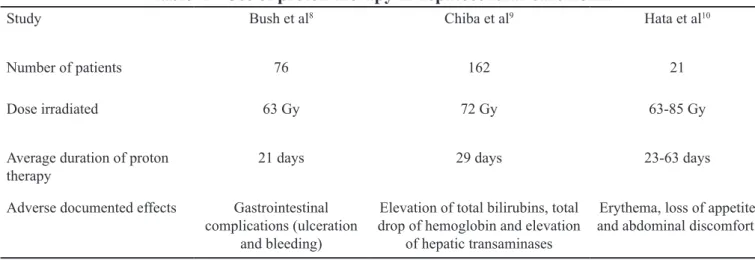
of high dose in this type of cancer. 76 patients were
evaluated between April 1998 and October 2006,
the age range varied from 40 to 83 years. The acute
toxicity of the treatment was minimal and included
a slight fatigue and cutaneous reactions consisting
of erythema (grade 1), no acute toxicity caused the
interruption of the treatment, the most important
toxicities were gastrointestinal complications
(when the primary tumor was located near the small
intestine there was an incidence of ulceration and
bleeding GI). Levels of liver enzymes, bilirubin
and albumin were used to evaluate liver disease
induced by radiation, no significant change was
observed after 6 months of treatment. The média
de sobrevivência livre de progressão
(Progression-Free Survival [PFS]) for the whole group was 36
months, with an average of PFS of 03 years of
60% for patients within the criteria of Milan. The
patients received 63 degrees (Gy) of radiation over
a period of 3 weeks with proton therapy
8Other uses of the therapy with protons to
local HCC were reported by treatment centers in
Japan. In a retrospective study conducted with 162
patients with HCC 192 treated from November 1985
to July 1998, Chiba et al
9reached the conclusion that
the therapy of protons beam for patients with HCC
is not resectable, it is effective, safe, well-tolerated
and repeatable. This is the mode of treatment useful
for cure or palliation for patients with non- resectable
HCC regardless of the size of the tumor, location
of the tumor in the liver, insufficient supply of the
tumor with arteries, presence of vascular invasion,
liver failure and coexistent intercurrent diseases.
The total median dose of irradiation of protons in
this study was 72 Gy in 16 fractions over 29 days.
9Hata et al
10observed similar results in retrospective
information on its use.
USES:
The peak Bragg effect enables the use of
proton therapy in tumors located near vital organs
and radiosensitive tissues, such as tumors of head,
neck and pelvic region, in addition to the pediatric
use, reducing the chance of injury to healthy and
developing adjacent tissues. Some studies have
been carried out to prove the effectiveness of this
type of therapeutic approach in certain tumors.
One of the most well-documented and
explored use of the radiation therapy with protons
beam is the uveal cancer. Wang et al
7(2012), in a
systematic and meta-analysis review, sought to
compare the use of therapy with particles radioactively
charged in uveal cancer with brachytherapy or
enucleation, analyzing variables such as the
recurrence of tumors, impact on mortality, adverse
clinical effects, among other factors. The particles
radioactively charged consisted mainly of protons
beam, but also, to a lesser number, therapy with
helium ions and ions of carbon. In total, 27 studies
and 8809 patients were included. The authors found
no statistically significant differences in mortality
between the two groups. However, the therapy with
particles radioactively charged (which includes the
proton therapy) was superior in preventing local
recurrence of tumor, retinopathy of radiation and
formation of cataracts. As to the risk of enucleation,
metastasis or the patients’ life expectancy, there
was no statistically significant difference between
the two groups. The authors, however, classified as
a low degree of recommendation of the conclusions
of the analysis, with a view to the heterogeneity
of the samples of included studies and the risk of
bias.
7Patients with hepatocellular carcinoma
(HCC) and prior cirrhosis, diagnosed by biopsy
or abdominal imaging exams (CT or magnetic
Proton beam radiotherapy: the use in oncological therapy SOARES, P. H. A.; DIAS, H. G.; AMARAL, F. R.; RABELO, M. O.; SOARES, L. G. B.; CAVACAMI, E. ISSN 2236-5257
245
REVISTA UNIMONTES CIENTÍFICA
review with 21 patients with HCC to whom other treatment modalities were contra-indicated or were not
viable due to cohabiting illness and unfavorable conditions, doses of 63 Gy until 84 Gy were used (mean
of 73 Gy) in 13 to 27 fractions (average of 18 fractions) for tumors treatments (Table 1).
10In another study, Makishimaet al11 (2015)
demonstrated decrease of doses of irradiation
at marginal tissues to tumor and, consequently,
of the adverse cardiac and pulmonary effects
when evaluating patients with esophageal cancer
undergoing therapy with protons, when compared
to those patients undergoing conventional
radiotherapic therapy with x-rays. It is worth
mentioning that these adverse effects are the major
concern when radiotherapy is used, more specifically,
in this type of tumor. The randomized clinical trial
with 44 patients with esophageal cancer, in which
19 patients received conventional radiotherapy
with x-rays and 25 patients received treatment
with protons. Despite the 44 patients in the study
present some degree of cardiopulmonary alteration
when evaluated, not all of them had these findings
with severity and/or clinical importance. In the 19
patients who underwent radiotherapy, the following
cases were reported: 10 cases of pericardial effusion,
four of pneumonia by radiation, two pleural lung,
one of the pharmacological pneumonitis and one
of pulmonary infection. Whereas in the group
Table 1 - Use of proton therapy in hepatocellular carcinoma
Study Bush et al8 Chiba et al9 Hata et al10
Number of patients 76 162 21
Dose irradiated 63 Gy 72 Gy 63-85 Gy
Average duration of proton
therapy 21 days 29 days 23-63 days
Adverse documented effects Gastrointestinal complications (ulceration
and bleeding)
Elevation of total bilirubins, total drop of hemoglobin and elevation
of hepatic transaminases
Erythema, loss of appetite and abdominal discomfort.
of patients undergoing proton therapy, only one
showed pericardial effusion of clinical relevance,
not being reported other adverse effects (Table 2).
The median dose of radiation was similar for both
groups. The patients had the same histological type
of cancer, squamous cell carcinoma, and all of them
also received chemotherapy during the first five
days of radiotherapy and subsequently additional
cycles, but none received a combination of x-rays
and beams of protons.
11Even though the surgical
treatment is the standard for the treatment of this
type of cancer, it is well-documented that when used
concurrently, chemotherapy and radiotherapy bring
additional benefits in relation to the prognosis, with
reduction of mortality and increasing the quality of
life of individuals after treatment. In this context, the
therapy with protons optimizes such benefits, with a
view to reducing the likelihood of complications of
normal tissues and the adverse effects of radiation
with its use. However, in the study of Makishima et
al
11(2015) the group undergoing radiotherapy with
conventional x-rays contained some patients with
more advanced disease than that has undergone the
therapy with protons, this may have affected the rates of morbidity, and it is important this consideration
in the interpretation of the results, besides the small sample of patients followed in the clinical trial. Still,
it was possible to observe that in patients with the same disease stage submitted to different therapies, the
adverse events were less frequent than in those who made use of proton therapy .
11Table 2 - Comparison between proton therapy and conventional radiation in esophagic cancer
Protons beams Conventional radiationNumber of patients 25 19
Dose irradiated 60-70 Gy 60 Gy
Average duration 24 months 20 months
Number of patients with
relevant adverse effects Pharmacological pneumonitis 00 01
Pulmonary infection 00 01
Radiation pneumonitis 00 04
Pulmonary stroke 00 02
Pericardial stroke 01 10
Ohriet al
12(2013) concluded, in a
meta-analysis of 20 studies, lower rates of late toxicity
in patients with prostate cancer treated with proton
therapy when compared to other types of treatment
that employs technique of external radiotherapy.
The systematic review included a total of 11.835
patients, seeking to assess the rates of late
gastrointestinal and genitourinary toxicities when
made use of therapy with protons or
intensity-modulated radiation and comparing with patients
who made use of conventional radiotherapy. The
assays used as assessment tool the RTOG (Late
Radiation Morbidity Scoring Schema). There was
a decrease in the rate of gastrointestinal toxicity in
patients who made use of proton therapy or
intensity-modulated radiation, but the same was not observed
for the genitourinary adverse events. It is important
to stress that the gastrointestinal and genitourinary
toxicities are events that can be caused by a number
of factors beyond the radiation, although symptoms
considered rare by the low incidence (5%), which,
however, are important due to their respective
clinical repercussions and severities presented..
12CONCLUSION
Because of the increase in the incidence
of malignant neoplasms in recent years and the
survival of patients when treated early, the use of an
effective therapy and which reduces adverse events
in the short and long term is essential in order to
decrease morbidity and improve the quality of
life of patients with cancer. Therapy with protons
beam, although still limited to a few countries due
to its high cost and its technological complexity,
has much to offer in the present and in the future
in the treatment of cancer patients. This is thanks to
its physical and technical characteristic that allows
to focus a high degree of radiation on some point
at the same time that saves the normal tissues. The
literature shows promising results with its use in
which there are many studies that corroborate the
fact that the reduction in adverse events with the
use of proton therapy in specific types of cancer,
however there are limitations in the work so far
carried out which should be taken into account in
Proton beam radiotherapy: the use in oncological therapy SOARES, P. H. A.; DIAS, H. G.; AMARAL, F. R.; RABELO, M. O.; SOARES, L. G. B.; CAVACAMI, E. ISSN 2236-5257
247
REVISTA UNIMONTES CIENTÍFICA