INTRODUCTION
The examination of endodontically treated teeth for restorative purposes requires a detailed radiographic analysis of root canal fillings because it seems to be the most accessible non-invasive method to obtain objective information that helps deciding on the need for revising the treatment. In the moment that the radiography is as-sessed, the recognition of voids caused by procedural inability is extremely important for the reason that these empty spaces have potential to promote leakage of bacteria and toxins to the periapex, thus compromising success of treatment (1).
Radiopacity is an essential attribute of root canal sealers and gutta-percha, since this property allows for clear distinction of root fillings and also for estimation of their extent and apparent condensation into root canals
Radiographic Analysis of Root Canal Fillings:
Influence of Two Sealers on the Perception of Voids
Augusto BODANEZI1
Etiene de Andrade MUNHOZ2
Norberti BERNARDINELI1
Ana Lúcia Álvares CAPELOZZA2
Ivaldo Gomes de MORAES1
Clovis Monteiro BRAMANTE1
1Department of Dentistry, Endodontics and Dental Materials,
Bauru Dental School, University of São Paulo, Bauru, SP, Brazil
2Department of Stomatology, Radiology and Oral Surgery, Bauru Dental School,
University of São Paulo, Bauru, SP, Brazil
The aim of this ex vivo was to investigate if two radiopaque root canal sealers with different formulations would influence the radiographic perception of root canal fillings. The root canals of 48 extracted maxillary canines were prepared and randomly assigned to 3 groups of 16 specimens each. In each group, the root canals were filled by lateral condensation of gutta-percha and one of the tested sealers: Endométhasone, Sealer 26, or a non-radiopaque sealer. A through-and-through void was simulated in half of the specimens from each group (n=8). The buccolingual radiographic images obtained were randomly interpreted for voids existence by a radiologist and an endodontist. The differences in sensitivity and specificity between groups and examiners were compared using, respectively, Fisher's Exact and McNemar tests at 5% significance level. Both radiopaque sealers caused a significant decrease in sensitivity at the coronal part of fillings. The use of Endométhasone increased specificity values for both coronal and apical portions of the root canal fillings. In conclusion, the tested sealers influenced the radiographic perceptions of laterally condensed root canal fillings in a different way.
Key Words: diagnosis, radiography, radiopacity, root canal filling, sealer.
(2-5). Routinely, radiopacity of commercially available sealers is ranked on in vitro tests, but the significance of this information to the radiographic appearance of root fillings is never mentioned (5-7). The image of an obturation on radiography consists of a two-dimensional, usually buccolingual, projection of filling materials confined in the root canal and superimposed with bone, dentin, and soft tissue. During lateral condensation, a sealer is applied to fill spaces between gutta-percha cones, which the bulk of fillings fails to reach and, for this reason, it may have an impact on the radiopacity of root fillings (4).
It is well established that intracanal distribution of sealers is determined by their particular physicochemical properties (8,9) and the filling technique adopted (10). Sealer 26 and Endométhasone are commercially avail-able root canal sealers that, in spite of remarkavail-able
ferences in formulations and physicochemical properties (11-13), have shown similar in vitro radiopacity when investigated alone or associated with gutta-percha (6). Given this configuration, the present study investigated if two root canal sealers with different formulations would influence the radiographic perception of defective (sensitivity) or adequate (specificity) root canal fillings performed with lateral condensation.
MATERIAL AND METHODS
Tooth Preparation
For this study, 48 maxillary permanent maxil-lary canines with single straight or slightly curved root canals (25 ± 2 mm) were selected and stored in 0.5% saline solution after approval of the research project by the University of São Paulo’s Research Ethics Commit-tee (Protocol #153/2005). After standard access, root canal lengths were assessed by placing a size 15 K-file (Dentsply-Maillefer, Ballaigues, Switzerland) inside the canal, until the tip has passed through the apical fora-men. The instrument was withdrawn 1 mm to record the working length. All canals were instrumented using a push-pull filing technique up to a size 55 K-file and, after every instrument withdrawal from the root canal, a full irrigation with 1% sodium hypochlorite was performed (Virex; Johnson Ltd., Jacarepaguá, RJ, Brazil). Apical foramen enlargement was obtained with a size 25 K-file. After a 3 min final irrigation with 17% EDTA (Biodin-âmica, Ibiporã, PR, Brazil), the root canals were rinsed with 0.5% saline and dried thoroughly with paper points. The specimens were randomly assigned to 3 groups (n=16). In each group, the root canals were filled by lateral condensation of gutta-percha
and one of the tested sealers: Endo-méthasone (Septodont, Saint-Maur-des-Fossés, Cedex, France), Sealer 26 (Dentsply Ind e Com., Petrópolis, RJ, Brazil), and a non-radiopaque sealer (NRS) which was used in the control group and formulated with epoxy resin, resin hardener and polyurethane polyol. The commercially available sealers were handled and mixed according to manufacturer’s recommendations (Table 1), whereas the non-radiopaque sealer compound was homogenized for 15 s.
Before being filled with lateral condensation, a radiolucent nylon monofilament string (0.25 mm in diameter and 20 cm in length) (Grilon Fishing Line; Mazzaferro Ltd., São Bernardo do Campo, SP, Brazil) was inserted into the root canal of half specimens from each group (n=8). Both ends of the string were tied in alignment to a device so that moving the tooth distally would stabilize the stretched fishing line in contact with the mesial wall of the root canal through the whole filling procedures, conducted by one operator.
The sealers were taken to the root canals by coat-ing with respective master gutta-percha cone. A size 30 master cone was exclusively used for root canals provided with nylon thread, while a size 55 cone was inserted into the remaining root canals. The master cone was laterally condensed with A and B finger spreaders (Dentsply-Maillefer), and 7 to 9 accessory cones (Tanari, Manacapuru, AM, Brazil) were added to the filling mass (Fig. 1). After obturation, coronally extruded excess sealer and gutta-percha were cleaned.
Radiographic Procedure
Radiographs of each canine were obtained in the buccolingual direction using the paralleling technique. The periapical E film (Insight; Kodak Co., Rochester, NY, USA) and each filled tooth were maintained in a constant position by inserting each root in the same canine socket of a phantom build from a dry human maxilla, which was fixed in a rigid position, in front of X-ray tube, with a specifically designed acrylic device. All radiographic exposures were completed with a dental X-ray unit (X 70; Dabi Atlante, Ribeirão Preto, SP, Brazil) at 70 kVp and 8 mA. The exposure time was
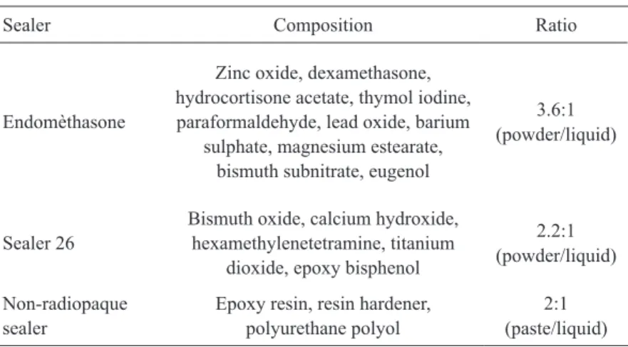
Table 1. Composition and powder/liquid or paste/liquid ratio of the tested materials.
Sealer Composition Ratio
Endomèthasone
Zinc oxide, dexamethasone, hydrocortisone acetate, thymol iodine, paraformaldehyde, lead oxide, barium
sulphate, magnesium estearate, bismuth subnitrate, eugenol
3.6:1 (powder/liquid)
Sealer 26
Bismuth oxide, calcium hydroxide, hexamethylenetetramine, titanium
dioxide, epoxy bisphenol
2.2:1 (powder/liquid)
Non-radiopaque sealer
Epoxy resin, resin hardener, polyurethane polyol
0.5 s, the focus-object distance was 40 cm, and the object to receptor distance was 2 cm.
The exposed films were immersed in the devel-oper solution for 3 min at 21ºC and then rinsed with water for 10 s before being submerged in the fixer solution for 8 min. The developed films were subsequently washed with running water for 10 min and allowed to dry in a dust free atmosphere.
Image Evaluation
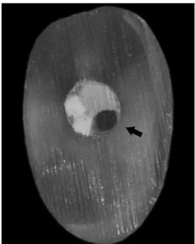
All radiographic evaluations were performed with the aid of an ethyl-vinyl-acetate mask positioned over a fluorescent cold light-box (Medalight LP-400; Hong Kong, China) in the same light-controlled room. Prior to image assessment, an endodontist and a radiologist received written instructions and were then calibrated with a pair of radiographs. The 48 radiographs were numbered, mounted in random order, and individually interpreted. The resulting images were analyzed for the presence or absence of voids in the coronal, medium, and/or apical third of the root canal filling (Fig. 2). The observers had no knowledge of the extent and distribu-tion of voids and were asked to ignore the distance of fillings to the apex or radiolucent areas in contact with the root. Analyses of images were recorded in a proper form and the viewing time was unlimited. A second evalu-ation was executed 4 weeks after the first assessment.
Statistical Analysis
The mean number of correct, false positive, incorrect and false negative results obtained from both sequential assessment stages was used as the basis for sensitivity and specificity calculations for each group. The differences in sensitivity and specificity values between groups and examiners were compared using categorical Exact Fisher’s and McNemar tests, respec-tively, which were both adjusted to the 95% confidence interval. The intra-observer agreement was determined by Cohen’s Kappa statistic method. Statistical evaluation for all tests was performed with SPSS database software program (SPSS v. 11.0 for Windows, Chicago, IL, USA).
RESULTS
Sensitivity
In the assessment by the endodontist, the experi-mental groups had statistically lower sensitivity values than the control group only for the coronal third of root canal fillings (Table 2). However, the differences in sen-sitivity between groups were not statistically significant in any part of fillings when they were evaluated by the radiologist. The coronal part of Endométhasone and Sealer 26 fillings exhibited statistically lower
sensitiv-Figure 2. Representative radiographic image of the root canal fillings assessed by the examiners. Note the presence of the simulated void in the apical third.
ity values than the apical part for both examiners. In the control group, this difference was only observed in the assessment by the radiologist. There were statisti-cally higher sensitivity values for the endodontist’s evaluation of control (cervical part) when compared to the radiologist’s examination (Table 2). The sensitivity values obtained in the middle of root fillings were not statistically different from the coronal and apical parts of fillings for the two examiners.
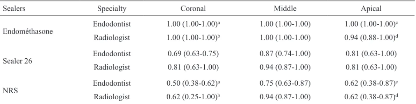
Specificity
The coronal and apical parts of Endométhasone root canal fillings exhibited statistically higher values of specificity than the control group for the radiologist’s and endodontist’s assessment (Table 3). The differences in specificity between the coronal and apical thirds of experimental and control groups were not statistically significant, even when compared to the specificity values obtained in the middle part of the root canal fillings. There were no statistically differences between the specificity values obtained from the different examiners.
The intra-observer Kappa values were 0.81 for the radiologist and 0.87 for the endodontist.
DISCUSSION
The analysis of periapical radiography is one of the most common diagnostic tests used in dentistry and, in this study, all efforts were conducted to reproduce as close as possible the radiographic image of maxillary canine root canal fillings normally obtained and assessed in clinical practice.
In previous investigations, an empty space of 0.25 mm in the obturation was considered the threshold to decide whether a root canal filling appeared well or was imperfectly condensed on radiography (3,14). Based on this representation, we adopted in this work a nylon string of equivalent size to simulate an empty space inside root canals. This maneuver permitted for some standardization of thickness, position, and even apical extension of the void through the entire obturation, which is a characteristic almost impossible to control in clinical studies (1,15). Despite the efforts to keep the
Table 2. Mean values and ranges of sensitivity in each third of the fillings assessed by the examiners.
Sealers Examiner Coronal Middle Apical
Endométhasone
Endodontist 0.13 (0.13-0.13)a§ 0.63 (0.63-0.63) 1.00 (1.00-1.00)a
Radiologist 0.06 (0.13-0.00)b 0.38 (0.38-0.38) 0.81 (0.88-0.75)b
Sealer 26 Endodontist 0.50 (0.50-0.50)
c* 0.57 (0.63-0.50) 0.81 (0.88–0.75)c
Radiologist 0.12 (0.25-0.00)d 0.50 (0.63-0.38) 0.81 (0.88-0.75)d
NRS Endodontist 0.82 (1.00-0.63)
§*F 0.69 (0.75-0.63) 0.94 (1.00-0.88)
Radiologist 0.19 (0.38-0.00)eF 0.44 (0.63-0.25) 0.75 (0.88-0.63)e
Same lowercase letters indicate statistically significant difference among the filling regions. Same symbols indicate statistically significant difference between groups. Same uppercase letters indicate statistically significant difference between examiners.
Table 3. Mean values and ranges of specificity in each third of the fillings assessed by the examiners.
Sealers Specialty Coronal Middle Apical
Endométhasone
Endodontist 1.00 (1.00-1.00)a 1.00 (1.00-1.00) 1.00 (1.00-1.00)c
Radiologist 1.00 (1.00-1.00)b 1.00 (1.00-1.00) 0.94 (0.88-1.00)d
Sealer 26
Endodontist 0.69 (0.63-0.75) 0.87 (0.74-1.00) 0.81 (0.63-1.00)
Radiologist 0.81 (0.63-1.00) 0.94 (0.87-1.00) 0.81 (0.63-1.00)
NRS
Endodontist 0.50 (0.38-0.62)a 0.75 (0.63-0.87) 0.62 (0.38-0.87)c
Radiologist 0.62 (0.25-1.00)b 0.94 (0.87-1.00) 0.62 (0.38-0.87)d
string attached to the mesial root canal wall, a central dislodgement due to subtle distal curvature of root apex must have occurred.
False positive assertions were surely originated from detection of inherent minor voids resulting from unfilled spreader tracks, insufficient sealer distribution or operational variability generated during lateral conden-sation (3,14,16), and this probably had some influence on the results. In order to estimate and neutralize this bias, natural root canal fillings were performed with a radiolucent sealer (control group).
According to the results presented, both sealers caused an equivalent reduction in sensitivity, especially at the coronal part of fillings. This phenomenon may have resulted from the thicker mass of gutta-percha added to sealer accumulation at this point, which is a natural characteristic of lateral condensation (9). The oval-shaped configuration of canine root canals (3,10) and the buccolingual direction of radiographic expo-sure may have also contributed for the radiographic superimposition that probably hid the simulated void. Conversely, in similar studies the root level did not af-fected void detection in canine fillings (17,18), but major methodological differences such as filling techniques, the use of filling materials separately and simulated void accomplishment with wires should be considered.
In the apical region of fillings, the highest sen-sitivity levels were independent of the sealer used, indicating that screening for defects at this area offers the most significant opportunity to correctly detect voids in root canals manually prepared and filled with lateral condensation. Probably the lower content of sealer and the large thickness of the simulated void both contrib-uted to the absence of statistical difference between the experimental groups. The identification of voids in the apical part of fillings by means of radiography is im-portant because, though distant from coronal potential microleakage, the apex may harbor bacteria that can grow when they receive nutrients from periapical region or lateral canals, thus compromising treatment (1).
When the coronal and apical regions of root fill-ings were evaluated, the root canal fillfill-ings performed with Endométhasone presented a statistically lower number of false positive results (higher specificity) than Sealer 26. In an attempt to explain the reasons for this phenomenon, technical or operational deficiencies as-sociated with lateral condensation procedures (3,14,16) and the difficulties in standardizing the volume of sealer reaching the apical region (16) should be considered as
possible causes for an uneven distribution of undesirable filling defects between groups. Similarly, the differences in sealers’ formulations and mixing characteristics could also influence on physicochemical properties that were able to configure the intracanal distribution of sealers (8-10). Almeida et al. (13) reported that Endométhasone has a significant lower flow (higher viscosity) when compared to Sealer 26. This property is directly related to the amount of radiopaque sealer’s accumulated between gutta-percha cones in the moment of filling procedures (19) and, for this reason, may have had an influence on the final radiopacity of root canal fillings (20). The supposed thicker layers of Endométhasone probably superimposed a great number of inherent filling voids than Sealer 26 did and, concomitantly, increased the general radiographic density of root fillings to a level which was recognized as a sign of good quality by the examiners (3,14). However, it should be clear that no physicochemical properties of sealers were evaluated in this study.
The dissonance detected between examiners, in the assessment of control group (defective fillings), resulted in no conclusive evidence regarding whether the radiopacity provided by gutta-percha alone would conceal the simulated void in the coronal and thickest portion of fillings. Likewise, the endodontist considered a higher number of voids in root canal fillings performed with Endométhasone. Since diagnostic decision is influ-enced by personal experience and previous knowledge of the subject analyzed (4,17,18), a suspicious atti-tude of the endodontist towards root canal treatments performed by unknown professionals could possibly explain the higher proportion of voids detected by this professional compared to the radiologist. In addition, it should be clear that only one specialist of each area was compared, thus these results are neither scientifically nor statistically powerful to support generalizations on specialties’ decision-making during radiographic judg-ment of fillings.
dissociate the sealers’ radiopacity effect from that gen-erated by the entire filling on the radiographic image. Therefore, it should be assumed that sealers are capable of influencing only the overall radiopacity of the filling, as already stated by Baksi et al. (4).
Although digital intraoral techniques seemed to perform better than the analogue intraoral images for small void detection in canine fillings (18), the influ-ence of sealers’ formulation on radiographic perception of root canal fillings presented in this study advises that diagnostic decision should never rely solely upon radio-graphic image. The act of gathering information from a detailed history and clinical examination as well as the awareness that root canal shape and filling techniques exert some influence on the diagnostic performance of periapical radiographs should be carefully considered in order to reduce the chances of misjudgment in the prognosis of difficult cases.
Based on the results of this study it seems that the tested sealers influenced the radiographic perception of laterally condensed root canal fillings in different ways.
RESUMO
O propósito deste estudo ex vivo foi investigar se dois ci-mentos obturadores de fórmulas diferentes influenciariam a percepção radiográfica de obturações de canais radiculares. Os canais radiculares de 48 caninos superiores extraídos foram preparados e divididos em 3 grupos. Em cada grupo os ca-nais foram preenchidos através da condensação lateral da guta-percha e de um dos cimentos testados (Endométhasone, Sealer 26 e cimento não-radiopaco), e um defeito de ponta a ponta foi simulado em metade dos espécimes de cada grupo (n=8). As imagens radiográficas vestíbulo-linguais obtidas foram aleato-riamente interpretadas quanto à existência de defeitos por um radiologista e um endodontista. As diferenças em sensibilidade e especificidade entre os grupos e examinadores foram comparadas respectivamente usando-se os testes Exato de Fisher e McNemar ajustados ao nível de significância de 5%. Ambos os cimentos radiopacos ocasionaram uma redução significativa da sensibili-dade na parte cervical das obturações. O uso do Endométhasone aumentou os valores de especificidade para as porções cervical e apical das obturações dos canais radiculares. Concluiu-se que os cimentos testados influenciaram de maneira distinta a percepção radiográfica de obturações endodônticas executadas com condensação lateral.
REFERENCES
1. Sundqvist G, Figdor D, Persson S, Sjögren U. Microbiologic analysis of teeth with failed endodontic treatment and the outcome of conservative re-treatment. Oral Surg Oral Med Oral Pathol Oral
Radiol Endod 1998;85:86-93.
2. Beyer-Olsen EM, Ørstavik D. Radiopacity of root canal sealers. Oral Surg Oral Med Oral Pathol 1981;51:320-328.
3. van der Sluis LWM, Wu M-K, Wesselink PR. An evaluation of the quality of root fillings in mandibular incisors and maxillary and mandibular canines using different methodologies. J Dent 2005;33:683-688.
4. Baksi BG, Eyüboğlu TF, Şen BH, Erdilek N. The effect of three
different sealers on the radiopacity of root fillings in simu-lated canals. Oral Surg Oral Med Oral Pathol Oral Radiol Endod 2007;103:138-141.
5. Carvalho-Junior JR, Correr-Sobrinho L, Correr AB, Sinhoreti MAC, Consani S, Souza-Neto MD. Radiopacity of root filling materials using digital radiography. Int End J 2007;40:514-520. 6. Ferreira FBA, Silva e Souza PAR, Bevilaqua MV, Tavano O.
Di-gital radiographic assesment of endodontic cements radiopacity J Appl Oral Sci (Formerly Rev Fac Odontol Bauru) 1999;7:55-60. 7. Tanomaru-Filho M, Jorge EG, Tanomaru JMG, Gonçalves M.
Evaluation of the radiopacity of calcium hydroxide- and glass-ionomer-based root canal sealers. Int End J 2008;41:50-53. 8. McMichen FR, Pearson G, Rahbaran S, Gulabivala K. A
compara-tive study of selected physical properties of five root-canal sealers. Int End J 2003;36:629-635.
9. Facer SR, Walton RE. Intracanal distribution patterns of sealers after lateral condensation. J Endod 2003;29:832-834.
10. Wu M-K, Özok AR, Wesselink PR. Sealer distribution in root canals obturated by three techniques. Int End J 2000;33:340-345. 11. Souza-Neto MD, Marchesan MA, Pécora JD, Junior AB, Silva-Souza YT, Saquy PC. Effect of Er:YAG laser on adhesion of root canal sealers. J Endod 2002;28:185-187.
12. Gomes BP, Pedroso JÁ, Jacinto RC, Vianna ME, Ferraz CC, Zaia AA, et al.. In vitro evaluation of the antimicrobial activity of five root canal sealers. Braz Dent J 2004;15:30-35.
13. Almeida JF, Gomes BP, Ferraz CC, Souza-Filho FJ, Zaia AA. Filling of artificial lateral canals and microleakage and flow of five endodontic sealers. Int Endod J 2007;40:692-699.
14. Kersten HW, Wesselink PR, Thoden van Velzen SK. The diagnos-tic reliability of the buccal radiograph after root canal filling. Int End J 1987;20:20-24.
15. Eckerbom M, Flygare L, Magnusson T. A 20-year follow-up study of endodontic variables and apical status in a Swedish population. Int End J 2007;40:940-948.
16. Souza EM, Wu M-K, van der Sluis LW, Leonardo RT, Bonetti-Filho I, Wesselink PR. Effect of filling technique and root canal area on the percentage of gutta-percha in laterally compacted root fillings. Int End J 2009;42:719-726.
17. Kositbowornchai S, Hanwachirapong D, Somsopon R, Pirmsin-thavee S, Sooksuntisakoonchai N. Ex vivo comparison of digital images with conventional radiographs for detection of simulated voids in root canal filling material. Int End J 2006;39:287-292. 18. Huybrechts B, Bud M, Bergmans L, Lambrechts P, Jacobs R. Void
detection in root fillings using intraoral analogue, intraoral digital and cone beam CT images. Int End J 2009;42:675-685.
19. Weisman MI. A study of the flow rate of ten root canal sealers. Oral Surg Oral Med Oral Pathol 1970;29:255-261.
20. Orfali S, Lilley JD, Molokhia A. The radiopacity of some endodon-tic sealer cements [Abstract 368]. J Dent Res 1987;66:876.