INTRODUCTION
Success in Endodontics depends on the complete removal of canal content, followed by apical closure using a material of adequate compatibility to avoid possible irritation to the remaining pulp and periapical tissues. The constant evolution of concepts is brought along with new methods, new techniques and the development of more effective materials and instruments. In pediatric dentistry, pulp therapy is performed due to pulp exposure or necrosis and aims at keeping the primary teeth in place until the exfoliation period (1,2). Although the concepts of endodontic treatment of primary and permanent teeth are different (1), biological behaviors of pulp and periapical tissues of primary teeth are similar to those of the permanent dentition, except for the physiological root resorption. In pediatric dentistry, biocompatibility with periapical tissues is of utmost importance, since
Histological Evaluation of Bone Response to
Pediatric Endodontic Pastes: An Experimental
Study in Guinea Pig
Andréa Mara LACATIVA Adriano M. LOYOLA Cassio José Alves SOUSA
Department of Endodontics, Dental School, UFU - Federal University of Uberlândia, Uberlândia, MG, Brazil
This study aimed to evaluate by the intra-osseous implant technique the most commonly used materials for pulp therapy in pediatric dentistry: calcium hydroxide (CH), Guedes Pinto paste and CTZ paste, according to FDI (1980) and ANSI/ADA(1982) recommendations. Thirty guinea pigs, 10 for each material, divided into experimental periods of 4 and 12 weeks received one implant on each side of the lower jaw symphysis. The external lateral tube wall served as control for the technique. At the end of the observation periods, the animals were euthanized and specimens were prepared for routine histological examination. It was observed that CH and CTZ paste induced severe inflammation, a large amount of necrotic tissue, lymphocytes, foreign body cells and bone resorption, while Guedes Pinto Paste induced little or no inflammation in the 4-week observation period. After 12 weeks, the reactions to CH and Guedes Pinto paste were also absent/mild, presenting a general pattern of replacement by recently formed bone tissue while a moderate to severe inflammatory response was observed with CTZ paste. Guedes Pinto paste presented acceptable biocompatibility levels in both analyzed periods; CH only showed acceptable biocompatibility in the 12-week period while CTZ paste showed no biocompatibility in both periods. Among the tested materials, only Guedes Pinto paste presented an acceptable biocompatibility.
Key Words: biocompatibility, dental apex, pediatric dentistry, pulp therapy, root filling.
any material which is not biologically acceptable may be responsible for failures (2,3).
A large number of materials have been proposed to achieve this goal; however, there is a lack of consensus concerning to when pulp therapy is required in primary teeth. Several techniques and materials have been empirically used for more than a century, often considering only the clinical aspects (1,2,4,5).
Particularly, iodophorm-based and calcium hydroxide (CH)-based pastes have been usually and successfully used in endodontic therapy for primary teeth, based specially on clinical application (3-5) and cytotoxicity assays (1). However, few studies focused on bone response to these materials (2).
Considering the biological properties of endodontic materials, there is a broad range of characteristics which may be considered and methodologies available for this purpose, including initial tests, secondary tests and usage
studies. The initial evaluation comprises some basic ways of assessing biological properties with in vitro methods. The secondary assessments are performed in vivo in laboratory animals and implantation as an experiment belongs to this category. Usage studies are applications of materials in therapeutic procedures on primates or humans (6,7).
The aim of this study was to compare the biocompatibility of CH, Guedes Pinto paste and CTZ paste with the intra-osseous implant technique.
MATERIAL AND METHODS
The materials tested in this study were CH, Guedes Pinto paste (composition: iodoform, PMCC and rifocort) and CTZ paste (composition: zinc oxide, tetracycline, chloramphenicol and eugenol). All materials were prepared according to the manufacturers’ instructions for its clinical use and inserted in a polytetrafluorethylene cup (Teflon®; DuPont, HABIA, Knivsta, Sweden),
ensuring that air was not entrapped.
The intra-osseous implant in the guinea pig mandible proposed by Spångberg (8,9) was used in this study, following standardized methods to evaluate the biological properties recommended by Technical Report #9 (6) and by Document #41 (7).
Thirty guinea pigs weighing about 800 g were used in the experiment. Each animal received two implants of the same material, with a total of 10 specimens for each material and observation period. Additionally, the connective tissue response along the lateral wall outside the cup served as a negative control for the technique. According to Technical Report #9 (6) and Document #41 (7) the reactions along the external periphery of the Teflon® cup reflect the trauma caused by surgical
procedures required for the introduction of the Teflon®
cup and its contents. Teflon® itself was proven to cause
only insignificant irritation to tissues(10).
Animals were anesthetized intramuscularly with 0.6 mL of ketamine chlorohydrate (100 mg/ mL; Francotar; Virbac do Brasil Indústria e Comércio Ltda. Santo Amaro, SP, Brazil) and 0.5 mg/mL of 2% xylazine chlorhydrate (Dorcipec; Vallée S/A Produtos Veterinários, Montes Claros, MG, Brazil). Local anesthesia was administered in the mucobuccal fold, in the area of the mandibular incisors, with 0.6 mL of 2% xylocaine with epinephrine (1:100,000) to prevent any local discomfort. The guinea pigs were shaved in the submandibular area and the skin was disinfected with a
5% tincture of iodine. An incision of about 15 mm was made in the midline along the symphysis in the triangle periosteal elevator used to expose the mandibular bone. On the side of the symphysis in the triangle between the symphysis and the constantly developing incisor, 2x2 mm diameter cylindrical holes were prepared with special burs under saline irrigation. The cups filled with the materials under sterile conditions were inserted into the bone cavities. The periosteum was pulled over the cup and the incision was sutured with 3-0 black silk. The observation periods were 4 and 12 weeks. After the guinea pigs were euthanized, the mandible was dissected out and the bone adjacent to the cups in situ was cut into 10 mm blocks. The specimens were immersed in 10% neutral buffered formalin and prepared for routine histological examination. The slides were stained with hematoxylin-eosin (HE) for cellular recognition.
The interface at the opening of the cup, between the material and the bone, was examined and evaluated for the intensity of inflammation, the presence or absence of necrosis and/or resorption, possible replacement of the tested material by bone and osteolytic, osteosclerotic or osteoclastic activity. The tissue reactions were classified as: absent to slight tissue reaction, moderate tissue reaction or severe tissue reaction, according to the criteria defined by FDI (6) and ANSI/ADA (7) protocols. It was considered as biologically acceptable the material that showed none to slight reaction at both experimental periods of 4 and 12 weeks, or a moderate reaction at 4 weeks that diminished at 12 weeks.
The present investigation was developed according to the Ethical Principles on Animal Research as directed by the Brazilian National Advice of Control and Animal Experimentation (CONCEA) and approved by the Ethics Committee on the Use of Animals (CEUA) from the Federal University of Uberlândia, Brazil.
RESULTS
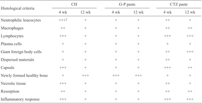
The histological evaluations of the materials at 4 and 12 weeks are summarized in Table 1 and illustrated in Figures 1 to 6.
Negative Control
4 Weeks
CH and CTZ Paste: The reaction was considered severe, showing bone necrosis, hemorrhage and a neutrophil inflammatory infiltrate in close contact with the material (Figs. 1 and 2).
Guedes Pinto Paste: The inflammatory response was classified as absent to slight. There was formation of new healthy bone in close contact with the material. In some cases, there was a narrow layer of connective tissue between the implanted material and the newly formed bone tissue with no presence of inflammatory response (Fig. 3).
12 Weeks
CH and Guedes Pinto Paste: The inflammatory response was classified as absent to slight. There was new formation and apposition of healthy bone which tended to enclose the entire implant. In some cases, the connective tissue around the cup showed abundant fibroblasts and vessels, with no inflammatory cells (Figs. 4 and 5).
CTZ Paste: The reaction was still considered severe, showing a mononuclear inflammatory infiltrate with a large number of lymphocytes, macrophages and giant foreign body cells. The presence of clastic
cells containing material in the cytoplasm and necrotic tissue was common. There was more collagen fiber deposition near the bone tissue with the presence of many inflammatory cells. The irregularities at the bone surface demonstrated osteoclastic activity (Fig. 6).
DISCUSSION
Compatibility with living tissues in the periapical region is one of the most important properties of a material used in root filling of primary teeth because it will be in permanent contact with these tissues during the physiological root resorption. Furthermore, a possible injury to the germ of permanent teeth is a constant concern.
Most scientific investigations providing the experimental basis for these therapeutic procedures began in the last three decades. Considering the amount of materials recommended for pulp therapy in primary teeth and the lack of consensus as to their indications, this work aimed to evaluate the biocompatibility of some of them.
The implant test in guinea pig bone tissue, a secondary test recommended by the FDI Technical Report #9 (6), allows testing of a material as it is used in clinical situations and prepared following the manufacturer’s recommendations. Although the
Table 1. Histological evaluation of tested materials according to the experimental chronology (4 weeks and 12 weeks).
Histological criteria
CH G-P paste CTZ paste
4 wk 12 wk 4 wk 12 wk 4 wk 12 wk
Neutrophilic leucocytes +++2 + + + ++ +
Macrophages ++ + + + ++ ++
Lymphocytes +++ + + + +++ +++
Plasma cells + + + + + +
Giant foreign body cells + + + + ++ +++
Dispersed materials + + + + ++ +
Capsule +++ + + + +++ ++
Newly formed healthy bone + +++ +++ +++ + +
Necrotic tissue +++ + + + ++ +
Resorption ++ + + + ++ ++
Inflammatory response +++ + + + +++ +++
results cannot be directly extrapolated to humans, the test is standardized and allows for a direct comparison between materials. The literature in this field includes research done in different laboratories at different times
with the same materials, thus the results of the present study could be compared and confirmed (11-14). The obtained results confirmed the findings of those authors and indicate that any material placed in contact with
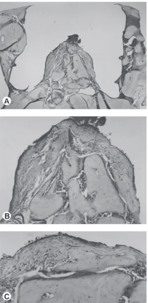
Figure 1. Host tissue reaction to calcium hydroxide after 4 weeks of implantation into the mandibular bone. A: Overview of the implanted area (virtual image of Teflon® cup with the tested material) into the host bone tissue (HE, original magnification ×40). B:
Figure 2. Host tissue reaction to Guedes Pinto paste after 4 weeks of material implantation into the mandibular bone. A: Overview of the implant showing an area occupied by the tested material and bone-material interface. Integration of the Teflon® cup (virtual
image) with the surrounding bone and growth of the latter into the lateral grooves of the cup (HE, original magnification ×40). B: A higher magnification of the bone-material interface area showing a thin strip of connective tissue and adjacent bone tissue (HE, original magnification ×200); C: Detail of panel B (left side) highlighting close contact of bone tissue and fibrous connective tissue with the area occupied by the material. It can be noticed that the inflammatory infiltrate is absent and osteoblasts are present in the peripheral area of the bone tissue. There is no evidence of bone resorption (HE, original magnification ×400); D: another detail of Panel B (right side) showing similar aspects to the Panel C. A thicker strip of connective tissue is shown, with no evidence of inflammation either. Bone tissue with osteoblasts in the periphery and apparent absence of resorption (HE, original magnification ×400). E: A higher magnification of the area in the Panel A (arrow) emphasizing the Teflon® cup-bone interface; area used for control (HE, original
magnification ×100). F: Detail of Panel E, showing a thin strip of dense fibrous connective tissue, interposed between the bone tissue and the Teflon®. Notice the absence of inflammatory infiltrate and bone resorption. Healthy bone presenting medullary spaces (lower
Figure 3. Host tissue reaction to CTZ paste implantation into the mandibular bone. A: Overview of the implantation area into the mandibular bone tissue showing the virtual area of the Teflon® cup. Notice the total integration of the Teflon® cup and the relation
of the tested material with the contiguous tissue (HE, original magnification ×40). B: A higher magnification of the tested material, connective tissue interface highlighting the material consisting of zinc oxide crystals, thick strip of connective tissue presenting dense inflammatory infiltrate and bone tissue with irregular surfaces (HE, original magnification ×100). C: Larger detail of Panel B (upper right side), where necrotic bone tissue fragments can be seen, mononuclear inflammatory infiltrate (HE, original magnification ×200). D: Larger detail of Panel B (left side) showing a strip of connective tissue with aspect of granulation tissue, with dense mononuclear inflammatory infiltrate, a large number of vessels. In addition, bone tissue with resorption can be noticed (arrow). (HE, original magnification ×400). E: Higher magnification of Panel C, where it can be noticed, in more details, fragments of necrotic bone tissue next to the tested material (HE, original magnification ×400).
vital tissues could elicit the development of a foreign body reaction. The bone in contact with the walls along the cup did not show inflammatory reaction in any of the observation periods, although in some cases there
was a slender connective fibrous capsule and giant cells between the bone and the cup. The grooves were filled by healthy bone.
Figure 4. Host tissue reaction to calcium hydroxide after 12 weeks implantation into the mandibular bone. A: Overview of the implant area: it can be noticed the replacement of the implanted material by connective tissue and bone. Total integration between the Teflon®
cup and the surrounding bone tissue (HE, original magnification ×40). B: Detail of the connective tissue inside the Teflon® cup, rich
in fibroblasts and fibrilar structures of newly formed collagen, vessels; absence of edema and inflammatory infiltrate (HE, original magnification ×400). C: View of the entire tissue inside the implant, where it is possible to notice areas of greater hyalinization, suggesting osteoid formation. No areas of inflammatory infiltrate are defined (HE, original magnification ×100). D: Detail of the opening area of the Teflon® cup, where it is possible to notice new connective tissue, rich in fibroblasts with hyperemic vessels and
apparent absence of inflammation. Presence of new bone tissue remodeling (HE, original magnification ×400).
contains eugenol elicits a severe tissue reaction because of the depression of cellular respiration. It has also been shown that the severity of inflammation increases with time because the reaction between the material and tissue fluids forms hydroxyl ions that release eugenol
Figure 5. Host tissue reaction to the Guedes Pinto paste after 12 weeks of bone implantation. A: Overview of the implant where the total integration is noticed between the implant and the adjacent bone and replacement of the material by newly formed bone (HE, original magnification ×40). B: Magnification of Panel A showing the newly formed bone tissue penetrating dense connective tissue matrix, with apparent absence of inflammatory infiltrate. New bone formation evolving to a structural pattern, similar to the normal bone (HE, original magnification ×100). C: Detail of Panel B showing the bone-implant interface, where may be noticed osteogenesis adjacent to the connective tissue (HE, original magnification ×400).
interface. Among all tested materials, the inflammatory response to CTZ paste was significantly higher for both observation periods in this study. Such a severe response to CTZ paste was previously described in the literature (20,21). It is thought to be due to the eugenol in the
composition of CTZ paste.
Several studies demonstrated the acceptable use of CH in permanent and deciduous dentition (2,18,22). The results of this study corroborate the findings of a previous investigation (24) regarding tissue response to CH. These authors tested CH in vivo, demonstrating its biocompatibility potential. The present results indicate that CH showed a high degree of toxicity within the 4-week experimental period, but this changed significantly at 12 weeks, when it presented biocompatibility characteristics with no significant inflammatory response.
Previous clinical studies have demonstrated the efficacy of Guedes Pinto paste in the endodontic treatment of deciduous teeth (2,3,24). However, there are no publications using intra-osseous implant methods with Guedes Pinto paste. The obtained results confirm the previously published clinical findings. The Guedes Pinto paste presented the best results among all tested materials in the two experimental periods analyzed in the present study. This could be explained because the presence of Rifocort corticosteroid promotes a decrease in the inflammatory reaction to the other antiseptic drugs (3,24). Previous studies have shown that iodoform-based materials are easily absorbed and do not cause a foreign body reaction when compared with ZOE-based materials (17,20).
When a new material is introduced to the market, or an existing material is proposed for a different application, its properties must be investigated and the results should, if possible, be compared with tests performed by other authors. It was concerning that not all of the studies referred to in this paper emphasized the long-term potential systemic effect of these materials. In this way, within the technical limitations and the acceptability of the technique, it was possible to compare the tested materials in the same situation and to verify the degree of the bone tissue response in guinea pigs in a characteristic location (the alveolar ridge near the periodontal and dental structures), thus providing evidence for the intensity of the inflammatory response to these materials.
Figure 6. Host tissue reaction after 12 weeks of the CTZ implantation into the mandibular bone. A: Overview of the Teflon® cup/CTZ
implant into the mandibular bone tissue. Presence of dense connective tissue right below the material, sustained by bone tissue. Further below, the medullar bone space is noticed (HE, original magnification ×40). B: and C: Higher magnifications of the connective tissue-material interface highlighting the tested tissue-material, formed by numerous zinc oxide crystals. A thick strip of connective tissue presenting dense inflammatory infiltrate. Bone tissue with peripheral irregularities and no evidence of osteoclasts (HE, original magnifications ×100 and ×200, respectively). D: Higher magnification of Panel C central area highlighting the chronic nature of the inflammatory infiltrate, with predominantly lymphohistiocytic composition. The arrow points to an area of close contact between mononuclear and multinuclear macrophages and the tested material (HE, original magnification ×400). E: Panel D details (smaller arrow, upper left side) where foreign body giant cells (bigger arrow) and mononuclear macrophages (smaller arrow) can be noticed (HE, original magnification ×1000). F: Detail of Panel C (upper right side) where it can be noticed foreign body giant cell in the intimacy of the implanted material, presenting material in its cytoplasm and a discrete strip of fibrous connective tissue right below (curly bracket) (HE, original magnification ×1000).
evidence of biocompatibility; CTZ paste presented no acceptable biocompatibility levels in the two analyzed periods. According to the FDI(6) and ANSI/ADA (7)
RESUMO
A pesquisa teve como objetivo avaliar a biocompatibilidade através da técnica de implantes intra-ósseos dos materiais utilizados em odontopediatria para tratamento pulpar: hidróxido de cálcio, pastas Guedes Pinto e CTZ, de acordo com as recomendações da FDI (1980) e ANSI/ADA(1982). Trinta guinea pigs, dez para cada material, divididos em períodos experimentais de 4 e 12 semanas receberam um implante em cada lado da sínfise mandibular. A parede lateral externa do copo serviu como controle para a técnica. No final dos períodos experimentais, os animais foram sacrificados e os espécimes preparados para o exame histológico de rotina. Observou-se que o hidróxido de cálcio e a pasta CTZ mostraram reação inflamatória severa, grande quantidade de tecido necrosado, linfócitos, células de corpo estranho e reabsorção óssea; enquanto a pasta Guedes Pinto induziu pouca ou nenhuma inflamação no período de 4 semanas. Após 12 semanas as reações para o hidróxido de cálcio e pasta Guedes Pinto foram ausentes/suaves apresentando um padrão geral de substituição por tecido ósseo neoformado, enquanto uma resposta inflamatória de moderada a severa foi observada para a pasta CTZ. A pasta Guedes Pinto apresentou níveis aceitáveis de biocompatibilidade nos dois períodos analisados; hidróxido de cálcio apresentou biocompatibilidade aceitável somente no período de 12 semanas e a pasta CTZ não mostrou biocompatibilidade em ambos os períodos. Entre estes, apenas a pasta Guedes Pinto apresentou níveis de biocompatibilidade nos dois períodos analisados.
ACKNOWLEDGEMENTS
The authors wish to thank the Conselho Nacional de Desenvolvimento Científico e Tecnológico (CNPq) and Fundação de Amparo a Pesquisa do Estado de Minas Gerais (FAPEMIG) for the financial support.
REFERENCES
1. Huang TH, Ding SJ, Kao CT. Biocompatibility of various formula root filling materials for primary teeth. J Biomed Mater Res Part B: Appl Biomater 2007;80:486-490.
2. Silva LAB, Leonardo MR, Oliveira DSB, Silva RAB, Queiroz AM, Hernadez PG, et al.. Histopathological evaluation of root canal filling materials for primary teeth. Braz Dent J 2010;21:38-45.
3. Cerqueira DF, Mello-Moura ACV, Santos EM, Guedes Pinto AC. Cytotoxicity, histopathological, microbiological and clinical aspects of an endodontic iodoform-based paste used in pediatric dentistry: a review. J Clin Pediatr Dent 2007;32:105-110. 4. Ramar K, Mungara J. Clinical and radiographic evaluation of
pulpectomies using three root canal filling materials: an in-vivo study. J Indian Soc Pedod Prevent Dent 2010;28:25-29. 5. Chawla HS, Setia S, Gupta N, Gauba K, Goyal A. Evaluation of
a mixture of zinc oxide, calcium hydroxide, and sodium fluoride as a new root canal filling material for primary teeth. J Indian Soc Pedod Prev Dent 2008;26:53-58.
6. FDI Technical Report No. 9. Langeland, K. & Cotton, W.R.: Recommended standard practices for biological evaluation of dental materials; 1980.
7. ANSI/ADA Biological evaluation of dental materials. American Dental Association, New York, Specification No. 41;1982. 8. Spangberg L. Biological effects of root-canal-filling materials.
Part 7. Reaction of bony tissue to implanted root-canal-filling material in guinea-pigs. Odontol Tidskr1969;77:133-159. 9. SpångbergLSW. Experimental endodontics. 1st ed. CRC Press,
Boca Raton;1990.
10. Stanley HR. Toxicity testing of dental materials, 1st ed. CRC Press, Miami;1985.
11. Andreana S, Pascon EA, Langeland K. Bone tissue response to hemofibrine [Abstract]. J Dent Res 1989;68:381.
12. Pascon EA, Spangberg L. In vitro cytotoxicity of root canal filling materials. J Endod 1991;16:429-433.
13. Sousa CJA, Loyola AM, Versiani MA, Biffi JCG, Oliveira RP, Pascon EA. A comparative histological evaluation of the biocompatibility of materials used in apical surgery. Int Endod J 2004;37:738-748.
14. Sousa CJA, Montes CR, Pascon EA, Loyola AM, Versiani MA. Comparison of the intraosseous biocompatibility of AH Plus, EndoREZ, and Epiphany root canal sealers. J Endod 2006;32:656-662.
15. Hume WR. The effect of eugenol on respiration and division on human pulp, mouse fibroblasts, and liver cells in vitro. J Dent Res 1984;63:1262-1265.
16. Batista RF, Hidalgo MM, Hernandes L, Consolaro A, Velloso TR, Cuman RK, et al..Microscopic analysis of subcutaneous reactions to endodontic sealer implants in rats. J Biomed Mater Res A 2007;81:171-177.
17. Trairatvorakul C, Chunlasikaiwan S. Success of pulpectomy with zinc oxide-eugenol vs hydroxide/iodoform paste in primary molars: a clinical study. Pediatr Dent2008;30:303-308.
18. Pinto DN, Sousa DL, Araújo RSR, Moreira-Neto JJS. Eighteen-month clinical and radiographic evaluation of two root canal filling materials in primary teeth with pulp necrosis secondary to trauma. Dent Traumatol2011;27:221-224.
19. Bernabé PFE (1994). Histological study of pulpless teeth after apicoectomy and retrograde root canal treatment: influence of the obturation level and filling material (Thesis). São Paulo State University, Brazil.
20. Maryon SD, Brook AM. In vitro comparison of the citotoxicity of twelve endodontic materials using a new technique. Int Endod J 1990;23:203-210.
21. Gulati N, Chandra S, Aggarwal PK, Singh M. Citotoxicity of eugenol in sealer containing zinc-oxide. Endod Dent Traumatol 1991;7:181-185.
22. Estrela C, Estrela CRA, Hollanda ACB, Decúrcio DA, Pécora JD. Influence of iodoform on antimicrobial potential of calcium hydroxide. J Appl Oral Sci2006;14:33-37.
23. Jerrel GR, Courts JF, Stanley RH. A comparison of two calcium hydoxide agents in direct pulp capping of primary teeth. J Dent Child 1984;51:34-38.
24. Guedes Pinto AC, Paiva JG, Bozzola JR. Endodontic treatment of deciduous teeth with nonvital pulp. Rev APCD1981;35:240-244.