The distributio n o f pre sum ptive
tho racic paraganglio nic tissue in the
co m m o n m arm o se t (
Callithrix jacchus
)
Department of Physiology, Royal Free and University College Medical School, London, UK
J.A. Clarke and M. de B. Daly†
Abstract
The aortic-pulmonary regions (APR) of seven adult marmosets (Callithrix jacchus) and the region of the right subclavian artery of a further three marmosets were diffusion-fixed with 10% buffered formol-saline solution. In both regions serial 5-µm sections were cut and stained by the Martius yellow, brilliant crystal scarlet and soluble blue method. Presumptive thoracic paraganglionic (PTP) tissue was only observed in the APR. PTP tissue was composed of small groups of cells that varied in size and number. The distribution of the groups of cells was extremely variable, so much so that it would be misleading to attempt to classify their position; they were not circumscribed by a connective tissue capsule, but were always related to the thoracic branches of the left vagus nerve. The cells lay in loose areolar tissue characteristic of this part of the mediastinum and received their blood supply from small adjacent connective tissue arterioles. Unlike the paraganglionic tissue found in the carotid body the cells in the thorax did not appear to have a profuse capillary blood supply. There was, however, a close cellular-neural relationship. The cells, 10-15 µm in diameter, were oval or rounded in appearance and possessed a central nucleus and clear cytoplasm. No evidence was found that these cells possessed a companion cell reminiscent of the arrangement of type 1 and type 2 cells in the carotid body. In conclusion, we found evidence of presumed paraganglionic tissue in the APR of the marmo-set which, however, did not show the characteristic histological features of the aortic body chemoreceptors that have been described in some primate mammals. A survey of the mediastina of other non-human primates is required to establish whether this finding is atypical for these animals.
Co rre spo nde nce
J.A. Clarke
Department of Physiology Royal Free and University College Medical School
Royal Free Campus Rowland Hill Street London, NW3 2PF UK
Fax: + 44-020-7472-6476 E-mail: f.leitao@ rfc.ucl.ac.uk
Research supported by the Welcome Trust.
†Deceased March 1, 2002
Received O ctober 9, 2001 Accepted February 6, 2002
Ke y wo rds
·Presumptive paraganglionic cells
·Thorax ·Marmoset
Intro ductio n
As far as we are aware, only Krahl (1) has made observations on paraganglionic tissue in the mediastinum of a non-human primate. In chimpanzees he described small masses of glomus tissue alongside the adventitia of the dorsal aspect of the bifurcation of the
observa-tions were made by him in humans, cats and cows (1). This raised the possibility that chemoreceptors existed in the mediastinum that responded to changes in the partial pres-sures of oxygen (PaO2) and carbon dioxide (PaCO2) and pH of mixed venous blood and that the glomus pulmonale may have con-tributed to the hyperpnea of muscular exer-cise. However, the idea received little sup-port; numerous other workers have failed to identify a pulmonary blood supply to glomus tissue in the mediastinum of adult non-pri-mate animals (2-6).
The aim of the present study was to re-examine the problem as to whether thoracic paraganglionic tissue exists in a non-human primate. The common marmoset (Callithrix jacchus) was used and serial sections of the mediastinum were examined in the area oc-cupied by the aorta, pulmonary trunk and hilus of the left lung, and origin of the left subclavian artery. Since the functions of the groups of cells to be described are not known, they will be referred to as presumptive tho-racic paraganglionic (PTP) cells (7). At present there is doubt as to whether the established physiological role of the aortic bodies and perhaps subclavian bodies in some non-primate species such as the cat and dog can be applied to primates, including man, in which the distribution of such chemorecep-tor tissue is unknown. Thus, we hoped that the outcome of this study would help to resolve the question of the exact distribution of this tissue in a non-human primate model that would be suitable for physiological in-vestigation at a future date.
Mate rial and Me tho ds
All experiments were carried out in ac-cordance with UK legislation governing ex-periments on animals. We would emphasise that we encountered some difficulty in ob-taining suitable material specifically for this study in that the nature of some of the mate-rial investigated has perforce to be part of
another study which has a minimal effect on the interpretation of our results. This also explains the relatively small numbers of ani-mals studied. Mediastinal tissue from the marmosets was examined at two distinct sites: the aortic-pulmonary region and the area surrounding the origin of the right sub-clavian artery.
Marm o se t: ao rtic-pulm o nary re gio n
This part of the mediastinal region was examined in two adult male common mar-mosets (Callithrix jacchus) weighing 370 and 385 g, respectively (Table 1). In addi-tion, five marmosets of either sex, weighing 309-338 g, were used which had been pre-treated with MPTP (1-methyl-4-phenyl-1,2,3,6-tetrahydropyridine; 8) as part of a parallel study. MPTP is a dopamine neuro-toxic agent which is selective for neurones in the substantia nigra. All animals were anes-thetized with sodium pentobarbitone (Sa-gatal, Rhöne Merieux, Harlow, Essex, UK), 50 mg/kg intraperitoneally and then decapi-tated. The chest was opened by a median sternotomy, and an injection of 10 ml of 0.1 M sodium phosphate in a sodium chloride solution (154 mM Na+) was made into the
systemic circulation via the left ventricle. Blood and injected fluid were allowed to escape freely from a large incision in the right atrium. After death, the thoracic vis-cera were removed.
habitat of paraganglionic tissues (Figures 1 and 2). The tissues were then fixed in 10% buffered formol-saline solution (the classi-cal solution used for routine histopathologi-cal investigations) within 20 min from death for 48 h, prepared routinely for light micros-copy, and embedded in paraffin wax (British Drug Houses, Ltd./VWR International Ltda., Lutterworth, UK) at 56ºC. Ribbons of para-sagittal serial 5-µm sections were cut and mounted on glass. Every 20th section (i.e., at intervals of 100 µm) was stained using the one-stage MSB method (Martius yellow, bril-liant crystal scarlet and soluble blue; 9) with Harris hematoxylin and eosin counterstain. Identification of PTP tissue in a section, together with subsequent staining of adja-cent sections from the serial section series where paraganglionic tissue had been found, provided a complete picture of the distribu-tion of such paraganglionic cells.
Marm o se t: o rigin o f right subclavian arte ry
A further three marmosets of either sex, weighing 340, 368 and 385 g, had been culled for husbandry reasons. The region of the right subclavian artery was exposed immediately after death and the clavicles, manubrium and strap muscles of the neck were removed. The first rib and origin of the right subclavian artery were identified. The first part of the right subclavian artery was removed, particular care being taken not to disturb perivascular con-nective tissues which might contain PTP tis-sue. The material was prepared, sectioned and stained as described above.
As pointed out in the Introduction, the distribution of non-primate thoracic chemore-ceptor tissue is, at least in some species, well established (e.g., in Felix domesticus) and for this reason the present study included a confirmatory examination of such tissues in one cat (Felix domesticus), about 6 months of age, male sex and 2.6 kg body weight. Again, the material was prepared, sectioned and stained as described above.
Re sults
Marm o se t: ao rtic-pulm o nary re gio n
Serial sections were cut in a parasagittal plane, starting at the left pulmonary hilus and cutting towards the median plane (Fig-ures 1 and 2), so as to observe the relation-ship of PTP tissue to the complex
arrange-Figure 1. Ventral view of the marmoset heart show ing the area removed (broken lines) for histological preparation. AA, aor-tic arch; LVN, left vagus nerve; RPA, LPA, right and left pulmo-nary arteries, respectively; SVC, superior vena cava. Pulmonary veins w ere omitted. The arrow denotes the parasagittal plane of sectioning.
leading to attempt to classify its position in terms only related to mediastinal vessels. It would be more satisfactory to record that mediastinal PTP tissue may be found mainly in close proximity to small terminal perivas-cular branches of the left vagus nerve. Such an approach conveniently overcomes two problems previously encountered by some authors (5,6,10), who regarded it as defini-tive chemoreceptor tissue: firstly, it provided a means for considering the tissues function in relation to its intrinsic cellular arrange-ment, and secondly, it overcame the problem of describing the position of a tissue, incon-stant in its own distribution, in an area where the vascular arrangements may be dissimilar within the same species and certainly so between different species.
Typically, the thoracic paraganglionic tis-sue was composed of small groups of cells and the size, number, and distribution of these groups varied in all seven marmosets (Table 1). In all the animals the groups of cells were always related to branches of the left vagus nerve, irrespective of their posi-tion in the mediastinum. The distribuposi-tion of all groups of paraganglionic cells in the seven animals is shown in Figure 2.
The cells of PTP tissue did not have an independent arterial supply in any animal. Moreover, in contrast to the PTP tissue found in the carotid bodies (7), the cells were not circumscribed by a connective tissue cap-sule. Instead, these mediastinal cells lay in loose areolar tissue characteristic of this part of the thorax (Figure 3) and received their blood supply from small adjacent con-nective tissue arterioles which did not origi-nate from any part of the course of the pul-monary arteries or other identifiable stem vessels. Although our material was of neces-sity diffusion-fixed (see Methods), the cells in the thorax, unlike the carotid body, did not appear to have a profuse capillary supply. In some sections groups of cells comprising paraganglionic tissue were associated with a small vein (Figure 4) and individual cells
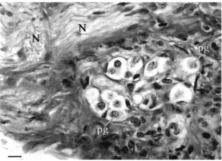
Figure 3. Photomicrograph of mediastinal paraganglionic tissue stained by the M artius scarlet, blue method. Collection of small clear presumptive paraganglionic (pg) cells lying in the mediastinal connective tissues adjacent to a small vein (not show n). N, branch of the left vagus nerve. Bar, 20 µm.
Table 1. Distribution of presumptive thoracic paraganglionic (PTP) tissue in the aortic-pulmonary region of marmosets.
Discrete islands of PTP tissue Animal
APR LSA
No. Sex Weight (g) No. Size (µm) No.
1 M 370 1 100 0
2 M 385 4 100, 200, 50, 150 0
3 M 312 2 500, 200 0
4 M 328 4 300, 50, 50, 50 0
5 F 338 4 150, 50, 50 0
6 F 309 2 100, 300 0
7 M 314 2 50, 100 0
Animals 1 and 2 w ere untreated controls; animals 3-7 treated w ith M PTP, a dopamine neurotoxic agent, as part of a parallel study (see M ethods). APR, aortic-pulmonary region; LSA, left subclavian artery.
ment of extrahilar left pulmonary vessels lying normal to the plane of section.
mis-even lay in close association with venous endothelium (Figure 4). In all the animals, a small nerve branched out around groups of these mediastinal cells and in some sections cells were interspersed along the path of the nerve (Figure 5). This neural arrangement occurred irrespective of the size and medias-tinal position of the PTP tissue, and particu-larly applied to cells observed in the aortic-pulmonary septum and in a periadventitial position adjacent to the aortic arch. Unfortu-nately, the limitations of light microscopy excluded a detailed description of the cellu-lar-neural relationship.
The appearance of the cells in the groups of PTP tissue was similar by light micro-scopic standards in all the animals. Each cell was oval or rounded in appearance, had a diameter between 10-15 µm and possessed a central nucleus and clear cytoplasm. The clear appearance of the cytoplasm in these cells in the thorax contrasted sharply with the orange-red tinctorial appearance of type 1 cells we have observed in the primate carotid body (11) and the carotid body of other non-primate species (12,13) using an identical MSB stain. In all the serial sections examined none of the cells in thoracic paraganglionic tissue possessed a compan-ion cell reminiscent of the arrangement of type 1 and type 2 cells found in the carotid body. In the larger arrangements of paragan-glionic tissue a fine matrix of mediastinal connective tissue embraced the cells so that to the casual observer fibroblast nuclei might be mistaken for type 2-like cell nuclei. A more careful examination of the tissue in many sections revealed that such an arrange-ment did not exist.
Marm o se t: o rigin o f le ft subclavian arte ry
With reference to Figure 2, it was pos-sible to examine in all seven animals the perivascular tissues in the region of the ori-gin of the left subclavian artery. No PTP tissue was found (Table 1).
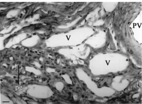
Figure 4. Collection of small clear paraganglionic (pg) cells lying adjacent to small veins (V) in the mediastinal connective tissues. PV, small extra-hilar pulmonary vein. Bar, 20 µm.
Figure 5. Small clear paraganglionic (PG) cells scattered in the course of a branch of the left vagus nerve (N) lying adjacent to the aortic w all (A). Bar, 50 µm.
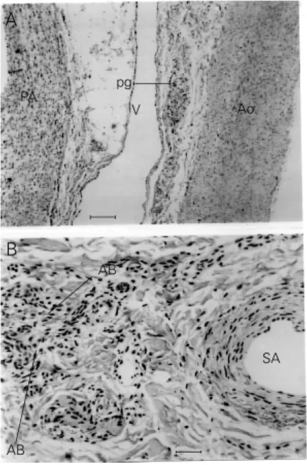
Cat (Felix d om esticus)
morpho-logical features similar to the paraganglionic cells described above for the marmoset (Fig-ure 6A), and the second group showed char-acteristics typical of aortic body chemore-ceptors consisting of both type 1 and type 2 cells (Figure 6B). It is to be noted that neither the mediastinal small clear cells in the cat
nor the type 1 cells of the cats aortic bodies stained particularly well with the MSB stain.
Marm o se t: o rigin o f right subclavian arte ry
No paraganglionic cells were found in the perivascular tissues surrounding the ori-gin of the right subclavian artery in any of the three animals studied.
D iscussio n
The original objective of the present study was to establish the distribution of aortic body chemoreceptor cells in a non-human primate, as a necessary preliminary to exam-ining under controlled conditions their re-flex control of the respiratory and cardiovas-cular systems. In fact the main finding of this study was somewhat surprising to us, since the small groups of paraganglionic cells found in the aortic-pulmonary region did not have classical characteristics associated with aor-tic body chemoreceptors in non-primate ani-mals (2,5,6,10,15-17, and present paper). Furthermore, no such cells were found in the perivascular tissues in the regions of the origins of the left and right subclavian arter-ies.
Our findings have therefore presented a major problem as to the identity of these paraganglionic cells. On purely morphologi-cal grounds the association of the marmoset cells with branches of the left vagus nerve would suggest that they have a similar origin to other paraganglionic cells and that they are neural crest in origin. Our observation that cells with similar characteristics to those noted in the marmoset were also observed in close proximity to typical type 1 cells in aortic bodies of the cat lends support to their common origin from the neural crest. Fur-ther morphological comparisons between marmoset paraganglionic and aortic body type 1 cells only emphasised differences between the two populations of cells. For instance the groups of paraganglionic cells
in the marmoset lacked a connective tissue capsule found in aortic bodies (2,5,18); the marmoset cells received their blood supply from connective tissue arterioles rather than from a stem vessel (2,10); the marmoset cells did not have the profuse capillary bed associated with chemoreceptor tissue, and there appeared to be an absence of type 2 cells in the animals we studied.
The clear cytoplasm associated with the marmoset mediastinal cells is probably an artefact due to diffusion-fixation. Unfortu-nately, we did not have sufficient freedom of access to the marmosets to prepare our mate-rial by the perfusion-fixation employed in our other studies (11-13). An ultrastructural study could probably demonstrate cytologi-cal characteristics shared with type 1 cells, but would be very difficult to carry out in view of the protean distribution of these cells. In the mediastina of non-primate ani-mals collections of small clear cells have also been observed along the course of the left vagus nerve and described as being simi-lar to small intensely fluorescent cells that are found in autonomic ganglia (19). The small clear cells we have observed are slightly larger than small intensely fluorescent cells. It could be that the small clear paragan-glionic cells are a morphological variant of the aortic bodies, as indeed there are struc-tural differences between the aortic bodies and the carotid bodies (20). In that study the blood supply to the subclavian body of the rat was described as arising from arterioles rather than from stem vessels, as we have described for the groups of paraganglionic cells in the marmoset. In this connection it has been shown in another marmoset not included in this study that some of the type 1 cells in the aortic region showed a similar morphology to those in the aortic-pulmo-nary area and gave a positive staining reac-tion to tyrosine hydroxylase typical of cells in the carotid bodies (Clarke JA, unpub-lished observation). Furthermore, in the cat, not only groups of cells whose structure was
similar to aortic body chemoreceptors have been described but also groups of paragan-glionic cells that lay on the surface of the atria and base of the heart (epicardial) with a different structure (17). This latter group of cells, however, is outside the region covered in the present investigation. Thus, in a situa-tion where the morphological identity of these small clear cells in the mediastina of marmosets is unclear, it would be premature to speculate on their exact function.
The results of physiological studies in some non-human primates (dog, cat), in which aortic chemoreceptors are abundant, indicate that this group of chemoreceptors differ quantitatively from those in the ca-rotid bodies with respect to both their re-sponses to specific stimuli and their reflex functions (14,21). Consequently, it is no longer possible to consider them functioning as a single entity in the control of the respira-tory and cardiovascular systems nor to apply the results of these studies to humans. Our present investigation revealing the apparent lack of classical aortic bodies in the marmo-set which might have been a suitable species for subsequent appropriate physiological studies, could be atypical for other non-human primates. A survey of the mediastina of other primates would therefore appear to be required to establish the thoracic chemoreceptor status of these animals.
negligible role, there is, in our opinion, no unequivocal evidence for any role of the aortic bodies in the control of the cardiovas-cular system (21).
Ackno wle dgm e nts
We wish to express our thanks to Dr. R.K.B. Pearce, Pharmacology Group, Bio-medical Sciences Division, Kings College London, to Drs. R.M. Baker and H.F. Baker,
Department of Experimental Psychology, Cambridge University for kindly supplying us with the specimens, and to Mr. Michael Jackson for technical assistance. We also thank Mrs. Beverley Wilson of the Depart-ment of Pathology, Addenbrooks Hospital, Cambridge, for her expert assistance in the histological preparation of the material, and Mr. C.F.J. Henn for his artistic contribution (Figures 1 and 2).
Re fe re nce s
1. Krahl VE (1962). The glomus pulmonale: its location and microscopic anatomy. In: de Reuck AVS & O’Connor M (Editors),
Pulmonary Structure and Function. Ciba Foundation Symposium, Churchill, Lon-don, UK.
2. Nonidez JF (1935). The aortic (depressor) nerve and its associated epithelioid body, the glomus aorticum. American Journal of Anatomy, 57: 259-301.
3. Knoche H & Schmitt G (1963). Über chemo- und Pressoreceptorenfelder am Coronarkreislauf. Zeitschrift für Zellfor-schung und M ikroskopische Anatomie, 61: 524-560.
4. Verity M A, Hughes T & Bevan JA (1964). Aortico-pulmonary glomus tissue distribu-tion and blood supply in the adult cat.
Science, 145: 172-173.
5. Coleridge HM , Coleridge JCG & How e A (1967). A search for pulmonary arterial chemoreceptors in the cat, w ith a com-parison of the blood supply of the aortic bodies in the new -born and adult animal.
Journal of Physiology, 191: 353-374. 6. Coleridge HM , Coleridge JCG & How e A
(1970). Thoracic chemoreceptors in the dog. A histological and physiological study of the location, innervation and blood sup-ply of the aortic bodies. Circulation Re-search, 26: 235-247.
7. Böck P (1982). The Paraganglia. Springer-Verlag, Berlin, Germany.
8. Pearce RKB, Jackson M , Smith L, Jenner P & M arsden CD (1994). Chronic L-dopa administration induces dyskinesias in the M PTP-treated marmoset. British Journal of Pharmacology, 112: 150P (Abstract). 9. Daw es RE & Hillier M H (1964). A
one-stage technique for differentiating the
al-pha and beta cells of the anterior pituitary.
Journal of M edical Laboratory Technol-ogy, 21: 62-63.
10. How e A (1956). The vasculature of the aortic bodies in the cat. Journal of Physiol-ogy, 134: 311-318.
11. Clarke JA, Daly M deB, Ead HW & Kreclo-vic G (1993). A morphological study of the size of the vascular compartment of the carotid body in a non-human primate (Cercopithicus ethiopus), and a compari-son w ith the cat and rat. Acta Anatomica, 147: 240-247.
12. Clarke JA & Daly M deB (1981). Distribu-tion of carotid body type-I cells and other periadventitial type-I cells in the carotid bifurcation regions of the rabbit. Cell and Tissue Research, 216: 603-614. 13. Clarke JA & Daly M deB (1983).
Distribu-tion of carotid body type-I cells and other periadventitial type-I cells in the carotid bifurcation regions of the cat. Anatomy and Embryology, 166: 169-189.
14. Lahiri S (1991). Oxygen biology of periph-eral chemoreceptors. In: Lahiri S, Cherni-ack NS & Fitzgerald RS (Editors), Re-sponse and Adaptation to Hypoxia. Ameri-can Physiological Society, New York, NY, USA, 95-106.
15. M uratori G (1935). Connessioni tra tes-suto paragangliare e zone recettrici aor-tiche in vari mammiferi. M onitore Zoolo-gico Italiano, 45: 300-310.
16. Nonidez JF (1937). Distribution of the aor-tic nerve fibers and the epithelioid bodies (supracardial ‘paraganglia’) in the dog.
Anatomical Record, 69: 299-313. 17. Goormaghtigh N & Pannier R (1939). Les
paraganglions du coeur et des zone vaso-sensibles carotidienne et cardio-aortique
chez le chat adulte. Archives de Biologie, 50: 455-533.
18. M cDonald DM & Blew ett RW (1981). Lo-cation and size of carotid body-like organs (paraganglia) revealed in rats by the per-meability of blood vessels to Evans blue dye. Journal of Neurocytology, 10: 607-643.
19. Kummer W & Neuhuber WL (1989). Vagal paraganglia of the rat. Journal of Electron M icroscopy, 12: 343-355.
20. Hansen JT (1981). Innervation of the rat aortic (subclavian) body: an ultrastructural study follow ing axonal degeneration.
Journal of Ultrastructure Research, 74: 83-94.
21. Daly M deB (1997). Peripheral Arterial Chemoreceptors and Respiratory-Cardio-vascular Integration. M onograph of the Physiological Society, No. 46. Clarendon Press, Oxford, UK.
22. Busacchi P (1912). I corpi cromaffini del cuore umano. Parte prima. I corpi cromaf-fini del tronco arterioso. Archivio Italiano di Anatomia e di Embriologia, 11: 352-376.
23. Penitschka W (1931). Paraganglion aorti-cum supracardiale. Zeitschrift für M ikros-kopisch-Anatomische Forschung, 24: 24-37.
24. Palme F (1934). Die Paraganglien über dem Herzen und im Endigungsgebiet des Nervus depressor. Zeitschrift für M ikros-kopisch-Anatomische Forschung, 36: 391-420.