Tatiana Kelly da Silva Fidalgo(a)
Roberta Barcelos(b)
Maristela Barbosa Portela(c)
Rosangela Maria de Araújo Soares(d)
Rogério Gleiser(c)
Fernando Costa e Silva-Filho(e)
(a) School of Dentistry of Rio de Janeiro, Federal University of Rio de Janeiro, Rio de Janeiro, RJ, Brazil.
(b) Department of Specific Formation, School of Dentistry, Fluminense Federal University, Nova Friburgo, RJ, Brazil.
(c) Department of Pediatric Dentistry and Orthodontics, School of Dentistry, Federal University of Rio de Janeiro, Rio de Janeiro, RJ, Brazil.
(d) Professor Paulo de Goes Institute of Microbiology, University of Rio de Janeiro, Rio de Janeiro, RJ, Brazil.
(e) Carlos Chagas Filho Biophysics Institute, Federal University of Rio de Janeiro, Rio de Janeiro, RJ, Brazil.
Corresponding author: Tatiana Kelly da Silva Fidalgo Rua Joaquim Távora 244/202 - Icarai Niterói - RJ - Brazil
CEP: 24230-541
E-mail: [email protected]
Received for publication on Feb 06, 2010 Accepted for publication on Aug 09, 2010
Inhibitory activity of root canal irrigants
against
Candida albicans
,
Enterococcus
faecalis
and
Staphylococcus aureus
Abstract: The present study evaluated the antimicrobial activity of three root canal irrigants against Enterococcus faecalis, Candida albicans, and Staphylococcus aureus. These microorganisms were incubated in the presence of citric acid (6 and 10%), EDTA (17%), and NaOCl (0.5, 1.0, 2.5, and 5.25%). Agar diffusion tests were performed and redox indi-cator resazurin was used to evaluate the inhibitory effect of the irrig-ants on the metabolic activity of these microorganisms. The mean diam-eters of the inhibition zones for the C. albicans cultures were 11.6 mm (17% EDTA), 5.5 mm (0.5% NaOCl), 12.9 mm (1% NaOCl), 22.1 mm (2.5% NaOCl), and 28.5 mm (5.25% NaOCl). The mean diameters of the inhibition zones for E. faecalis were 2.8 mm (1% NaOCl), 5.4 mm (2.5% NaOCl), and 8.3 mm (5.25% NaOCl). For S. aureus, the mean values were 8.0 mm (17% EDTA), 3.0 mm (1% NaOCl), 8.8 mm (2.5% NaOCl), and 10.0 mm (5.25% NaOCl). Most of the irrigant solutions presented effective antimicrobial activity against C. albicans. A high in-hibitory effect on the metabolic activity of E. faecalis was detected when the microorganisms were incubated with 17% EDTA. The same result was reached when S. aureus was incubated in the presence of ≥ 2.5% NaOCl. Altogether, these results indicate that 2.5% and 5.25% NaOCl are microbicides against S. aureus while 0.5% and 1% NaOCl are only microbiostatic against the tested bacteria. The 6% and 10% citric acid as well as 17% EDTA did not affect the viability of any of the assayed microorganisms.
Descriptors:Candida albicans; Enterococcus faecalis; Staphylococcus aureus; Root Canal Irrigants.
Introduction
Microorganisms are the major causative factor associated with end-odontic treatment failure.1,2 The success of endodontic treatment depends
on the reduction or elimination of bacteria present in the root canal sys-tem. Residual pulpal tissue, bacteria, and dentine debris may persist in the irregularities of root canal systems, even after meticulous mechanical preparation.3 Therefore, irrigant solutions should be used in
combina-tion with canal preparacombina-tion.4-6
canal.7,8 Chemomechanical preparation is one of
the most important phases of endodontic treatment and irrigants such as sodium hypochlorite (NaOCl), citric acid, and ethylene diamine tetra-acetic acid (EDTA) are commonly used. The eficacy of these procedures also depends upon the vulnerability of the species involved.9
Many in vitro studies relate the antimicrobial activity of root canal irrigants against microorgan-isms, but studies on the metabolic activity of micro-organisms after contact with these irrigants was not found in the literature.5,6 Therefore, this study irst
evaluated the effectiveness of NaOCl (0.5, 1, 2.5, 4, and 5.25%), EDTA (17%), and citric acid (6 and 10%) against Enterococcus faecalis, Candida albi-cans,and Staphylococcus aureus, and then assessed their metabolic activities under the same conditions.
Material and Methods
Microorganisms
The following microorganisms and their related ATCC strains were used throughout: Staphylo-coccus aureus (ATCC 6538), Enterococcus faeca-lis (ATCC 29212) and Candida albicans (ATCC 10231). All microorganisms were grown in BHI-agar medium (Difco, Rio de Janeiro, RJ, Brazil) supplemented with 10% sheep blood (24 h, 37°C, 5% CO2 atmosphere or in anaerobiosis). The mi-croorganisms were then collected with a Drigalsky spatula for standardization of quantities collected and resuspended in sterile 0.01 M phosphate buffer pH 7.2, containing 0.15 M NaCl (PBS pH 7.2). The quantity of all microorganisms was adjusted using McFarland’s scale (McFarland Standard no. 0.5) and spectrophotometrically adjusted to 105 CFU.
ml−1 through optical density measurement at 600 nm
(OD600).
Root canal irrigants
The root canal irrigants used were: citric acid (6 and 10% - VETEC, Rio de Janeiro, RJ, Brazil), EDTA (17% - Biodinâmica, Rio de Janeiro, RJ, Bra-zil), and NaOCl (0.50, 1.00, 2.50, and 5.25% - Fór-mula & Ação, São Paulo, SP, Brazil). All of these compounds were diluted in sterile PBS pH 7.2.
Agar Diffusion Test (ADT)
For this test, nitrocellulose membranes (13.0 mm - Catalogue No GSWP04700; Millipore Co.,
Bil-lerica, MA, USA) impregnated with the root canal irrigants (30 µl each), described above, were placed on the top of S. aureus, E. faecalis,and C. albicans
(0.1 ml, 105 CFU.ml−1, resuspended in PBS pH 7.2)
homogeneously spread onto separated Petri dishes (10 cm diameter) containing BHI agar medium. The microorganism cultures were kept in an incubator (24 h, 37°C, 5% CO2 atmosphere or in anaerobio-sis) before measuring the growth inhibition zones. Cell viability (negative control) was determined by incubating the bacteria with Bactrin (Sulfametoxa-zol-trimetoprima - RJ 0401, Roche, Rio de Janeiro, RJ, Brazil) and the fungus with Fluconazole (Ache, Rio de Janeiro, RJ, Brazil); and the positive controls consisted in incubation of the same microorgan-isms in PBS pH 7.2. The inhibition zone limit was measured from the edge of the membrane disk to the end of the inhibition zone. A millimeter mark-ing rule was used for this purpose.9 All assays were
done in triplicate.
Metabolic activity
In order to collect a standard quantity of sam-ples submitted to ADT, the authors developed the following method. Briely, micropipette tips (S1111-0006; TipOne; Blakelands, MK, UK) with 15 mm cut off from the thinner end were used to collect a ixed (standard) quantity of sample from the inhibi-tion zone. For each sample collecinhibi-tion, the tip was inserted 1 mm from the nitrocellulose membrane, and then the collected sample was resuspended in PBS (300 µl). Equivalent selection points to collect the microorganism are essential to maintain the same parameters for all microorganisms studied. To adjust the quantity of microorganisms to be tested, OD530 of the samples was taken using PBS as a blank solution. The OD530 of all samples was ~0.5.
Microorganisms (100 µl), in the above condi-tions, were reacted (3 h, 37°C) with resazurin10
spectro-metrically at 530 nm (OD530).
Statistical analysis
The data were analyzed by the SPSS 16.0 (SPSS Inc, Chicago, IL, USA) software using the analysis of variance (ANOVA) and the Tukey (p < 0.05) test.
Results
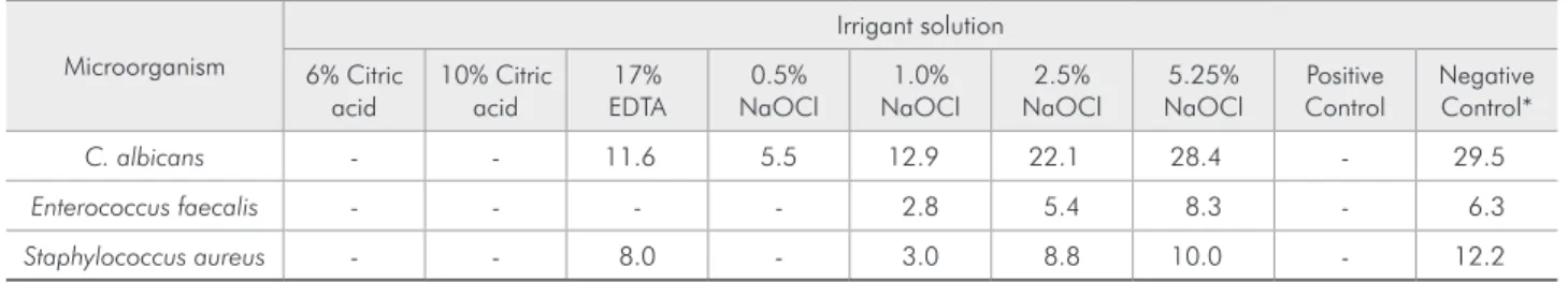
The data summarized in Table 1 show that in-cubation of the microorganisms with citric acid (6 or 10%) did not inhibit their growth, since inhibi-tion zones were not apparent (data not shown). Neither 17% EDTA nor 0.5% NaOCl inhibited the growth of Enterococcus fecalis, and the latter did not have any effect on Staphylococcus aureus either. In contrast, 17% EDTA presented activity against both Candida albicans and Staphylococcus aureus. NaOCl at higher concentrations (1, 2.5, and 5.25%) presented inhibitory activity against all microorgan-isms tested. Furthermore, the antimicrobial effect of 5.25% NaOCl was comparable to the results pre-sented by the negative controls (Bactrim and Fluco-nazole). EDTA (17%) presented higher antimicro-bial activity than 0.5% NaOCl, when tested against
Candida albicans and Staphylococcus aureus. The effect of EDTA (17%) on Candida albicans was similar to that exhibited by 1% NaOCl, but more effective on Staphylococcus aureus. Higher concen-trations of NaOCl (2.5 and 5.25%) were more ef-fective on Candida albicans than 17% EDTA, how-ever their activities on Staphylococcus aureus were similar.
EDTA (17%) presented more effective antimicro-bial activity than citric acid (6 and 10%) against all assayed microorganisms. It can also be noted that
the effectiveness of NaOCl on E. faecalis was dose-dependent. Furthermore, 5.25% NaOCl was lethal to all assayed microorganisms (Table 2).
To evaluate the microbicidal or microbiostatic activity of the root canal irrigants studied here, re-dox analyses of samples taken from the inhibition zones were carried out. Candida albicans and En-terococcus faecalis metabolic activities were de-tected in all tested root canal irrigants (that formed inhibition zones). However, no metabolic activity of
Staphylococcus aureus was detected for 2.5% and 5.25% NaOCl (Table 3 and Graph 1).
Discussion
Chemomechanical procedure plays an important role in reducing microorganisms in the root canal.11
Previous studies have shown that irrigation with 0.5% NaOCl eliminated bacteria in 50% to 75% of infected root canals at the end of the irst treat-ment.12 In the present study, only Candida albicans
was sensitive to 0.5% NaOCl. However, all micro-bial species tested were sensitive to 5.25% NaOCl. EDTA is an auxiliary substance that has a chelating action, biocompatibility with the periapical tissues13
and optimal cleansing abilities.14 In the current
study, 17% EDTA showed a superior antimicrobial effect in the inhibition zone test against Candida al-bicans when compared to0.5% NaOCl.
Enterococcus faecalis is considered one of the most resistant species in the oral cavity and a pos-sible cause of failure in root canal treatments. It can survive after instrumentation and irrigation with NaOCl up to 2.5%.15 The indings of the present
study showed that 5.25% NaOCl had a signiicantly better performance than 2.5% NaOCl against this
Table 1 - Inhibition zones (mm) detected when microorganisms were cultured in the presence of different concentrations (%) of citric acid, EDTA or NaOCl.
Microorganism
Irrigant solution
6% Citric acid
10% Citric acid
17% EDTA
0.5% NaOCl
1.0% NaOCl
2.5% NaOCl
5.25% NaOCl
Positive Control
Negative Control*
C. albicans - - 11.6 5.5 12.9 22.1 28.4 - 29.5
Enterococcus faecalis - - - - 2.8 5.4 8.3 - 6.3
Staphylococcus aureus - - 8.0 - 3.0 8.8 10.0 - 12.2
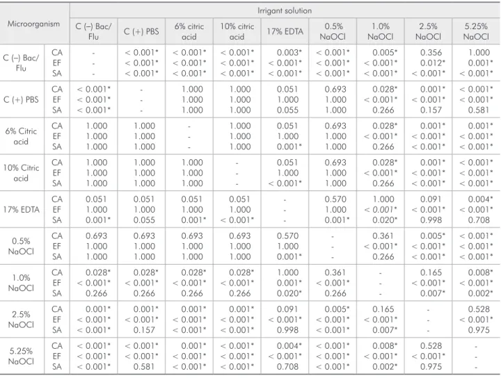
Table 2 - A comparative statistical analysis of the data obtained from assays carried out to generate inhibition zones by
in-cubating the microorganisms Staphylococcus aureus (SA), Enterococcus faecalis (EF), and Candida albicans (CA) with different
concentrations (%) of canal irrigant solutions
Microorganism
Irrigant solution C (−) Bac/
Flu C (+) PBS
6% citric acid
10% citric
acid 17% EDTA
0.5% NaOCl 1.0% NaOCl 2.5% NaOCl 5.25% NaOCl
C (−) Bac/ Flu CA EF SA -< 0.001* < 0.001* < 0.001* < 0.001* < 0.001* < 0.001* < 0.001* < 0.001* < 0.001* 0.003* < 0.001* < 0.001* < 0.001* < 0.001* < 0.001* 0.005* < 0.001* < 0.001* 0.356 0.012* < 0.001* 1.000 0.001* < 0.001*
C (+) PBS CA EF SA < 0.001* < 0.001* < 0.001* -1.000 1.000 1.000 1.000 1.000 1.000 0.051 1.000 0.055 0.693 1.000 1.000 0.028* < 0.001* 0.266 0.001* < 0.001* 0.157 < 0.001* < 0.001* 0.581 6% Citric acid CA EF SA 1.000 1.000 1.000 1.000 1.000 1.000 -1.000 1.000 1.000 0.051 1.000 0.001* 0.693 1.000 1.000 0.028* < 0.001* 0.266 0.001* < 0.001* < 0.001* 0.001* < 0.001* < 0.001* 10% Citric acid CA EF SA 1.000 1.000 1.000 1.000 1.000 1.000 1.000 1.000 1.000 -0.051 1.000 < 0.001* 0.693 1.000 1.000 0.028* < 0.001* 0.266 0.001* < 0.001* < 0.001* < 0.001* < 0.001* < 0.001* 17% EDTA CA EF SA 0.051 1.000 0.001* 0.051 1.000 0.055 0.051 1.000 0.001* 0.051 1.000 < 0.001* -0.570 1.000 0.001* 1.000 < 0.001*
0.020* 0.091 < 0.001* 0.998 0.004* < 0.001* 0.708 0.5% NaOCl CA EF SA 0.693 1.000 1.000 0.693 1.000 1.000 0.693 1.000 1.000 0.693 1.000 1.000 0.570 1.000 0.001* -0.361 < 0.001* 0.266 0.005* < 0.001* < 0.001* < 0.001* < 0.001* < 0.001* 1.0% NaOCl CA EF SA 0.028* < 0.001* 0.266 0.028* < 0.001* 0.266 0.028* < 0.001* 0.266 0.028* < 0.001* 0.266 1.000 0.001* 0.020* 0.361 < 0.001* 0.266 -0.165 < 0.001* 0.007* 0.008* < 0.001* 0.002* 2.5% NaOCl CA EF SA 0.001* < 0.001* < 0.001* 0.001* < 0.001* 0.157 0.001* < 0.001* < 0.001* 0.001* < 0.001* < 0.001* 0.091 < 0.001* 0.998 0.005* < 0.001* < 0.001* 0.165 < 0.001* 0.007* -0.528 < 0.001* 0.975 5.25% NaOCl CA EF SA < 0.001* < 0.001* < 0.001* < 0.001* < 0.001* 0.581 0.001* < 0.001* < 0.001* < 0.001* < 0.001* < 0.001* 0.004* < 0.001* 0.708 < 0.001* < 0.001* < 0.001* 0.008* < 0.001* 0.002* 0.528 < 0.001* 0.975
-*Values of statistical significance (p < 0.05). C (−) and C (+) are respectively related to microorganisms which were incubated in root canal irrigant-depleted medium (−) or PBS (+) before cultivation in BHI medium.
Microorganisms
Irrigant solution
C (−) 17% EDTA 0.5% NaOCl 1.0% NaOCl 2.5% NaOCl 5.25% NaOCl
Candida albicans 0.0015 0.0170 0.0370 0.0065 0.0095 0.0040
Enterococcus faecalis 0.0080 NA NA 0.0460 0.0225 0.0045
Staphylococcus aureus 0.0305 0.0015 NA 0.0010 0 0
No significant (p < 0.05) differences could be seen between groups. Numbers represent the diameters of the inhibition zones. NA = not applicable (In these cases, inhibition zones were not formed in the diffusion agar test, so the metabolic activity test was not applicable). C (−) = Negative control
Table 3 - Metabolic activity of the microorganisms before
[C (−)] and after their
incubation with different concentrations (%) of EDTA or NaOCl
microorganism.
Chelate and acidic solutions, including EDTA and citric acid, have been recommended for remov-ing the smear layer from instrumented root canals.16
same authors showed an inhibition zone using 10% citric acid against Enterococcus faecalis. Although citric acid presents low cytotoxity as an advantage,17
in the present study, 6% and 10% citric acid did not form any inhibition zone against any of the microor-ganisms tested. In agreement with Grawehr et al.,18
the current study demonstrated that 17% EDTA was more effective than 0.5% NaOCl against Candida albicans and Staphylococcus aureus. The mainte-nance of microorganisms in contact with EDTA over a prolonged period is as lethal as short periods of contact, increases the permeability of the outer membrane to hydrophobic molecules, and improves the action of antibacterial agents.19 Branin at al,20
reported that the metal chelator EDTA is known to have activity against bioilms of gram-positive bac-teria such as Staphylococcus aureus.
The agar diffusion test does not distinguish mi-crobiostatic and microbicidal properties of dental materials neither does it provide any information about the microorganisms viability after the test.21
The bacteria around the inhibition zone might grow back after some days. In clinical practice, it may be possible that after contact with root canal irrigants, microorganisms could still remain viable; this would depend on the irrigant and its concentration. For that reason it is important to associate the metabolic activity to the agar diffusion test, when evaluating the inhibitory activity of root canal irrigants.
Even after irrigation of the root canal with an antimicrobial solution, it may not be possible to eliminate all microorganisms from the root canal.22
The microorganisms may multiply rapidly in 2-4 days, almost returning to their original numbers, if the canal is not illed with an antimicrobial sub-stance between visits.12 In the present study, a low
metabolic activity that was not statistically signii-cant was noted. Perhaps the low activity could be explained by the short period of incubation, since the metabolic activity was evaluated after only 24 h. More studies are necessary to evaluate metabolic ac-tivity after longer periods. The high pH of NaOCl interferes in cytoplasmatic membrane integrity with irreversible enzymatic inhibition, biosynthetic al-terations in cell metabolism and phospholipids de-struction,9 and probably 24 h was not suficient to
allow bacteria recuperation, the same occurred with 17% EDTA against Candida albicans and Staphylo-coccus aureus.
In the inhibition zone test, Candida albicans,
Enterecoccus faecalis and Staphylococcus aureus
were sensitive to 1% NaOCl and higher concentra-tions. However, only 2.5 and 5.25% NaOCl were conirmed as microbicidal irrigants for Staphylococ-cus aureus after the metabolic activity test. The as-sociation of the two tests demonstrated that the oth-er irrigants tested had a microbiostatic effect against
Candida albicans and Enterecoccus faecalis. These results suggest that microorganism eradication in an endodontic infection may be obtained in association with other measures such as intracanal medication between sessions. Moreover, the results obtained by association of the two tests demonstrated the impor-tance of the metabolic activity test as a complemen-Graph 1 - Metabolic activity of
Candida albicans, Enterococcus
faecalis and Staphylococcus aureus
after their incubation for 24 h with EDTA and NaOCl.
0.05
0.045
C (−) EDTA 0.5%
NaOCl
1.0% NaOCl
2.5% NaOCl
5.25% NaOCl 0.04
0.035
0.03
0.025
0.02
0.015
0.01
0.005
0
Metabo
lic
A
ctiv
ity
(A
bs
)
Candida albicans
Enterococcus faecalis
Staphylococcus aureus
0
.0
0
1
5 0.0
0
8
0
.0
3
0
5
0
.0
1
7
0 0
.0
0
1
5
0
.0
3
7
0 0
0
.0
0
6
5
0
.0
4
6
0
.0
0
1
0
.0
0
9
5
0
.0
2
2
5
0
0
.0
0
4
0
.0
0
4
5
tary test to assess antimicrobial properties of root canal irrigants.
The present results support an initial hypothesis that there is a metabolic activity around the inhibi-tion zone after the inhibiinhibi-tion test, although without statistical signiicance. The short period of incuba-tion may contribute to this result. Further studies are necessary to evaluate metabolic activity over a prolonged period.
Conclusion
Citric acid did not present antimicrobial activity. Only 2.5% and 5.25% NaOCl presented
antimicro-bial activity against Staphylococcus aureus. In addi-tion, 17% EDTA, 0.5% and 1% NaOCl presented only microbiostatic activity against some of the mi-croorganisms tested. The highest concentration of NaOCl (5.25%) presented superior antimicrobial activity when compared with other irrigants used.
Acknowledgements
The authors would like to thank Dr. Catarina Akiko Miyamoto from CNRMN-UFRJ for review-ing this manuscript, for grammar and style. This study was supported by the following Brazilian agencies: FUJB-UFRJ, FAPERJ and INPeTAm.
References
1. Siqueira Jr JF. Endodontic infections: concepts, paradigms, and perspectives. Oral Surg Oral Med Oral Pathol Oral Radiol Endod. 2002 Sep;94(3):281-93.
2. Siqueira Jr JF, Rocas IN. Polymerase chain reaction-based analysis of microorganisms associated with failed endodon-tic treatment. Oral Surg Oral Med Oral Pathol Oral Radiol Endod. 2004 Jan;97(1):85-94.
3. Abou-Rass M, Piccinino MV. The effectiveness of four clinical irrigation methods on the removal of root canal debris. Oral Surg Oral Med Oral Pathol. 1982 Sep;54(3):323-8.
4. D’Arcangelo C, Di Nardo Di Maio F, Stracci N, Spoto G, Malagnino VA, Caputi S. Pulp-dissolving ability of several endodontic irrigants: a spectrophotometric evaluation. Int J Immunopathol Pharmacol. 2007 Apr;20(2):381-6.
5. Nudera WJ, Fayad MI, Johnson BR, Zhu M, Wenckus CS, Begole EA, et al. Antimicrobial effect of triclosan and triclosan with Gantrez on five common endodontic pathogens. J Endod. 2007 Oct;33(10):1239-42.
6. Oliveira DP, Barbizam JV, Trope M, Teixeira FB. In vitro antibacterial efficacy of endodontic irrigants against Entero-coccus faecalis. Oral Surg Oral Med Oral Pathol Oral Radiol Endod. 2007 May;103(5):702-6.
7. Siqueira Jr JF, Rocas IN, Santos SR, Lima KC, Magalhaes FA, de Uzeda M. Efficacy of instrumentation techniques and irrigation regimens in reducing the bacterial population within root canals. J Endod. 2002 Mar;28(3):181-4.
8. Fidalgo TKS, Barcelos R, Petrópolis DB, Azevedo BR, Primo LG, Silva FC. Citotoxidade de diferentes concentrações de hi-poclorito de sódio sobre osteoblastos humanos. RGO (Porto Alegre). 2009 Jul;57(3):317-21.
9. Estrela C, Ribeiro RG, Estrela CR, Pecora JD, Sousa-Neto MD. Antimicrobial effect of 2% sodium hypochlorite and 2% chlorhexidine tested by different methods. Braz Dent J. 2003;14(1):58-62.
10. Mariscal A, Lopez-Gigosos RM, Carnero-Varo M, Fernandez-Crehuet J. Fluorescent assay based on resazurin for detection of activity of disinfectants against bacterial biofilm. Appl Microbiol Biotechnol. 2009; 82(4): 773-8311.
11. American Academy of Pediatric Dentistry. Guideline on pulp therapy for primary and young permanent teeth. Pediatr Dent. 2004;26(7 Suppl):115-9.
12. Bystrom A, Sundqvist G. Bacteriologic evaluation of the effect of 0.5 percent sodium hypochlorite in endodontic therapy. Oral Surg Oral Med Oral Pathol. 1983 Mar;55(3):307-12. 13. Segura JJ, Calvo JR, Guerrero JM, Jimenez-Planas A,
Sampe-dro C, Llamas R. EDTA inhibits in vitro substrate adherence capacity of macrophages: endodontic implications. J Endod. 1997 Apr;23(4):205-8.
14. Garberoglio R, Becce C. Smear layer removal by root canal irrigants. A comparative scanning electron microscopic study. Oral Surg Oral Med Oral Pathol. 1994 Sep;78(3):359-67. 15. Gomes BP, Ferraz CC, Vianna ME, Berber VB, Teixeira FB,
Souza-Filho FJ. In vitro antimicrobial activity of several con-centrations of sodium hypochlorite and chlorhexidine gluco-nate in the elimination of Enterococcus faecalis. Int Endod. J 2001 Sep;34(6):424-8.
16. Arias-Moliz MT, Ferrer-Luque CM, Espigares-Rodriguez E, Liebana-Urena J, Espigares-Garcia M. Bactericidal activity of phosphoric acid, citric acid, and EDTA solutions against Enterococcus faecalis. Oral Surg Oral Med Oral Pathol Oral Radiol Endod. 2008 Aug;106(2):84-9.
17. Guimarães LF, Fidalgo TK, Menezes GM, Primo LG, Silva-Filho FC. Effects of citric acid on cultured human osteoblastic cells. Oral Surg Oral Med Oral Pathol Oral Radiol Endod. In press 2010.
19. Nikaido H. Molecular basis of bacterial outer membrane per-meability revisited. Microbiol Mol Biol Rev. 2003;67(4):593-656.
20. Banin E, Brady KM, Greenberg EP. Chelator-induced dispersal and killing of Pseudomonas aeruginosa cells in a biofilm. Appl Environ Microbiol. 2006 Dec;72(3):2064-9.
21. Estrela C, Estrela CRA, Bammann LL, Pecora JD. Two meth-ods to evaluate the antimicrobial action of calcium hydroxide paste. J Endod.2001 Dec;27(12):720-3.