online | memorias.ioc.fiocruz.br
The antimicrobial activity of lapachol
and its thiosemicarbazone and semicarbazone derivatives
Marina Azevêdo Souza1, Susana Johann1,2/+,Luciana Alves Rodrigues dos Santos Lima3, Fernanda Fraga Campos1,2, Isolda Castro Mendes4, Heloisa Beraldo5,
Elaine Maria de Souza-Fagundes6,Patrícia Silva Cisalpino2, Carlos Augusto Rosa2 , Tânia Maria de Almeida Alves1, Nívea Pereira de Sá2, Carlos Leomar Zani1
1Laboratório de Química de Produtos Naturais, Centro de Pesquisas René Rachou-Fiocruz, Belo Horizonte, MG, Brasil 2Departamento de Microbiologia 6Departamento de Fisiologia e Biofísica, Instituto de Ciências Biológicas 4Escola de Belas Artes
5Departamento de Química, Instituto de Ciências Exatas, Universidade Federal de Minas Gerais, Belo Horizonte, MG, Brasil 3Universidade Federal de São João Del Rei, Campus Centro Oeste Dona Lindu, Divinópolis, MG, Brasil
Lapachol was chemically modified to obtain its thiosemicarbazone and semicarbazone derivatives. These com-pounds were tested for antimicrobial activity against several bacteria and fungi by the broth microdilution meth-od. The thiosemicarbazone and semicarbazone derivatives of lapachol exhibited antimicrobial activity against the bacteria Enterococcus faecalis and Staphylococcus aureus with minimal inhibitory concentrations (MICs) of 0.05
and 0.10 μmol/mL, respectively. The thiosemicarbazone and semicarbazone derivatives were also active against
the pathogenic yeast Cryptococcus gattii (MICs of 0.10 and 0.20 μmol/mL, respectively). In addition, the lapachol thiosemicarbazone derivative was active against 11 clinical isolates of Paracoccidioides brasiliensis, with MICs
ranging from 0.01-0.10 μmol/mL. The lapachol-derived thiosemicarbazone was not cytotoxic to normal cells at the concentrations that were active against fungi and bacteria. We synthesised, for the first time, thiosemicarbazone and
semicarbazone derivatives of lapachol. The MICs for the lapachol-derived thiosemicarbazone against S. aureus,E. faecalis, C. gattii and several isolates of P. brasiliensis indicated that this compound has the potential to be developed into novel drugs to treat infections caused these microbes.
Key words: antimicrobial agents - lapachol - Paracoccidioides
Lapachol (2-hydroxy-3-(3-methylbut-2-enyl)naph-thalene-1,4-dione) is a natural pigment that was origi-nally isolated from species of the Bignoniaceae family, but is also found in other plant families (Araújo et al. 2002, Fonseca et al. 2003). This naphthoquinone exhib-its interesting antimicrobial, antitumor, antiplasmodial, antioxidant and trypanocidal properties (Araújo et al. 2002). There are many literature reports on lapachol de-rivatives. However, the thiosemicarbazone and semicar-bazone derivatives of lapachol have never been studied. Thiosemicarbazones and semicarbazones from sources other than lapachol, such as isatin thiosemicarbazone and benzaldehyde semicarbazone, have been widely studied and are known to have extensive pharmacologi-cal profiles, e.g., antimicrobial and anticonvulsant ac-tivities (Beraldo 2004, Vieira et al. 2010).
In this study, we searched for novel lapachol de-rivatives with greater activity and lower toxicity for the treatment of infectious diseases. Treating infectious dis-eases that are caused by bacteria or fungi remains an im-portant and challenging public health problem (Jones et
Financial support: FIOCRUZ, CNPq, CAPES, FAPEMIG, INCT-INOFAR
+ Corresponding author:[email protected] Received 27 June 2012
Accepted 19 September 2012
al. 2008). Although substantial advances in biomedical science and public health have facilitated the control of numerous infectious diseases over the past century, the world has witnessed an increasing incidence and geo-graphical expansion of emerging and re-emerging infec-tious diseases (Jones et al. 2008, Yang et al. 2012). Dis-covering new antimicrobial agents is important because several pathogenic microorganisms have acquired resis-tance. The prevalence of Staphylococcus aureus strains that are resistant to conventional antibiotics has greatly increased in some hospitals (Fluit et al. 2001, Hidron et al. 2008, Rosenthal et al. 2010).
et al. 2002, Paniago et al. 2003, Travassos et al. 2008). The increase in antibacterial resistance and the restricted number of commercially available antifungal drugs em-phasise the need for the development of novel and more effective antifungal and antibacterial agents.
In this work, we evaluated the antimicrobial activity of the thiosemicarbazone and semicarbazone derivatives of lapachol, which we synthesised for the first time, and tested against several clinically important microorgan-isms. Lapachol was also tested. Lapachol and its thi-osemicarbazone derivative were also tested against hu-man peripheral blood mononuclear cells (PBMCs).
SUBJECTS, MATERIALS AND METHODS
Synthesis - Lapachol was isolated from Tabebuia ochracea in a previous study by Zani et al. (1991). Thi-osemicarbazide (Sigma-Aldrich, Steinheim, Germany) was purified by crystallisation from water. Acetone (Vetec, RJ, Brazil), methanol (Sigma-Aldrich), semi-carbazide hydrochloride (Sigma-Aldrich) and sodium hydroxide (Sigma-Aldrich, Steinheim, Germany) were used without further purification. Column chromatogra-phy was performed using silica gel 60G (70-230 mesh). Thin-layer chromatography (TLC) was executed on pre-coated TLC silica gel 60F254 plates Merck (Darmstadt, Germany) and the spots were visualised under ultravio-let (UV) light at wavelengths of 254 and 366 nm after the plates had been sprayed with vanillin-H2SO4 and heated at 120ºC for 10 min. Analytical high-pressure liquid chromatography (Shim-Pak prep SiL, 4 µm, 4.6 x 250 mm, 1 mL/min) was performed on a Shimadzu chro-matography system equipped with an LC10AD pump and a UV detector set at λ210 nm and λ240 nm. Nuclear
magnetic resonance (NMR) spectra were obtained on a Bruker DRX 400 spectrometer at 400 MHz using tetram-ethylsilane as an internal standard. Mass spectrometry (MS) results were acquired on a Thermo Finnigan LCQ-Advantage spectrometer equipped with an electrospray ion (ESI) source. Solutions of the compounds at 200 μg/ mL in 1:1 MeOH-H2O were infused at a rate of 0.025 mL/min and positive and negative mass spectra were ac-quired with an m/z range of 50-1,000 daltons. The cone voltages were optimised for positive and negative ion analysis between 25-50 V. The capillary voltage was set at 4.5 kV in positive ion mode and 23.1 kV in negative ion mode. For the MS/MS experiments, the parent ion isolation width was 3.8 daltons and the normalised col-lision energy was set at 30% for both compounds. Fifty scans from 150-600 daltons were collected to generate the averaged spectra.
The X-ray diffraction data were collected on an Oxford-Diffraction GEMINI diffractometer (LabCri) using a graphite-enhanced MoKa radiation source (k = 0.71069 Å) at 293(2) K. The lapachol thiosemicarbazone twin crystal was mounted on a glass fibre and examined by structural X-ray diffraction methods. The data col-lection, integration and scaling of the reflections were performed with the CrysAlis suite programs (CrysAlis RED, Oxford Diffraction Ltd, version 1.171.32.38 and SCALE3 ABSPACK Scaling Algorithm, CrysAlis RED, Oxford Diffraction Ltd, version 1.171.32.38).
The final unit cell parameters were determined by fitting all the reflections. The structures were solved by direct methods with the program SHELXS-97 (Sheldrick 1997) and were refined by full-matrix least-squares methods against F2 using SHELXL-97 (Sheldrick 1997).
The positional and anisotropic atomic displacement pa-rameters of the compound were refined for non-hydro-gen atoms. Although all the hydronon-hydro-gen atoms could be identified in a Fourier difference map in the final model, the hydrogen atoms of lapachol thiosemicarbazone were included in the molecular model at stereo-chemical po-sitions and were refined with the riding method. The molecular graphic was obtained from ORTEP (Johnson 1965, Farrugia 1997, Farrugia & Win 1999). The studied material was endotoxin free.
Synthesis of lapachol thiosemicarbazone - A suspen-sion of 10 mmol lapachol in 100 mL of water was added to 100 mL of 0.1 M NaOH, yielding a dark red solution. An aqueous-methanolic (50%) solution of thiosemicar-bazide (12 mmol) was added dropwise to the above solu-tion with constant stirring. The mixture was stirred for approximately 21 h, after which time the solution was neutralised with 10% HCl. The crude precipitated prod-uct was filtered and washed with cold water. The result-ing solid was recrystallised from methanol and acetone (1:1), giving an orange crystalline thiosemicarbazone with a yield of 73% (Chikate et al. 2005). The lapachol thiosemicarbazone was analysed by melting point analy-sis, MS, NMR and X-ray diffraction.
Synthesis of lapachol semicarbazone - A solution of 30 mL of 0.1 M NaOH was added to a suspension of 1 mmol lapachol, yielding a dark red solution. An aqueous-methanolic (50%) solution of semicarbazide (2.5 mmol) was added dropwise to the above solution with constant stirring. The mixture was stirred for approximately 23 h, after which time the solution was neutralised with 10% HCl. The crude product was filtered and washed with cold water. The resulting solid was purified by column chromatography (silica gel 70-230 mesh) using dichlo-romethane and ethyl acetate as eluents in mixtures with increasing polarity to give the semicarbazone, which was isolated as a yellow solid in a 13% yield (Chikate et al. 2005). The lapachol semicarbazone was analysed by MS and NMR.
Antimicrobial assays - Microorganism targets - The antimicrobial activity was evaluated using the following microorganisms from the American Type Culture Col-lection (ATCC) (Rockville, MD, USA): S. aureus ATCC 25295, Escherichia coli ATCC 18804, Salmonella
typh-imurium ATCC 14028, Pseudomonas aeruginosa ATCC
49189, Enterococcus faecalis ATCC 19433, Candida
albicans ATCC 18804, Candida parapsilosis ATCC
Pb-4 (clinic isolates from chronic PCM patients, SP - MHH Forjaz/TIE Svidzinski), Pb-2 (Epm 60), Pb-1578, Pb-8, Pb-ED01 and Pb-11 (clinic isolates from acute PCM patients, state of Paraná, Brazil, TIE Svidzinski). The bacterial strains were maintained on brain heart infusion agar (Difco, USA). All the fungal strains were maintained on Sabouraud dextrose agar (Oxoid, Basing-stoke, UK). The P. brasiliensis strains were maintained in yeast-peptone-dextrose (YPD).
Culture media and inocula - Mueller-Hinton broth (HiMedia, India) was prepared in accordance with the Clinical and Laboratory Standards Institute (CLSI) document M7-A6 (NCCLS 2003) for minimal inhibi-tory concentration (MIC) bacterial assays. Inocula of all the bacteria were prepared using the spectrophotometric method according to CLSI M7-A6 (NCCLS 2003) at a fi-nal concentration of 5 x 10⁵ colony-forming unit/mL. The
fungal cultures of the Candida species and C. gattii were freshly grown at 35ºC and the inoculum suspensions were prepared by the spectrophotometric method accord-ing to the CLSI document M27-A3 (CLSI 2008)with a final concentration of 1.5 ± 1.0 x 10³ cells/mL for
suscep-tibility tests. A weekly passage on solid YPD medium at 37ºC was performed to grow P. brasiliensis. Yeast cells in the exponential phase were collected aseptically with a sterile loop and resuspended in a tube containing 5 mL of sterile saline. Large aggregates were allowed to settle for several minutes and the supernatants were collected. The suspensions were diluted in synthetic RPMI me-dium (Sigma-Aldrich, St. Louis, MO, USA) containing L-glutamine and buffered to pH 7.0 with 0.165 M mor-pholine propanesulfonic acid (Sigma-Aldrich). The sus-pensions were prepared according to the CLSI document M27-A3 to obtain a final inoculum size suitable for each strain (CLSI 2008). After homogenisation of the inocula by vortexing, the transmittance was measured at 520 nm and adjusted to 69-70% (Hahn & Hamdan 2000).
Susceptibility test - Broth microdilution testing was performed in accordance with the guidelines presented in the CLSI documents M7-A6 (NCCLS 2003) for bacteria and M27-A3 for fungi (CLSI 2008). The susceptibility to antimicrobial agents was determined by the microbroth dilution method and was performed in sterile flat-bottom 96-well microplates (Difco, Detroit, MI, USA).
The compounds were dissolved in dimethyl sulfox-ide (DMSO) after the addition of Mueller-Hinton broth for the bacterial assays or RPMI for the fungal assays. Subsequently, serial dilutions were prepared using the corresponding media as the diluent, maintaining a con-stant volume of 1 mL in each tube. The compounds were tested at eight concentrations from 0.006-0.84 μmol/ mL. For each dilution, aliquots of 0.1 mL were added to the wells of the microplates.
For a growth and sterility control, media was used without the addition of extract or solvent. As a control for the toxicity of the solvent, a culture was inoculated with DMSO. Chloramphenicol (2.41 x 10⁻³ to 0.31 μmol/
mL; Sigma-Aldrich) was used as a positive antibacterial control. Amphotericin B (3.25 x 10⁻⁵ to 0.03 μmol/mL;
Sigma-Aldrich) and trimethoprim/sulphamethoxazole (4.49 x 10⁻³ to 2.3 μmol/mL; Roche, state of Rio de Janei
-ro, Brazil) were utilised as positive antifungal controls. After the plates were prepared, the inocula of each bacterial and fungal strain were added and the plates were incubated at 37ºC for 24 h for the bacteria, 48 h for Can-dida spp, 72 h for C. gattii and 10 days for P. brasilien-sis. Each test was performed in triplicate. The endpoints were determined visually by comparison of the samples with the drug-free control well. The MIC was defined as the lowest compound concentration at which the well was optically clear and was expressed in μmol/mL.
Sorbitol protection assays - The MICs were deter-mined using P. brasiliensis strain Pb18 by the standard broth microdilution procedure described above. Dupli-cate plates were prepared: one contained lapachol thi-osemicarbazone and 0.8 M sorbitol as an osmotic sup-port and the other contained lapachol thiosemicarbazone alone. The MICs were determined after 10 days (Esca-lante et al. 2008).
Human PBMCs - PBMCs were prepared using a
modified version of the protocol previously described by Gazzinelli et al. (1983). Briefly, PBMC samples were obtained via an agreement with the Minas Gerais Hae-matology and Haemotherapy Centre Foundation (proto-col 105/2004). The PBMCs were obtained from healthy adult volunteers of both sexes by the centrifugation of heparinised venous blood over a Ficoll cushion (Sigma-Aldrich, St. Louis, MO). The mononuclear cells were collected from the interphase after Ficoll separation and were washed three times in RPMI-1640 medium before further processing. All the cultures were grown in RPMI-1640 medium (Sigma-Aldrich) supplemented with 5% (v/v) heat-inactivated, pooled AB (GIBCO/ BRL, Grand Island, NY) sera and 2 mM L-glutamine. An antibiotic/antimycotic solution containing 1,000 U/ mL penicillin, 1000 μg/mL streptomycin and 25 μg/mL fungizone (GIBCO/BRL, Grand Island, NY) was added to prevent fungal and bacterial contamination.
was added to dissolve the formazan crystals. The optical densities were measured with a spectrophotometer at a wavelength of 590 nm. The results were normalised to the DMSO control (0.01%) and were expressed as the per cent inhibition of cell viability. The interactions between the compounds and the media were estimated based on the variations between the drug-containing medium and the drug-free medium to control for false-positive and false-negative results. The data were analysed using Prism 5.0 (GraphPad Software, Inc).
Statistical analyses - All the PBMC results were ex-pressed as the mean ± standard deviation of three inde-pendent experiments that were performed at least in trip-licate. These data were analysed using Student’s t test for paired comparisons. A p value of less than 0.05 indicated statistical significance.
RESULTS
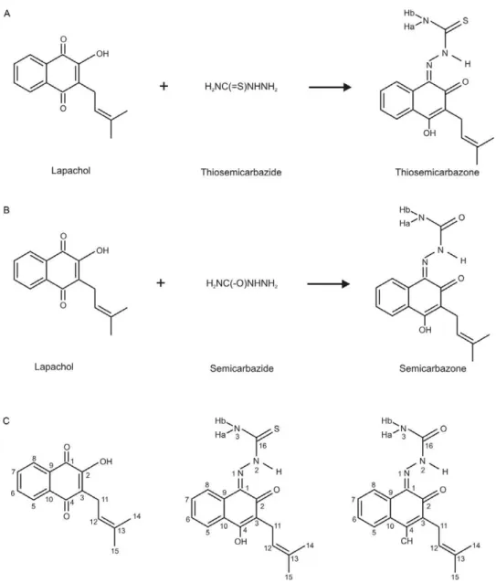
The reactions of lapachol (compound 1) with thi-osemicarbazide and semicarbazide gave a thiosemicar-bazone (compound 2) and a semicarthiosemicar-bazone (compound 3) with yields of 73% and 13%, respectively (Fig. 1). The ESI-MS data for compound 2 showed a positive ion peak at m/z 316, which was attributed to the quasi-molecular ion peak [M + H]+ and the ESI-MS of compound 3
exhib-ited a negative ion peak at m/z 298, which was attributed to the quasi-molecular ion peak [M - H]-; these
quasi-molecular ion peaks correspond with the expected mo-lecular weights of 315 and 299 g/mol for the compound 2 and the compound 3, respectively. The compound 2 had a melting point of 174.3-175.3ºC.
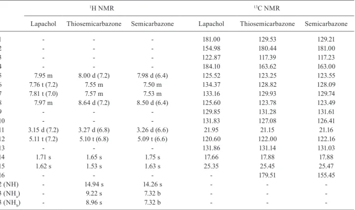
The comparison of the 1H spectra (Table I) between
lapachol and its derivatives (compounds 2 and 3)
ed that the signals from the side-chain at C-3 were virtu-ally unchanged. In addition, the N2-H chemical shifts at 14.94 and 14.26 in the spectra of 2 and 3 were indicative of conformations in which N2-H is hydrogen bonded to O-2. The N3-Ha and N3-Hb hydrogens of 2 were diaste-reotopic. The different chemical shifts likely result from a hydrogen bond between N3-Ha and N1, indicating that the same conformation that has been suggested in solu-tion is found in the solid state.
Changes were predominantly observed in the 13C
NMR spectrum (Table I). Shielding effects were ob-served at C-1 [δC 129.53 (2) and 129.21 (3)], C-4 [δC 163.62 (2) and 163.00 (3)] and C-3 [δC 117.39 (2) and 117.23 (3)] with the carbonyl group of lapachol being replaced with an imine group (C-1), a hydroxyl group (C-4) or a C3-C4 double bond in the obtained products. Changes in the values of the aromatic ring and a deshielding effect at C-2 [δC 180.44 (2) and 181.00 (3)] compared with lapa-chol (δC 154.98) suggested the formation of a carbonyl group at C-2. The 13C signals related to the
thiosemicar-bazone (compound 2) and semicarthiosemicar-bazone (compound 3) moieties were observed at δC 179.51 (C = S) for 2 and at
δC 155.45 (C = O) for 3 (Table I). The COSY, HMQC and HMBC spectra were used to confirm the structures and to unambiguously assign the chemical shifts for all the hydrogen and carbon atoms of 2 and 3 (Table I).
The presence of the thiosemicarbazone and semicar-bazone moieties at C-1 was confirmed by the observa-tion of N2-H∙∙∙O1 hydrogen bonds in (2) and (3) and by the heteronuclear long-range couplings of the hydrogen
and carbon atoms that were revealed by the cross-peaks in the HMBC spectra. These cross-peaks corresponded to the spin-spin interactions of H-11 [δH 3.27 (2) and 3.26 (3)] with C-2 [δC 181.00 (3)] and C-4 [δC 163.62 (2) and 163.00 (3)] and of H-5 with C-4 [δC 163.62 (2)].
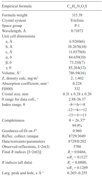
Fig. 2 presents a perspective view of the lapachol thi-osemicarbazone. The crystal data and refinement results are listed in Table II. Selected intramolecular bond dis-tances and angles in the structure are presented in Table III. The bond lengths and angles for the lapachol moiety are in good agreement with the values that were reported for Hooker’s “lapachol peroxide” (Júnior et al. 2009).The most significant differences are the C-O bonds. The C-O distance varies from 1.364-1.431 Å for the single bonds and from 1.209-1.227 Å for the double bonds in Hooker’s lapachol structure. In lapachol thiosemicarbazone, the C4-O2 (1.350(2) Å) single bond is shorter and the C2-O1 [1.236(2) Å] double bond is longer. The bond distances and angles in the thiosemicarbazone framework are sim-ilar to the bonds and angles in other thiosemicarbazones (Beraldo et al. 2001, Lessa et al. 2010).
Lapachol thiosemicarbazone adopts the E conforma-tion with respect to the N2-C16 bond in the solid state. A weak intramolecular N2-H∙∙∙O1 hydrogen bond (Table IV) is present in the structure, but is not likely to facilitate rotation around the N2-C16 bond. The same conformation occurs in 2 in a DMSO solution, as was discussed earlier. In the compound packing, weak intermolecular O2-H∙∙∙S1 and N3-H∙∙∙O2 hydrogen bonds result in the formation of linear arrangements in the solid state (Fig. 3).
TABLE I
1H nuclear magnetic resonance (NMR) [400 MHz, dimethyl sulfoxide (DMSO), δ, J (Hz)]
and 13C NMR (100 MHz, DMSO, δ) data for lapachol, thiosemicarbazone and semicarbazone
1H NMR 13C NMR
Lapachol Thiosemicarbazone Semicarbazone Lapachol Thiosemicarbazone Semicarbazone
1 - - - 181.00 129.53 129.21
2 - - - 154.98 180.44 181.00
3 - - - 122.87 117.39 117.23
4 - - - 184.10 163.62 163.00
5 7.95 m 8.00 d (7.2) 7.98 d (6.4) 125.52 123.25 123.55
6 7.76 t (7.2) 7.55 m 7.50 m 134.37 128.82 128.09
7 7.81 t (7.0) 7.57 m 7.53 m 133.16 129.93 129.74
8 7.97 m 8.64 d (7.2) 8.50 d (6.4) 125.60 123.78 123.49
9 - - - 129.85 131.28 131.61
10 - - - 131.83 127.08 126.41
11 3.15 d (7.2) 3.27 d (6.8) 3.26 d (6.6) 21.95 21.15 21.16
12 5.11 t (7.2) 5.10 t (6.8) 5.09 t (6.6) 120.60 122.00 122.16
13 - - - 131.86 131.14 131.03
14 1.71 s 1.65 s 1.75 s 17.66 17.88 17.88
15 1.62 s 1.53 s 1.63 s 25.35 25.45 25.47
16 - - - - 179.51 155.45
2 (NH) - 14.94 s 14.26 s - -
-3 (NHa) - 9.22 s 7.32 b - -
-The lapachol thiosemicarbazone and semicarbazone derivatives were tested against five pathogenic bacteria (E. coli, S. aureus, P. aeruginosa, S. typhimurium and E. faecalis). The most effective antimicrobial activities were observed against two pathogenic Gram-positive bacteria, S. aureus and E. faecalis (Table V). These compounds were inactive against Gram-negative bacte-ria. The lapachol thiosemicarbazone and semicarbazone derivatives had MICs of 0.10 μmol/mL against S. aureus and 0.05 μmol/mL against E. faecalis. Lapachol inhibit-ed the growth of S. aureus and E. faecalis at a concentra-tion of 0.52 μmol/mL. The semicarbazide and thiosemi -carbazide reagents were tested against five pathogenic bacteria and were active against E. faecalis, inhibiting its growth at a concentration of 0.83 and 0.69 μmol/mL, respectively (Table V).
Lapachol was tested against opportunistic Candida sp., C. gattii and P. brasiliensis. It had superior activity only against P. brasiliensis; the MIC was 0.13 μmol/ mL for most of the isolates and 0.26 μmol/mL for P. brasiliensis 01.
The lapachol thiosemicarbazone derivative exhibited the best antifungal activity, with MICs ranging from 0.01-0.10 μmol/mL for the isolates of P. brasiliensis. The lapachol semicarbazone derivative was less active than the thiosemicarbazone derivative against the P. brasili- ensis isolates (MICs 0.42 vs. 0.84 μmol/mL, respec -tively). C. albicans and C. tropicalis were resistant to the thiosemicarbazone and semicarbazone derivatives at the tested concentrations. C. gattii was susceptible to the thiosemicarbazone and semicarbazone derivatives, with MICs of 0.10 and 0.21 μmol/mL, respectively. The semi -carbazide and thiosemi-carbazide reagents were inactive against all fungi tested except P. brasiliensis isolates, the growth of which was inhibited by the thiosemicarbazide reagent at a concentration of 1.37 μmol/mL.
In this study, the lapachol thiosemicarbazone de-rivative had the greatest activity against P. brasiliensis;
therefore, its effect on the fungal cell wall was deter-mined using the sorbitol assay. A distinctive feature of specific inhibitors of fungal cell wall synthesis is that the antifungal effect is reversed in media containing an osmotic stabiliser such as sorbitol (Frost et al. 1995). However, the lapachol thiosemicarbazone derivative did not affect the fungal cell wall because the MIC for P. brasiliensis did not change upon the addition of sorbitol to the culture medium.
Compared with the control (0.01% DMSO), the lapa-chol-derived thiosemicarbazone did not reduce cell vi-ability over the concentration range of 9.52 x 10⁻⁴ to 0.32 μmol/mL (0.3-100 μg/mL), but at 0.64 μmol/mL (200 μg/ mL), this compound significantly reduced cell viability, indicating cytotoxicity (Fig. 4). The MICs for this com-pound against the isolates of P. brasiliensis were in the range of 0.10-5.40 x 10⁻³ μmol/mL (31.2-1.7 μg/mL). For
Fig. 2: molecular plot of lapachol thiosemicarbazone showing the la-belling scheme of the non-H atoms and their displacement ellipsoids at the 50% probability level.
TABLE II
Crystal data and refinement results for lapachol thiosemicarbazone
Empirical formula C16H17N3O2S
Formula weight 315.39
Crystal system Triclinic
Space group P-1
Wavelength, Å 0.71073
Unit cell dimensions
a, Å 6.9260(6)
b, Å 10.2676(18)
c, Å 11.0379(8)
α, 0 84.659(10)
β, 0 73.210(7)
γ, 0 85.264(12)
Volume, Å3 746.94(16)
Z, density calc, mg/m3 2, 1.402
Absorption coefficient, mm-1 0.228
F(000) 332
Cristal size, mm 0.31 x 0.28 x 0.20
θ range for data coll., o 2.88-26.37
Index range, θ -8<=h<=8
-12<=k<=12 -13<=l<=13
Completeness θ = 26.37º
99.9%
Goodness-of-fit on F2 0.960
Reflec. collect./unique 8729/3049 Data/restraints/parameters 8729/0/202
Observed reflections, I>2σ(I) 5780
E. faecalis and S. aureus, the MICs were between 0.10-0.05 μmol/mL (between 31.2-15.6 μg/mL). These results indicate that the lapachol-derived thiosemicarbazone is not cytotoxic against normal cells at the concentrations that were active against fungi and bacteria. Conversely, lapachol had a biphasic dose-response curve, exhibiting toxicity at both the maximal concentration of 0.83 μmol/ mL (200 μg/mL) and the intermediate concentration of 0.01 μmol/mL (3 μg/mL). At 0.01 mmol/mL (3 mg/mL), there was a significant reduction in cell viability com-pared with the control (0.01% DMSO). At the other tested concentrations, this compound was not toxic. The MICs for lapachol against P. brasiliensis and the two tested bacterial strains were in the range of 0.52-0.13 μmol/mL (124-31.2 μg/mL) and 0.52-0.06 μmol/mL (125-15.6 μg/ mL), respectively, and no toxicity was observed against the PBMCs at these concentrations.
Supplementary material - CCDC reference 844492 for 2 contains the supplementary crystallographic data. These data can be obtained free of charge from the CCDC via ccdc.cam.ac.uk/data_request/cif.
DISCUSSION
In this study, we determined the MICs for lapachol and its derivatives against clinical pathogens and the re-sults indicate that these compounds were active against C. gattii, several isolates of P. brasiliensis and Gram-positive bacteria, including S. aureus and E. faecalis.
TABLE III
Selected bond distances (Å) for lapachol thiosemicarbazone
Atoms
Bond distance
(Å) Atoms
Angle (°)
C1-N1 1.307(2) C1N1N2 119.36(15)
N1-N2 1.342(2) N1N2C16 119.01(15)
N2-C16 1.365(2) N2C16N3 115.96(15)
C16-N3 1.312(2) N2C16S1 118.81(13)
C16-S1 1.668(2) N3C16S1 125.23(14)
C2-O1 1.236(2) C1C2O1 120.47(15)
C4-O2 1.350(2) C3C2O1 120.79(16)
C3-C11 1.510(2) C10C4O2 112.30(15)
C12-C13 1.317(2) C3C4O2 123.81(17)
Fig. 3: molecular packing of lapachol thiosemicarbazone showing the scheme of hidrogen bonding.
TABLE IV
Hydrogen bonds distances (Å) and angles (°) for lapachol thiosemicarbazone with d(H..A) < r(Å) + 2.00 Å and <D–H∙∙∙A > 110º
D-H∙∙∙A d(D-H) d(H∙∙∙A) d(D∙∙∙A) (D-H∙∙∙A) Symmetry operation
N3-H3B∙∙∙O2 0.86 2.32 3.0805(18) 147.4 [x,y,z-1]
O2-H2∙∙∙S1 0.82 2.49 3.1556(14) 139.3 [x,y,z+1]
N2-H2A∙∙∙O1 0.86 1.90 2.5682(18) 133.2 intramolecular
The lapachol semicarbazone derivative had weaker an-timicrobial activity than the lapachol thiosemicarbazone. Although semicarbazones have extensive pharmacologi-cal profiles, many activities are lost or diminished by the substitution of the sulphur for oxygen (Beraldo 2004).
The activity against E. faecalis is promising because this bacterium has adapted such that it can survive and prevail in the bacterial flora that colonise the gastroin-testinal tract of critically ill, immunocompromised and/ or neutropenic patients. Consequently, the number of se-vere enterococci infections is increasing, especially in tertiary hospitals (McBride et al. 2007). The thiosemi-carbazone and semithiosemi-carbazone derivatives of lapachol
Ac
tiv
ity o
f l
ap
ac
ho
l a
nd i
ts d
eri
vat
ive
s • M
ari
na A
zê
ve
do S
ou
za e
t a
l.
3
49
Antimicrobial activity (μmol/mL) of lapachol and its derivatives tiosemicarbazone and semicarbazone against several opportunistic microorganisms
Lapachol Thiosemicarbazone lapachol Semicarbazone lapachol Sulfamethoxazole + trimethoprim Anphotericin B Chloramphenicol
Escherichia coli > 1.03 > 0.80 > 0.84 - - 0.05
Pseudomonas aeruginosa > 1.03 > 0.80 > 0.84 - - 0.10
Salmonella typhimurium > 1.03 > 0.80 > 0.84 - - 0.10
Staphylococcus aureus 0.52 0.10 0.10 - - 0.10
Enterococcus faecalis 0.52 0.05 0.05 - - 0.10
Candida albicans > 1.03 > 0.80 > 0.84 - 1.08 x 10⁻³
-Candida tropicalis > 1.03 > 0.80 > 0.84 - 2.70 x 10⁻⁴
-Cryptococcus gattii > 1.03 0.10 0.2- - 1.30 x 10⁻³
-Paracoccidioides brasiliensis Pb18 0.13 0.04 0.84 1.15 6.71 x 10⁻⁵
-P. brasiliensis ED01 0.13 0.10 0.84 0.29 3.35 x 10⁻⁵
-P. brasiliensis 2 0.13 0.02 0.84 0.58 3.35 x 10⁻⁵
-P. brasiliensis 11 0.13 0.04 0.84 0.58 3.35 x 10⁻⁵
-P. brasiliensis 01 0.26 0.04 0.84 1.15 1.30 x 10⁻⁴
-P. brasiliensis B339 0.13 0.02 0.42 0.29 6.71 x 10⁻⁵
-P. brasiliensis 3 0.13 0.04 1.03 1.15 1.62 x 10⁻⁵
-P. brasiliensis 14 0.13 0.04 0.42 0.29 1.35 x 10⁻⁴
-P. brasiliensis 8 0.13 0.10 0.84 1.15 6.71 x 10⁻⁵
-P. brasiliensis 1578 0.13 0.01 0.84 0.29 6.71 x 10⁻⁵
-P. brasiliensis 4 0.13 0.04 0.84 0.58 6.71 x 10⁻⁵
were two-fold more active than chloramphenicol against E. faecalis and had MICs similar to that of chloram-phenicol when tested against S. aureus. S. aureus causes staphylococcal infections and following the introduction of methicillin, there has been a steady increase in me-thicillin-resistant S. aureus isolates that are resistant to vancomycin, even in Brazil (Palazzo et al. 2005).
The initial studies, conducted at the Department of Antibiotics at the Federal University of Pernambuco in 1973, demonstrated the robust activity of lapachol against Gram-positive bacteria (Lima & Weigert 1972, Nagata et al. 1998). These studies revealed that lapachol was active against Helicobacter pylori, Staphylococcus, Streptococcus, Enterococcus, Bacillus and Clostridium, with MICs ranging from 1,560-25,000 μg/mL (Almeida 2009). In general, thiosemicarbazones obtained from other sources, in particular, α-(N)-heterocyclic thi -osemicarbazones and their metal complexes, inhibit the growth of Gram-positive bacteria, such as Neisseria
gonorrhoeae, Neisseria meningitides, Staphylococcus faecalis, Streptococcus faecalis and Enterococcus, but do not effectively inhibit Gram-negative bacteria, such as Pseudomonas, Klebsiella, Enterobacter, Shigella, E. coli and Proteus (Beraldo 2004).
In this study, the lapachol-derived thiosemicarbazone had superior antimicrobial activity against clinical iso-lates of P. brasiliensis. Our data indicated that the lapa-chol thiosemicarbazone derivative was more active than sulphonamides, the first class of drugs available for treat-ing patients with PCM. This findtreat-ing is important because the use of sulphonamides for more than two years may be required to treat PCM. Moreover, there is increasing con-cern about drug toxicity and treatment cost (Brummer et al. 1993, Paniago et al. 2003, Travassos et al. 2008).
The lapachol-derived semicarbazone and thiosemi-carbazone also exhibited antimicrobial activity against C. gattii (Table V). These results are significant because this pathogenic yeastprimarily infects healthy individu-als and has a high mortality rate (Chaturvedi et al. 2005). Amphotericin B, alone or in combination with flucyto-sine, remains the standard antifungal therapy for these infections, despite the toxicity of both drugs (Lima & Weigert 1972). Other drugs, such as fluconazole and itraconazole, are used as oral maintenance or consolida-tion therapy for cryptococcosis (Perfect & Casadevall 2002). However, resistance to fluconazole has arisen in recent years (Sabbatani et al. 2004).
Lapachol was previously tested against C. albicans and Cladosporium cucumerinum on silica gel plates and it exhibited antimicrobial activity at minimal concen-trations of 0.04 and 2.48 x 10⁻³ μmol/mL, respectively
(Gafner et al. 1996). However, the MICs for lapachol were not determined. In addition, it has been reported that analogues of furanonaphthoquinone from Tecoma ipe have MICs of 1-8 μg/mL against several pathogenic fungi (Nagata et al. 1998), indicating a therapeutic po-tential for naphthoquinones against fungi.
Lapachol and its thiosemicarbazone derivative had dramatic antifungal activity. Lapachol is known for its antimicrobial activity, but its thiosemicarbazone
de-rivative has never been described. In this study, this derivative was found to be superior to lapachol in terms of antifungal activity.
Our data demonstrate that these derivatives were less toxic than lapachol, which exhibited a biphasic response with toxicity at higher concentrations and hormesis at 0.01 μmol/mL (3 μg/mL). Hormesis de -scribes a dose-response curve with opposite effects at low doses vs. high doses (Hoffman 2009). Therefore, the in vitro cytoxicity assays performed using immune system cells revealed a potentially greater immunotox-icity for lapachol than for its derivatives.
The MICs for the lapachol-derived thiosemicarbazone against pathogenic bacteria (S. aureus and E. faecalis) and fungi (mainly P. brasiliensis) indicate that these com-pounds are excellent choices for the development of novel drugs to treat microbial infections. Further studies are necessary to verify the effectiveness of these compounds in treating infections caused by microorganisms.
REFERENCES
Almeida ER 2009. Preclinical and clinical studies of lapachol and beta-lapachone. The Open Natural Products Journal2: 42-47. Araújo EL, Alencar JRB, Rolim Neto PJ 2002. Lapachol: segurança e
eficácia na terapêutica. Rev Bras Farmacogn 12: 57-59. Beraldo H 2004. Semicarbazonas e tiossemicarbazonas: o amplo
per-fil farmacológico e usos clínicos. Quim Nova 27: 461-471. Beraldo H, Lima R, Teixeira LR, Moura AA, West DX 2001.
Crys-tal structures and IR, NMR and UV spectra of 4-formyl and 4-acetylpyridine N(4)-methyl and N(4)-ethylthiosemicarbazones.
J Mol Struct 559: 99-106.
Brummer E, Castaneda E, Restrepo A 1993. Paracoccidioidomycosis: an update. Clin Microbiol Rev 6: 89-117.
Chaturvedi S, Dyavaiah M, Larsen RA, Chaturvedi V 2005. Crypto-coccus gattii in AIDS patients, southern California. Emerg Infect Dis 11: 1686-1692.
Chikate RC, Belapure AR, Padhye SB, West DX 2005. Transition metal quinone-thiosemicarbazone complexes 1: evaluation of EPR covalency parameters and redox properties of pseudo-square-planar copper (II)-naphthoquinone thiosemicarbazones.
Polyhedron 24: 889-899.
CLSI - Clinical and Laboratory Standards Institute 2008. Reference method for broth dilution antifungal susceptibility testing of yeast. Approved Standard M27-A3, CLSI, Wayne, 25 pp. Coutinho ZF, Silva D, Lazera M, Petri V, Oliveira RM 2002.
Para-coccidioidomycosis mortality in Brazil (1980-1995). Cad Saude Publica 18: 1441-1454.
Escalante A, Gattuso M, Pérez P, Zacchino S 2008. Evidence for the mechanism of action of the antifungal phytolaccoside B isolated from Phytolacca tetrâmera Hauman. J Nat Prod71: 1720-1725. Farrugia LJ 1997. ORTEP-3 for Windows - a version of ORTEP-III with
a graphical user interface (GUI). J Appl Crystallogr 30: 565. Farrugia LJ, Win GX 1999. Suite for single crystal small molecule
crystallography. J Appl Crystallogr 32: 837.
Fonseca SGC, Braga RMC, Santana DP 2003. Lapachol: chemistry, pharmacology and assay methods. Rev Bras Farm 84: 9-16. Frost D, Brandt K, Cugier D 1995. A whole-cell Candida albicans
assay for the detection of inhibitors towards fungal cell wall syn-thesis and assembly. J Antibiot (Tokyo) 48: 306-310.
Gafner S, Wolfender JL, Nianga M, Stoeckli-Evans H, Hostettmann K 1996. Antifungal and antibacterial naphthoquinones from
Newbouldia laevis roots. Phytochemistry 42: 1315-1320. Gazzinelli G, Katz N, Rocha RS, Colley DG 1983. Immune responses
during human schistosomiasis mansoni. X. Production and stan-dardization of an antigen-induced mitogenic activity by periph-eral blood mononuclear cells from treated, but not active cases of schistosomiasis. J Immunol 130: 2891-2895.
Hahn RC, Hamdan JS 2000. Effects of amphotericin B and three azole derivatives on the lipids of yeast cells of Paracoccidioides brasili- ensis. Antimicrob Agents Chemother 44: 1997-2000.
Hidron AI, Edwards JR, Patel J, Horan TC, Sievert DM, Pollock DA, Fridkin SK, National Healthcare Safety Network Team, Par-ticipating National Healthcare Safety Network Facilities 2008. NHSN annual update: antimicrobial-resistant pathogens associ-ated with healthcare-associassoci-ated infections: annual summary of data reported to the National Healthcare Safety Network at the Centers for Disease Control and Prevention, 2006–2007. Infect Control Hosp Epidemiol 29: 996-1011.
Hoffman GH 2009. A perspective on the scientific, philosophical and policy dimensions of hormesis. Dose Response 7: 1-51.
Johnson CK 1965. ORTEP-I Report ORNL-3794, Oak Ridge National Laboratory, Oak Ridge, Tennessee, USA.
Jones KE, Patel NG, Levy MA, Storeygard A, Balk D, Gittleman JL, Daszak P 2008. Global trends in emerging infectious diseases.
Nature 451: 990-993.
Júnior ENS, Pinto MCFR, Moura KCG, Simone CA, Nascimento CJ, Andrade CKZ, Hooker’s PAV 2009. Lapachol peroxide’ revis-ited. Tetrahedron Lett 50: 1575-1577.
Lessa JA, Mendes IC, da Silva PR, Soares MA, dos Santos RG, Spe-ziali NL, Romeiro NC, Barreiro EJ, Beraldo H 2010. 2-Acetylpyri-dine thiosemicarbazones: cytotoxic activity in nanomolar doses against malignant gliomas. Eur J Med Chem 45: 5671-5677. Lima OG, Weigert E 1972. Atividade antimicrobiana e antineoplásica
de juglona, lapachol e plumbagina. Rev Ints Antibiot 12: 3-12. McBride SM, Fischetti VA, LeBlanc DJ, Moellering Jr RC, Gilmore
MS 2007. Genetic diversity among Enterococcus faecalis. PLoS ONE 2: e582.
Mosmann T 1983. Rapid colorimetric assay for cellular growth and survival: application to proliferation and cytotoxicity assays.
J Immunol Methods 65: 55-63.
Nagata K, Hirai KI, Koyama J, Wada Y, Tamura T 1998. Antimicro-bial activity of novel furanonaphthoquinone analogs. Atimicrob Agents Chemother 42: 700-702.
NCCLS - National Committee for Clinical Laboratory Standards 2003. Methods for dilution antimicrobial susceptibility tests for bacteria that grow aerobically, NCCLSI document M7-A6, 6th ed., NCCLS, Wayne, 53 pp.
Palazzo ICV, Araujo MLC, Darini ALC 2005. First report of vanco-mycin-resistant Staphylococci isolated from healthy carriers in Brazil. J Clin Microbiol 43: 179-185.
Paniago AMM, Aguiar JIA, AguiarES, Cunha RV, Pereira GROL, Londero AT, Wanke B 2003. Paracoccidioidomicose: estudo clínico e epidemiológico de 422 casos observados no estado de Mato Grosso do Sul. Rev Soc Bras Med Trop 36: 455-459. Perfect JR, Casadevall A 2002. Cryptococcosis. Infect Dis Clin North
Am 16: 837-874.
Rosenthal VD, Maki DG, Jamulitrat S, Medeiros EA, Todi SK, Gomez DY, Leblebicioglu H, Abu Khader I, Miranda Novales MG, Berba R, Ramírez Wong FM, Barkat A, Pino OR, Dueñas L, Mitrev Z, Bijie H, Gurskis V, Kanj SS, Mapp T, Hidalgo RF, Ben Jaballah N, Raka L, Gikas A, Ahmed A, Thu le TA, Guzmán Siritt ME, INICC Members 2010. International nosocomial infection con-trol consortium report, data summary for 2002-2007, issued June 2009. Am J Infect Control 38: 95-104.
Sabbatani S, Manfredi R, Pavoni M, Consales A, Chiodo F 2004. Voriconazole proves effective in long-term treatment of a cere-bral cryptococcoma in a chronic nephropathic HIV-negative pa-tient, after fluconazole failure. Mycopathologia 158: 165-171. Sheldrick GM 1997. SHELXL-97 - Program for Crystal Structure
Re-finement, University of Göttingen, Germany.
Travassos LR, Taborda CP, Colombo AL 2008. Treatment options for paracoccidioidomycosis and new strategies investigated. Expert Rev Anti Infect Ther 6: 251-262.
Vieira RP, Rocha L, Teixeira LR, Sinisterra RD, Coelho MM, Beral-do H 2010. BenzaldeíBeral-do semicarbazona: um candidato a fármaco que alia simplicidade estrutural a um amplo perfil de atividades.
Revista Virtual de Química 2: 2-9.
White TC, Marr KA, Bowden RA 1998. Clinical, cellular and mo-lecular factors that contribute to antifungal drug resistance. Clin Microbiol Rev 11: 382-402.
Wills EA, Redinbo MR, Perfect JR, Poeta MD 2000. New potential targets for antifungal development. Expert Opin Ther Targets 4: 265-296.
Yang K, LeJeune J, Alsdorf D, Lu Bo, Shum CK, Liang S 2012. Global distribution of outbreaks of water-associated infectious diseases.
PLoS Negl Trop Dis 6: e1483.