Luana Carneiro Diniz SOUZA(a)
Vanise Barros Rodrigues da MOTA(b)
Alícia Valéria dos Santos Zaranza de CARVALHO(b)
Rita da Graça Carvalhal Frazão CORRÊA(b)
Silvana Amado LIBÉRIO(c)
Fernanda Ferreira LOPES(c)
(a) Universidade Federal do Maranhão – UFMA, Postgraduate Dentistry Program, São Luís, Maranhão, Brazil.
(b) Universidade Federal do Maranhão – UFMA, University Hospital, São Luís, Maranhão, Brazil.
(c) Universidade Federal do Maranhão – UFMA, Department of Dentistry, São Luís,
Maranhão, Brazil.
Association between pathogens from
tracheal aspirate and oral biofilm of
patients on mechanical ventilation
Abstract:
The aim of this study was to detect possible associations
between respiratory pathogens from tracheal aspirate and oral bioilm
samples in intubated patients in an intensive care unit (ICU), and to
identify the most common respiratory pathogens in oral bioilm,
particularly in patients that developed ventilator-associated pneumonia
(VAP). Two oral bioilm samples were collected from the tongue of
intubated patients (at admission and after 48 hours) and analyzed by
culture with the Antibiotic Sensitivity Test. The results from the tongue
bioilm samples were compared with the tracheal secretions samples.
A total of 59.37% of patients exhibited the same species of pathogens
in their tracheal aspirate and oral bioilm, of which 8 (42.1%) developed
VAP, 10 (52.63%) did not develop pneumonia and one (5.26%) had
aspiration pneumonia. There was a statistically signiicant association
between presence of microorganisms in the tracheal and mouth
samples for the following pathogens:
Klebsiella pneumoniae, Candida
albicans, Pseudomonas aeruginosa, Enterobacter gergoviae, Streptococcus spp
and
Serratia marcescens
(p < 0.05). Pathogens that are present in tracheal
aspirates of intubated patients can be detected in their oral cavity,
especially in those who developed VAP or aspiration pneumonia.
Thus, the results indicate that an improved oral care in these patients
could decrease ICU pneumonia rates.
Keywords:
Bioilms; Intubation; Pneumonia, Ventilator-Associated;
Intensive Care UnitS.
Introduction
The oral cavity is host to many microorganisms, bearing almost half
of the entire microlora of the human body, including the most prevalent
species of Streptococcus, Gemella, Eubacterium, Selenomonas, Veillonella,
Actinomyces, Atopobium, Rothia, Neisseria, Eikenella, Campylobacter,
Porphyromonas, Prevotella, Capnocytophaga, Fusobacterium and
Leptotrichia.
1,2,3The oral bioilm is a permanent reservoir of microorganisms
and may determine infections in distant body sites.
4,5,6Respiratory pathogens are not usually found in the oral microbiota of
healthy people, but hospitalized patients are susceptible to oral bioilm
colonization by these microorganisms.
7Moreover, bioilm components can
prevent the penetration of chemotherapeutic agents in the bioilm, making
Declaration of Interests: The authors certify that they have no commercial or associative interest that represents a conflict of interest in connection with the manuscript.
Corresponding Author: Luana Carneiro Diniz Souza E-mail: luana.dinizz@hotmail.com
https://doi.org/10.1590/1807-3107BOR-2017.vol31.0038
Submitted: Dec 08, 2015
those pathogens more resistant to antimicrobial
agents and hindering their elimination.
8,9In addition,
patients at intensive care units (ICUs) frequently
exhibit poor oral hygiene, with substantial increase
of the oral bioilm.
10Ventilator-associated pneumonia (VAP) is an
important cause of morbidity and mortality in
ICUs.
11,12Recent studies have suggested that VAP may
be associated with microbial colonization of dental
plaque and oropharynx; however, these evaluations
were conducted with different methodologies and
attained no conclusive results.
4,5,6,7A high prevalence of respiratory pathogens, such
as
Pseudomonas spp.
and
Acinetobacter ssp
., was found
in saliva and dental bioilm of hospitalized patients.
13,14Signiicant levels of
Staphylococcus aureus, Klebsiella
pneumoniae
and
Enterobacter cloacae
have also been
found in the dental bioilm of ICU patients.
10Due to the diversity of pathogens detected in the
oral bioilm of patients with VAP, it is important to
investigate which respiratory pathogens can colonize
the oral bioilm of intubated patients in ICUs. Thus, the
aim of this study was to explore possible associations
between respiratory pathogens from tracheal aspirate
and oral bioilm in intubated patients in an ICU and
to identify the most common respiratory pathogens
present in the oral bioilm, particularly in patients
that develop VAP.
Methodology
This research was approved by the Ethics
Research Committee of the University Hospital
of Federal University of Maranhão - HUUFMA
(Protocol No. 251 610). This short-term longitudinal
descriptive study used a convenience sample of
patients admitted to the General ICU of the “Presidente
Dutra” University Hospital (HUUPD). The sample
size was deined using the formula for descriptive
studies, assuming a 4% rate of the variable of interest
(intubated patients), a sampling error of 5%, a 90%
confidence level, a study design effect of 1.0 and
the total of 110 ICU patients, from a period of six
months. Thus, the minimum required sample size
was 31 intubated patients. Only intubated patients
with tracheal secretions were included. Patients
with previous episodes of gastric content aspiration,
those who underwent thoracic surgery, with chronic
obstructive pulmonary disease and carriers of the
acquired immune deiciency syndrome virus (AIDS)
were excluded
7. The inal sample was 32 patients.
Two lingual bioilm samples were collected from
each patient, always in the morning. The irst collection
was performed at admission, at the same time as the
collection of tracheal aspirate samples, and the second
collection was performed after exactly 48 hours.
Bioilm was collected with a sterile swab and sent for
immediate processing for bacterial culture using the
antibiotic sensitivity test (AST). Oral sample collections
were performed by a single dentist. The collection
of tracheal secretion samples was performed using
a suction catheter in a sterile bottle, according to the
routine of the nursing staff.
The ICU nursing team was trained in accordance
with the oral hygiene protocol deined in the Dentistry
Department Meeting of the Brazilian Association for
Intensive Medicine (Associação de Medicina Intensiva
Brasileira, AMIB) of 2011, as follows
15:
a.
Execution: Nurse technician, nurse, and dentist.
b.
Supervision: Dentist.
c.
Materials needed: Personal Protection
Equipment (PPE), soft infant toothbrush, tongue
depressor / tongue scraper, dental loss, 10 mL
of antiseptic mouthwash (cetylpyridinium
chloride or essencial oils), suction probe 12-14,
oil-based essential fatty acids /5% dexpanthenol
cream, artiicial saliva, gauze.
d.
Description: 1. Wash hands; 2. Organize
necessary materials; 3. Explain the procedure to
the patient; 4. Position the patient respecting his
or her limitations; 5. Put on PPE as needed; 6. In
the presence of endotracheal tube: Ensure that
the endotracheal tube is appropriately secured,
check the cuff inlation pressure (with the help
of a Physiotherapist); 7. Perform oropharyngeal
aspiration; 8. Moisten the toothbrush in the
antiseptic mouthwash and perform oral
hygiene with back and forth movements in
the tongue, vestibule, cheek mucosa, palate,
tooth surfaces (buccal, lingual and occlusal)
and gingival; 9. Aspirate saliva and antiseptic
the end of the procedure; 10. Dry with gauze;
11. Perform intra-oral hydration with artiicial
saliva and perioral lubricants (oil-based
essential fatty acids/dexpanthenol cream
5% - assess dryness of mucous membranes and
reduced salivary low); 12. Clean and organize
the setting: discard gloves, masks and gauze in
the contaminated waste bin, wash hands, write
a report of the procedure in the clinical ile.
e.
NOTE 1: Always monitor the mucosa for allergy
or sensitivity.
f.
NOTE 2: For non-cooperating patients (or with
jaw trismus) ask for help from the dentist.
Oral bioilm and tracheal aspirate samples were
grown on the following media: MacConkey Agar,
Blood Agar, Sabouraud Agar, and BHI broth (Brain
Heart Infusion). With the use of a sterile handle,
the cultured samples were transferred to an oven
(34.5ºC to 36.5ºC) and kept in aerobic conditions for
24 to 48 hours. After visual bacteria growth, colonies
were submitted to Gram staining and viewed under
the optical microscope for morphology assessment
(gram-positive, gram-negative and yeast).
After, inoculums were prepared by adding
microbial colonies to 3 mL of saline solution, with
subsequent adjustment for the McFarland turbidity
(0.5–0.63 McF for bacteria and 1.8–2.2 McF for yeast)
and taken to the Vitek 2 (bioMérieu) equipment, for
microbial identiication using identiication cards
and Advanced Colorimetric
TMtechnology.
16Patients were monitored while in the ICU
and progressed as follows: development of VAP,
development of aspiration pneumonia, discharge
or death. VAP was diagnosed by the medical staff
based on the recent diagnostic algorithm for VAP
published by the Centers for Disease Control and
Prevention / National Health Care Safety Network
(CDC / NHSN) in 2012.
17The results were tabulated in an Excel spreadsheet
(version 2010) and later analyzed by BioEstat statistical
software version 5.3 (Optical Digital Technology, Belém,
PA, Brazil). Descriptive statistics was initially performed
providing absolute and relative frequencies. The Fisher’s
exact test was used to verify the association of a pathogen
in the two different collection sites, oral bioilm and
trachea. Statistical signiicance was set at 5% (p < 0.05).
Results
The mean age of the 32 patients was 56 years,
and 59.4% were females and 40.6% males. The most
common cause of hospitalizations was neurological
disorders (34.4%). Of the 32 patients, 40.6% evolved to
VAP and 9.4% to aspiration pneumonia, while 50%
did not develop pneumonia. Only 37.5% of patients
were discharged and the other 62.5% died. Of the 13
patients who developed VAP, 53.84% (seven) died.
Tables 1, 2 and 3 show the distribution of the
pathogens identiied in the tracheal aspirate and
in the two oral samples, according to the patient’s
pulmonology evolution. There was a decrease in the
amount of species detected in the second collection
(16 species) compared with the first (20 species).
The second oral collection was not performed in
four patients because they died within the 48
hour-period from the irst collection.
Table 4 shows the signiicant associations found
between tracheal and oral samples for the following
pathogens:
Klebsiella pneumoniae, Candida albicans,
Pseudomonas aeruginosa, Enterobacter gergoviae,
Streptococcus spp
and
Serratia marcescens
. Nineteen of
the 32 patients exhibited the same species in tracheal
aspirate and oral bioilm, of which 8 developed VAP,
accounting for 25% of the sample. The following
pathogens detected in both sites were investigated:
Acinetobacter baumannii, Pseudomonas aeruginosa,
Enterobacter cloacae, Klebsiella pneumoniae, Enterobacter
gergoviae, Candida albicans, Coagulase-negative
Staphylococcus spp.
and
Citrobacter koseri.
Among patients who developed aspiration
pneumonia, 33.3% harbored
Streptococcus spp.
The most
frequent pathogen in patients that did not progress
to pneumonia was
Candida albicans
, found in the
tracheal aspirate (25%), and in both oral collections
(25% in the irst and 37.5% in the second collection).
Discussion
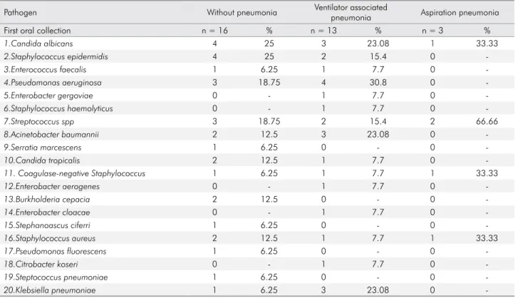
Table 2. Distribution of pathogens identified in the first (at admission) oral collection of patients on mechanical ventilation in the adult intensive care unit according to the pulmonology evolution of the patient.
Pathogen Without pneumonia Ventilator associated
pneumonia Aspiration pneumonia
First oral collection n = 16 % n = 13 % n = 3 %
1.Candida albicans 4 25 3 23.08 1 33.33
2.Staphylococcus epidermidis 4 25 2 15.4 0
-3.Enterococcus faecalis 1 6.25 1 7.7 0
-4.Pseudomonas aeruginosa 3 18.75 4 30.8 0
-5.Enterobacter gergoviae 0 - 1 7.7 0
-6.Staphylococcus haemolyticus 0 - 1 7.7 0
-7.Streptococcus spp 3 18.75 2 15.4 2 66.66
8.Acinetobacter baumannii 2 12.5 3 23.08 0
-9.Serratia marcescens 1 6.25 0 - 0
-10.Candida tropicalis 2 12.5 1 7.7 0
-11. Coagulase-negative Staphylococcus 1 6.25 1 7.7 1 33.33
12.Enterobacter aerogenes 0 - 1 7.7 0
-13.Burkholderia cepacia 2 12.5 0 - 0
-14.Enterobacter cloacae 0 - 1 7.7 0
-15.Stephanoascus ciferri 1 6.25 0 - 0
-16.Staphylococcus aureus 2 12.5 1 7.7 1 33.33
17.Pseudomonas fluorescens 1 6.25 0 - 0 -
18.Citrobacter koseri 0 - 1 7.7 0 -
19.Steptococcus pneumoniae 1 6.25 0 - 0
-20.Klebsiella pneumoniae 1 6.25 3 23.08 0 -
Source: University Hospital of UFMA (April-September, 2013).
Table 1. Distribution of pathogens identified in the tracheal aspirate of mechanically ventilated patients in the adult intensive care unit according to the pulmonology evolution of the patient.
Pathogen in tracheal aspirate Without pneumonia
Ventilator associated
pneumonia Aspiration pneumonia
n = 16 % n = 13 % n = 3 %
1. Klebsiella pneumoniae 1 6.25 2 15.4 1 33.33
2.Candida albicans 4 25 1 7.7 0
-3.Staphylococcus epidermidis 1 6.25 2 15.4 0
-4.Enterococcus faecalis 0 - 1 7.7 0
-5.Pseudomonas aeruginosa 2 12.5 2 15.4 0
-6.Enterobacter gergoviae 0 - 1 - 0
-7.Staphylococcus haemolyticus 1 6.25 0 - 0
-8.Streptococcus spp 1 6.25 1 7.7 1 33.33
9.Acinetobacter baumannii 1 6.25 4 30.77 0 -
10.Serratia marcescens 1 6.25 0 - 0 -
11.Candida tropicalis 1 6.25 0 - 0 -
12. Coagulase-negative Staphylococcus 2 12.5 2 15.4 0 -
13.Stenotrophomonas maltophilia 1 6.25 1 7.7 0 -
14.Burkholderia cepacia 1 6.25 0 - 0
-15.Enterobacter cloacae 0 - 2 15.4% 0 -
16.Escherichia coli 0 - 0 - 1 33.33
17.Staphylococcus aureus 1 6.25 0 - 0
-18.Elizabethkingia meningoseptica 0 - 1 7.7 0 -
19.Citrobacter koseri 0 - 1 7.7 0 -
Total pathogens 18 - 21 - 3 -
were detected in the oral cavity of mechanically
ventilated patients. This result conirms that patients
in ICU may present a signiicant level of respiratory
pathogens in their microbiota.
10Acinetobacter baumannii
is the most frequently
isolated bacterial species in tracheal secretion cultures
of patients with VAP
19. In the present study, a high
proportion of patients who developed VAP presented
Acinetobacter baumannii
(30.77%) in the tracheal aspirate.
However, Barbier et al
20found that the pathogens
most often associated with VAP were
Staphylococcus
aureus, Pseudomonas aeruginosa
and
Enterobacteriaceae.
In our study,
Pseudomonas aeruginosa
and
Enterobacter
cloacae
were also detected in patients who developed
VAP, but less frequently than
Acinetobacter baumannii
.
At the time of hospital admission, the presence of
Acinetobacter baumannii, Pseudomonas aeruginosa, Klebsiella
pneumoniae and Enterobacter cloacae
was observed in
the oral bioilm of patients. After 48 hours, 25% of
the patients developed VAP, exhibiting the same
pathogens in their tracheal aspirate. These pathogens
are reported in other studies as the most frequently
associated with VAP.
19,20However, other pathogens
such as
Citrobacter koseri, Proteus mirabilis, Pseudomonas
aeruginosa
and
Pseudomonas luorescence
have also been
detected in both oral and tracheal samples of intubated
or tracheotomy patients at ICUs
21, indicating their
important role in the pathogenesis of VAP.
22Tracheal aspirate collection is routinely performed
in patients with clinical signs of infection at admission
before starting antibiotic therapy.
23,24An association
of
Klebsiella pneumoniae
and
Pseudomonas aeruginosa
was found between tracheal aspirate and oral bioilm
from the irst collection only. One explanation for
these indings is that the antibiotics chosen at the
patient’s admission, which were speciic for those
pathogens due to their association with VAP etiology,
could have eliminated them from the oral cavity
within the 48-hour interval from the first to the
second collection
19, 20.
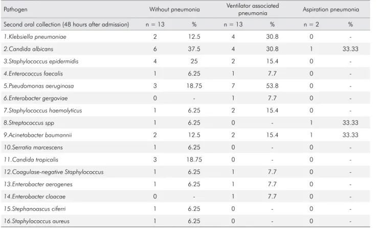
Table 3. Distribution of pathogens identified in the second (48 h) oral collection of patients on mechanical ventilation in the adult intensive care unit according to the pulmonology evolution of the patient.
Pathogen Without pneumonia Ventilator associated
pneumonia Aspiration pneumonia
Second oral collection (48 hours after admission) n = 13 % n = 13 % n = 2 %
1.Klebsiella pneumoniae 2 12.5 4 30.8 0
-2.Candida albicans 6 37.5 4 30.8 1 33.33
3.Staphylococcus epidermidis 4 25 2 15.4 0
-4.Enterococcus faecalis 1 6.25 1 7.7 0
-5.Pseudomonas aeruginosa 3 18.75 7 53.8 0
-6.Enterobacter gergoviae 0 - 1 7.7 0
-7.Staphylococcus haemolyticus 1 6.25 2 15.4 0
-8.Streptococcus spp 1 6.25 0 - 1 33.33
9.Acinetobacter baumannii 2 12.5 2 15.4 1 33.33
10.Serratia marcescens 1 6.25 0 - 0
-11.Candida tropicalis 3 18.75 0 - 0
-12.Coagulase-negative Staphylococcus 1 6.25 1 7.7 0
-13.Enterobacter aerogenes 1 6.25 1 7.7 0
-14.Enterobacter cloacae 0 - 1 7.7 0
-15.Stephanoascus ciferri 1 6.25 0 - 0
-16.Staphylococcus aureus 1 6.25 0 - 0
A relevant aspect of this study was the collection
of oral samples in two stages: on admission and after
48 hours. Thus, the change in the oral lora between
the two time-points could be evaluated, following the
course of the disease. Although a quantitative analysis
of individual species was not done, a small decrease
in the number of species was observed from the
irst (20 species) to the second collection (16 species).
Some studies have shown a quantitative increase of
pathogens that colonize the oral bioilm of patients
in ICU with time.
10,25However, the patients of this
study had oral hygiene performed by trained nurse
Table 4. Distribution of study participants according to the presence of the same species of microorganisms in both oral collections, as well as tracheal aspirate of mechanically ventilated patients in the adult intensive care unit.Pathogen in oral sample
Tracheal sample
p-value
Present Absent
n % n %
Klebsiella pneumoniae (1st. collection) 0.01*
Present 4 50.0 4 50.0
Absent 2 7.2 26 92.8
Klebsiella pneumoniae (2nd. collection) 0.57
Present 0 0 6 -100
Absent 3 15.8 19 84.2
Candida albicans (1st. collection) < 0.01*
Present 5 62.5 3 37.5
Absent 0 0 24 -100
Candida albicans (2nd collection) 0.01*
Present 4 36.4 7 63.6
Absent 0 0 17 -100
Pseudomonas aeruginosa (1st. collection) 0.02*
Present 3 14.2 4 42.8)
Absent 1 4.0 24 96.0
Pseudomonas aeruginosa (2nd. collection) 0.11
Present 3 30.0 7 70.0
Absent 1 5.6 17 94.4
Enterobacter gergoviae (1st. collection) 0.03*
Present 1 -100 0 0
Absent 0 0 31 -100
Enterobacter gergoviae (2nd. collection) 0.03*
Present 1 -100 0 0
Absent 0 0 27 -100
Streptococcus spp (1st. collection) 0.35
Present 2 28.6 5 71.4
Absent 1 4.0 24 96.0
Streptococcus spp (2nd. collection) < 0.01*
Present 2 -100 0 0
Absent 1 3.8 25 96.2
Serratia marcescens (1st. collection) 0.03*
Present 1 -100 0 0
Absent 0 0 31 -100
Serratia marcescens (2nd. collection) 0.03*
Present 1 -100 0 0
Absent 0 0 27 -100
technicians under the supervision of a dentist, which
might have decreased the reservoir of respiratory
pathogens in the dental plaque between sample
collections.
22Although the ICU nursing staff had been
calibrated for the oral hygiene of intubated patients,
the absence of oral hygiene products that should
be supplied by the hospital, such as chlorhexidine,
may have contributed for the high number of
pathogens found in the collected samples. The
use of chlorhexidine in oral hygiene signiicantly
decreases the oropharyngeal colonization with
gram-negative and gram-positive microorganisms
and the incidence of VAP.
26,27,28,29,30,3-32Moreover,
in our study, oral hygiene (mechanical removal of
bioilm) was done twice a day at most, which may
partly explain the high rate of respiratory pathogens
found in the oral bioilm of patients and the high
prevalence of VAP (40.6%) in the period of data
collection. The chemical control of oral pathogens
by 0.12% chlorhexidine seems to be more effective
in preventing VAP than the mechanical removal
by toothbrushing.
33Fifty-nine percent of patients exhibited the
same species of pathogens in their tracheal aspirate
and oral bioilm, of which 42.1% evolved to VAP.
VAP is an infection caused predominantly by aerobic
microorganisms, with an unclear role of anaerobic
pathogens.
34VAP is a nosocomial infection that
causes signiicant morbidity and mortality in ICUs
and prolongs hospitalization.
22Candida spp
rarely
lead to pneumonia. Its
isolation in respiratory material is usually due
to the colonization, until proven otherwise.
35Candida colonization of the respiratory tract occurs
in up to 80% of critical ICU patients,
36,37unlike
invasive candidiasis, which is less than 10%.
38,39Patients who did not progress to pneumonia
exhibited high
Candida albicans
concentration
in the tracheal aspirate, as well as in the oral
biofilm, especially in the second sample collection.
However, in another study,
Candida albicans
was
the most frequent microorganism detected in the
tracheal aspirate of patients diagnosed with VAP
after cardiac surgery.
40Thus, it is necessary to
carefully analyze the hypothesis that fungi are
the 2nd most common etiological agents of VAP
after gram-negative non-fermenting bacilli.
40In our
study, there was only 1 patient (7.7%) positive
for
Candida albicans
among the 13 patients who
developed VAP, suggesting that the widespread
use of broad-spectrum antibiotics can lead to
fungal emergence in patients with VAP.
40This
emphasizes the importance of having guidelines
for Candida colonization and invasive candidiasis
assessment and thus establishing early antifungal
treatment, according to the diagnosis.
41The incidence of aspiration pneumonia has been
associated with dysphagia and with oral colonization
by respiratory pathogens. Effective prevention for
this pathology is achieved with the elimination of
respiratory pathogens through oral hygiene and
improvement of oral functions such as swallowing.
42In the present study,
Streptococcus spp
was observed
in the oral bioilm and tracheal aspirate of patients
with aspiration pneumonia, which confirms the
theory that oral bacteria that colonize the oropharynx
can be aspirated into the lower respiratory tract,
particularly in individuals with high risk of infection
such as hospitalized patients. Nevertheless, there is no
consensus in the literature regarding the hypothesis
that oral bacteria may contribute to the etiology of
respiratory diseases.
43Conclusion
Based on the follow up of patients on mechanical
ventilation in an adult ICU, this study reported
on the disease evolution according to the bacteria
detected in the respiratory tract and oral bioilm,
1. Aas JA, Paster BJ, Stokes LN, Olsen I,
Dewhirst FE. Defining the normal bacterial flora of the oral cavity. J Clin Microbiol. 2005; 43(11):5721-32. https://doi.org/10.1128/JCM.43.11.5721-5732.2005 2. Dewhirst FE, Chen T, Izard J, Paster BJ, Tanner AC,
Yu WH et al. The human oral microbiome. J Bacteriol. 2010;192(19):5002-17. https://doi.org/10.1128/JB.00542-10 3. Palmer RJ Jr. Composition and development of oral
bacterial communities. Periodontol 2000. 2014;64(1):1-24. https://doi.org/10.1111/j.1600-0757.2012.00453.x 4. Scannapieco FA, Rethman MP. The relationship between
periodontal diseases and respiratory diseases. Dent Today. 2003;22(8):79-83.
5. El-Solh AA, Pietrantoni C, Bhat A, Okada M, Zambon J, Aquilina A et al. Colonization of dental plaques: a reservoir of respiratory pathogens for hospital-acquired pneumonia in institutionalized elders. Chest. 2004;126(5):1575-82. https://doi.org/10.1016/S0012-3692(15)31374-X 6. Amaral SM, Cortês AQ, Pires FR. Pneumonia
nosocomial: importância do microambiente oral. J Bras Pneumol. 2009;35(11):1116-24.
http://dx.doi.org/10.1590/S1806-37132009001100010 7. Oliveira LBS, Carneiro PPM, Fischer RG, Tinoco BEM.
A presença de patógenos respiratórios no biofilme bucal de pacientes com pneumonia nosocomial. Rev Bras Ter Intensivo. 2007;19(4):428-33. 07X. http://dx.doi.org/10.1590/S0103-507X2007000400004 8. Smith AJ, Jackson MS, Bagg J. The ecology
of Staphylococcus species in the oral cavity. J Med Microbiol. 2001;50(11):940-6. https://doi.org/10.1099/0022-1317-50-11-940 9. Huang R, Li M, Gregory RL. Bacterial interactions
in dental biofilm. Virulence. 2011;2(5):435-44. https://doi.org/10.4161/viru.2.5.16140
10. Sachdev M, Ready D, Brealey D, Ryu J, Bercades G, Nagle J et al. Changes in dental plaque following hospitalisation in a critical care unit: an observational study. Critical Care. 2013;17(5R189):R189.
https://doi.org/10.1186/cc12878
11. Hingston CD, Cole JM, Hingston EJ, Frost PJ, Wise MP. Oral hygiene and nosocomial pneumonia in critically iII patients. Chest Journal. 2010;137(1):237-8. https://doi.org/10.1378/chest.09-1319
12. DeKeyser Ganz F, Fink NF, Raanan O, Asher M, Bruttin M, Nun MB et al. ICU nurses’ oral-care practices and the current best evidence. J Nurs Scholarsh. 2009;41(2):132-8. https://doi.org/10.1111/j.1547-5069.2009.01264.x 13. Zuanazzi D, Souto R, Mattos MB, Zuanazzi MR,
Tura BR, Sansone C et al. Prevalence of potential bacterial respiratory pathogens in the oral cavity of
hospitalised individuals. Arch Oral Biol. 2010;55(1):21-8. https://doi.org/10.1016/j.archoralbio.2009.10.005 14. Paju S, Scannapieco FA. Oral biofilms, periodontitis,
and pulmonary infections. Oral Dis. 2007;13(6):508-12. https://doi.org/10.1111/j.1601-0825.2007.01410a.x
15. Associação de Medicina Intensiva Brasileira – AMIB. Procedimento operacional padrão: higiene bucal (HB) em UTI (adulto). São Paulo: Associação de Medicina Intensiva Brasileira; 2014. 16. Clinical Laboratory and Standards Institute – CLSI.
Performance standards for antimicrobial susceptibility testing; twnty-fifth informational supplement. Wayne: Clinical Laboratory and Standards Institute; 2013.
17. Centers for Disease Control and Prevention – CDC. Ventilator-Associated Event (VAE) surveillance for adults special edition. Atlanta: Centers for Disease Control and Prevention; 2012.
18. Sands KM, Wilson MJ, Lewis MA, Wise MP, Palmer N, Hayes AJ et al. Respiratory pathogen colonization of dental plaque, the lower airways, and endotracheal tube biofilms during mechanical ventilation. J Crit Care. 2017;37:30-7. https://doi.org/10.1016/j.jcrc.2016.07.019
19. Medell M, Hart M, Duquesne A, Espinosa F, Valdés R. Nosocomial ventilator-associated pneumonia in cuban intensive care units: bacterial species and antibiotic resistance. MEDICC Rev. 2013;15(2):26-9.
20. Barbier F, Andremont A, Wolff M, Bouadma L. Hospital-acquired pneumonia and ventilator-associated pneumonia: recent advances in epidemiology and management. Curr Opin Pulm Med. 2013;19(3):216-28. https://doi.org/10.1097/MCP.0b013e32835f27be 21. Santos PSS, Mariano M, Kallas MS, Vilela MCN. Impacto
da remoção de biofilme lingual em pacientes sob ventilação mecânica. Rev Brás Ter Intensiva. 2013;25(1):44-8. http://dx.doi.org/10.1590/S0103-507X2013000100009 22. Karatas M, Saylan S, Kostakoglu U, Yilmaz G. An assessment
of ventilator-associated pneumonias and risk factors identified in the Intensive Care Unit. Pak J Med Sci. 2016;32(4):817-22. http://dx.doi.org/10.12669/pjms.324.10381.
23. Reinhart K, Brunkhorst FM, Bone HG, Bardutzky J, Dempfle CE, Forst H et al. Prevention, diagnosis, therapy and follow-up care of sepsis: 1 st revision of S-2k Guidelines of the German Sepsis Society (Deutsche Sepsis-Gesellschafte.V. (DSG)) and the German Interdisciplinary Association of Intensive Care and Emergency Medicine (Deutsche InterdisziplinäreVereinigungfürIntensiv- und Notfallmedizin (DIVI)). German Med Sci. 2010;8:1-43. 24. Dellinger RP, Levy MM, Rhodes A, Annane D, Gerlach H,
25. Munro CL, Grap MJ. Oral health and care in the intensive care unit: state of the science. Am J Crit Care. 2004;13(1):25-33.
26. Koeman M, Ven AJ, Hak E, Joore HC, Kaasjager K, Smet AG et al. Oral decontamination with chlorhexidine reduces the incidence of ventilator-associated pneumonia. Am J Respir Crit Care Med. 2006;173(12):1348-55. https://doi.org/10.1164/rccm.200505-820OC 27. Chlebicki MP, Safdar N. Topical chlorhexidine for
prevention of ventilator-associated pneumonia:
a meta-analysis. Crit Care Medicine. 2007;35(2):595-602. https://doi.org/10.1097/01.CCM.0000253395.70708.AC 28. Chan EY, Ruest A, Meade MO, Cook DJ. Oral
decontamination for prevention of pneumonia in mechanically ventilated adults: systematic review and meta-analysis. BMJ. 2007;334(7599):889. https://doi.org/10.1136/bmj.39136.528160.BE 29. Munro CL, Grap MJ, Jones DJ, McClish DK,
Sessler CN. Chlorhexidine, toothbrushing, and preventing ventilator-associated pneumonia in critically ill adults. Am J Crit Care.2009;18(5):428-37. https://doi.org/10.4037/ajcc2009792
30. Oliveira MS, Borges AH, Mattos FZ, Semenoff TA,
Segundo AS, Tonetto MR et al. Evaluation of different methods for removing oral biofilm in patients admitted to the intensive care unit. J Int Oral Health. 2014;6(3):61-4.
31. Nicolosi LN, Carmen Rubio M, Martinez CD, González NN, Cruz ME. Effect of oral hygiene and 0.12% chlorhexidine gluconate oral rinse in preventing ventilator-associated pneumonia after cardiovascular surgery. Respir Care. 2014;59(4):504-9. https://doi.org/10.4187/respcare.02666 32. Liao YM, Tsai JR, Chou FH. The effectiveness of an oral
health care program for preventing ventilator-associated pneumonia. Nurs Crit Care. 2015;20(2):89-97. https://doi.org/10.1111/nicc.12037
33. Vilela MC, Ferreira GZ, Santos PS, Rezende NP. Oral care and nosocomial pneumonia: a systematic review. Einstein (São Paulo). 2015;13(2):290-6. https://doi.org/10.1590/S1679-45082015RW2980 34. Park DR. The microbiology of ventilator-associated
pneumonia. Respir Care. 2005;50(6):742-63.
35. Sociedade Paulista de Infectologia. Diretrizes sobre pneumonia associada a ventilação mecânica (PAV). São Paulo: Office; 2006. 36. Vincent JL, Rello J, Marshall J, Silva E, Anzueto A,
Martin CD et al. International study of the prevalence and outcomes of infection in intensive care units. JAMA. 2009;302(21):2323-9. https://doi.org/10.1001/jama.2009.1754 37. Pfaller M, Neofytos D, Diekema D, Azie N, Meier-Kriesche HU,
Quan SP et al. Epidemiology and outcomes of candidemia in 3648 patients: data from the Prospective Antifungal Therapy (PATH Alliance®) registry, 2004-2008. Diagn Microbiol Infect Dis. 2012;74(4):323-31. https://doi.org/10.1016/j.diagmicrobio.2012.10.003 38. Guery BP, Arendrup MC, Auzinger G, Azoulay E,
Borges Sá M, Johnson EM et al. Management of invasive candidiasis and candidemia in adult non-neutropenic intensive care unit patients: part I. Epidemiology and diagnosis. Intensive Care Med. 2009;35(1):55-62. https://doi.org/10.1007/s00134-008-1338-7
39. Kett DH, Azoulay E, Echeverria PM, Vincent JL, Extended Prevalence of Infection in ICU Study (EPIC II) Group of Investigators. Candida bloodstream infections in intensive care units: analysis of the extended prevalence of infection in intensive care unit study. Crit Care Med. 2011;39(4):665-70. https://doi.org/10.1097/CCM.0b013e318206c1ca
40. Serban RI, Dan M, Pânzaru CV, Anghel D, Dăscălescu D, Ciucu L et al. [Fungi as emergent
etiologic agents in ventilator-associated pneumonia after cardiac surgery]. Rev Med Chir Soc Med Nat Iasi. 2010;114(4):1077-82. Romarian.
41. Bruyère R, Quenot JP, Prin S, Dalle F, Vigneron C, Aho S, et al. Empirical antifungal therapy with an echinocandin in critically-ill patients: prospective evaluation of a pragmatic Candida score-based strategy in one medical ICU. BMC Infect Dis. 2014;14(1):385. https://doi.org/10.1186/1471-2334-14-385
42. Tada A, Miura H. Prevention of aspiration pneumonia (AP) with oral care. Arch Gerontol Geriatr. 2012;55(1):16-21. https://doi.org/10.1016/j.archger.2011.06.029