Juliana Nunes BOTELHO(a) Mario VILLEGAS-SALINAS(b) Pía TRONCOSO-GAJARDO(b) Rodrigo Andrés GIACAMAN(b) Jaime Aparecido CURY(a)
(a)University of Campinas – UNICAMP, Piracicaba Dental School, Department of Physiological Sciences, Piracicaba, SP, Brazil.
(b)University of Talca – UTALCA, Department of Oral Rehabilitation, Cariology Unit, Talca, Región del Maule, Chile.
Enamel and dentine demineralization
by a combination of starch and sucrose
in a biofilm – caries model
Abstract: Sucrose is the most cariogenic dietary carbohydrate and starch is considered non-cariogenic for enamel and moderately cariogenic for dentine. However, the cariogenicity of the combination of starch and sucrose remains unclear. The aim of this study was to
evaluate the effect of this combination on Streptococcus mutans bioilm
composition and enamel and dentine demineralization. Bioilms of
S. mutans UA159 were grown on saliva-coated enamel and dentine slabs in culture medium containing 10% saliva. They were exposed (8 times/day) to one of the following treatments: 0.9% NaCl (negative control), 1% starch, 10% sucrose, or 1% starch and 10% sucrose (starch + sucrose). To simulate the effect of human salivary amylase
on the starch metabolization, the bioilms were pretreated with
saliva before each treatment and saliva was also added to the culture
medium. Acidogenicity of the bioilm was estimated by evaluating
(2 times/day) the culture medium pH. After 4 (dentine) or 5 (enamel)
days of growth, bioilms (n = 9) were individually collected, and the
biomass, viable microorganism count, and polysaccharide content
were quantiied. Dentine and enamel demineralization was assessed by determining the percentage of surface hardness loss. Bioilms
exposed to starch + sucrose were more acidogenic and caused higher demineralization (p < 0.0001) on either enamel or dentine than those
exposed to each carbohydrate alone. The indings suggest that starch
increases the cariogenic potential of sucrose.
Keywords: Amylases; Bioilms; Dental Caries; Dietary Carbohydrates;
Tooth Demineralization.
Introduction
Dental caries is a sugar bioilm-dependent disease,1 and sucrose is
the most cariogenic dietary carbohydrate.2 Starch, a major source of
dietary carbohydrate, is considered non- or slightly cariogenic when
used as the sole source of dietary carbohydrate.3 However, starch is
currently consumed simultaneously or interspersed with sucrose,4 and
this combination could inluence the bioilm composition, modulating
the pathogenesis of dental caries.5
The increased cariogenic potential of this combination of starch and sucrose (starch + sucrose) has been explained by the fact that these two carbohydrates, in the presence of the enzymes salivary
Declaration of Interests: The authors certify that they have no commercial or associative interest that represents a conflict of interest in connection with the manuscript.
Corresponding Author: Jaime Aparecido Cury E-mail: [email protected]
DOI: 10.1590/1807-3107BOR-2016.vol30.0052
Submitted: Sep 14, 2015
α-amylase and glycosyltransferases, enhance
the formation of highly insoluble extracellular polysaccharides (EPS) and structurally change the biofilm matrix. This would result in the accumulation of strong, cohesive, and adherent
bioilms on dental surfaces.5 The cariogenic potential
of this combination was suggested by in vitro
studies evaluating the compositions of Streptococcus
mutans bioilms formed on hydroxyapatite discs.6,7
Furthermore, starch + sucrose caused a greater number of enamel caries in rats8,9 and induced higher
in situ demineralization on deciduous enamel10
than sucrose. However, the greater cariogenicity of starch + sucrose was not confirmed by two subsequent studies, one using a multispecies
bioilm model formed on enamel slabs and another
evaluating caries in rats.11 Moreover, regarding root
dentine, starch + sucrose was not signiicantly more
cariogenic than sucrose, when evaluated in situ.12
These inconsistencies could be explained by the mechanism of starch hydrolysis in the mouth. Salivary amylase, which is required to metabolize
starch,13 is responsible for approximately 75% of
the total amylase activity in bioilms.14 Therefore,
to evaluate the cariogenic potential of starch + sucrose,
we used a validated S. mutans bioilm model15 that
was previously tested to evaluate the cariogenicity of milk.16,17 This model was modiied by the addition
of saliva to simulate the key role of salivary amylase in starch metabolism. This model also simulates the “fast and famine” exposure to dietary sugars to which
dental bioilm is subjected in the mouth.
Methodology
Experimental design
Independent studies were conducted using
slabs of bovine enamel or dentine. S. mutans
UA159 bioilms were grown on these slabs using
a validated model15 that was modiied to simulate
the action of salivary amylase. Biofilms were
grown in ultrailtered (10-kDa-cutoff membrane;
Prep/Scale; Millipore, Billerica,USA), buffered tryptone-yeast extract broth (UTYEB), and exposed 8 times/day to one of the following treatments: 0.9% NaCl, 1% starch, 10% sucrose, and 1% starch
plus 10% sucrose (starch + sucrose). Each experiment
was performed 3 times, each in triplicate (n = 9).
To simulate the effect of salivary amylase, saliva was
added to the culture medium, and the bioilms were
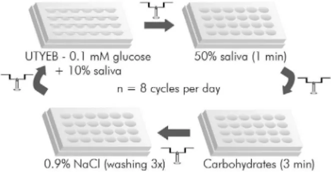
also pretreated with saliva before being exposed to the treatments described above. Culture medium was changed two times per day, at the beginning and at the end of the treatments (Figure 1), and
its pH was determined as an indicator of bioilm
acidogenicity. After 4 days for dentine and 5 days for enamel, the biomass (dry weight), viable bacteria count, and polysaccharide composition of the
bioilm samples were determined. Demineralization
induced on enamel and dentine slabs was assessed as the percentage of surface hardness (SH) loss. For statistical analyses, each biofilm/slab was considered as an experimental unit, with the data for enamel and dentine analyzed independently.
Enamel and dentin slabs preparation
Flattened and polished enamel and root dentine slabs (4 × 7 × 1 mm) were obtained from bovine
incisors.15 Baseline SH of the slabs was measured
using a Knoop microhardness tester coupled to FM-ARS 900 software (Future-Tech Corp., Kawasaki, Japan). Three indentations, spaced 100 µm apart, were made using a load of 50 g for the enamel and 5 g for the dentine for 5 seconds. Slabs with SH 323.1 ± 8.7 and 40.5 ± 2.0 kg/mm² for enamel and dentine, respectively, were used in the study, after sterilization with ethylene oxide.
Figure 1. Diagram of the treatments administered 8 times/day (9:00, 10:30, 12:00, 13:00, 14:30, 16:00, 17:00, and 18:30 h) to the biofilms formed on slabs of enamel or dentine
( ). The medium was changed twice/day, at the beginning
Saliva collection and preparation
Whole saliva was collected on ice from two healthy
volunteers (22 and 24 years old) who chewed parafin ilm (Parailm M; American Can Co., Neenah, USA).
They had not used antimicrobials, mouthwashes, or any other medication known to affect salivary
composition and low during the preceding 3 months.
Both participants provided written informed consent and the protocols were previously approved by the
Research and Ethics Committee of Piracicaba Dental
School (Protocol No. 104/2011).
Saliva was used: (1) to form an acquired pellicle on the enamel and dentine surfaces, (2) to pretreat the slabs before treatments, and (3) as an additive to the
culture medium in which the bioilms were grown.
Saliva collection was performed daily in the morning before any meal and in the afternoon after 2 h of fasting. For acquired pellicle formation, saliva was diluted 1:1 with adsorption buffer and supplemented with the protease inhibitor phenylmethylsulfonyl
luoride (1.0 mmol/L inal concentration)18 and then
centrifuged at 3,800 g for 10 min at 4 °C. Saliva used
to pretreat the bioilms and that added to culture
medium was collected daily and immediately centrifuged as described above. Both supernatants
were collected and individually iltered (Filtermax 0.2 µm Vacuum System, TPP, St. Louis, USA). The clariied, ilter-sterilized saliva was added to the
culture medium in a 1:10 (v/v) proportion. Saliva
used to pretreat the bioilm was diluted 1:1 (v/v)
with 0.9% NaCl. The amylase activity of the saliva source was assessed using the lugol test (positive after 15 min of incubation).
S. mutans biofilm growth
For the acquired pellicle formation, slabs were maintained in a 24-well plate and incubated with
iltered saliva in an orbital shaker at 60 rpm and
37 °C for 30 minutes. The slabs coated with human salivary pellicle were individually positioned in
a new 24-well plate containing 2.0 ml of S. mutans
UA159 inoculum (OD 1.6 at 600 nm) prepared
in a ratio of 1:500 in UTYEB supplemented with 1% sucrose. After 8 h at 37 °C in an atmosphere
containing 10% CO2, the slabs were transferred to
another plate where they were immersed in 2.0 mL
UTYEB containing 0.1 mM glucose (basal salivary concentration) and 10% saliva.
After 24 h of bioilm growth, slabs were treated
8 times/day, 3 days for dentine and 4 days for enamel. The culture medium was changed twice, before the
irst treatment of the day and after the last treatment
of the day. The pH of each change of medium was
measured as indicator of bioilm acidogenicity.
Treatments (Figure 1)
The carbohydrate solutions were the same as those
previously used.10 The starch solution was prepared
from soluble starch (S9765, 80% amylopectin and 20%
amylose; Sigma Chemical Co., St. Louis, USA), and the
sucrose solution was prepared from powdered sucrose
(107651, Merck Millipore, Darmstadt, Germany).
To prepare 1% starch and 1% starch + 10% sucrose, the suspensions were boiled until dissolution was complete. All solutions were autoclaved and stored
at room temperature. During the experiments,
the solutions were aseptically transferred to the 24-well plates to be used.
The bioilms on the enamel and dentine slabs were individually treated 8 times/day at deined
times (9:00, 10:30, 12:00, 13:00, 14:30, 16:00, 17:00, and 18:30 h). Before each treatment, the slabs were removed from the UTYEB medium containing 0.1 mM glucose and 10% saliva and transferred to a new plate containing saliva for the pretreatment. After 1 min, they were transferred to another plate
containing the speciied treatments (0.9% NaCl,
1% starch, 10% sucrose, or 1% starch + 10% sucrose). After 3 min, the slabs were washed 3 times with 0.9% NaCl and returned to the culture plate containing the medium described above.
Biofilm collection and analysis
After 4 and 5 days of bioilm growth for dentine and
enamel, respectively,16,17 the slabs were individually
washed 3 times with 0.9% NaCl, transferred to
microcentrifuge tubes containing 1 mL of 0.9% NaCl, and sonicated for 30 s at 7 W (Branson, Soniier 150, Danbury, USA) to detach the bioilm from the slabs.15
Biomass
Aliquots (150 µl) of the suspension were centrifuged (10 min at 5,000 g and 4 °C); the pellets were dried in a Speed-Vac concentrator (Savant Instruments Inc., Hicksville, USA) for 2 h, and then the pellets were
weighed (± 0.01 mg) to obtain the bioilm dry weight,
which was used as a biomass indicator.
Viable microorganisms
Aliquots (100 µl) of the suspensionwere serially
diluted in 0.09% NaCl and then used to inoculate BHI
agar (BD, Sparks, USA), in triplicate, to determine the
number of viable microorganisms.19 The plates were
incubated for 48 h at 37 °C and 10% CO2. Colonies of
S. mutans were counted and expressed as the number
of CFU/mg of bioilm dry weight.
Polysaccharides
Aliquots (400 µl) of the suspension were used to extract the polysaccharides and determine the concentrations of soluble polysaccharides (SEPS), insoluble extracellular polysaccharides (IEPS), EPS,
and intracellular polysaccharides (IPS) in the bioilm.12
The results were normalized by bioilm dry weight and
expressed as micrograms per milligram of biomass.
Enamel and dentine demineralization
The inal SH of each slab was measured a second
time using 3 indentations 100 µm apart from the initial indentations or in the center of the slabs if the initial indentations were not visible. Baseline
and inal values were used to obtain the percentage of surface hardness loss – %SHL: ((baseline SH value – inal SH value) × (100/baseline SH value)),
which was used as an indicator of enamel20 and
dentine21 demineralization.
Statistical analysis
The assumptions of equality of variances and normal distribution of errors were checked using Shapiro–Wilk’s test for all response variables tested. Variables that did not satisfy these assumptions were transformed and analyzed using an analysis of variance followed by Tukey’s test. Enamel and dentine data were analyzed separately. SAS 9.0 software (SAS Institute, Cary, USA) was used to perform the
analyses, with a signiicance level ixed at 5%.
Results
Compared to the other groups, starch + sucrose
showed a signiicantly (p < 0.0001) more pronounced decrease in medium pH at 32, 56, 80, and 104 h of
bioilm growth for enamel (Figure 2A) and at 32, 56, and 80 h of bioilm growth for dentine (Figure 2B).
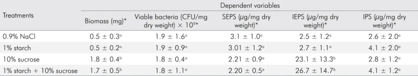
The bioilms treated with starch + sucrose did
not differ from those treated with sucrose alone with respect to the variables biomass, viable bacteria, SEPS, and IPS for either enamel (Table 1) or dentine (Table 2). The IEPS produced by the biofilms treated with
starch + sucrose were signiicantly higher (p < 0.0001) than those by bioilms treated with sucrose alone for
dentine (Table 2) but not for enamel (Table 1).
Figure 2. Acidogenicity of the biofilms (medium pH) formed on enamel (A) and dentine (B) slabs (mean ± SD, n = 9) according to time (h) and treatment (0.9% NaCl, 1% starch, 10% sucrose, 1% starch + 10% sucrose). *Significant differences between starch + sucrose and sucrose group (p < 0.0001).
Regarding demineralization, treatment using
starch + sucrose caused signiicantly (p < 0.0001) greater %SHL both for enamel and dentine (Figure 3)
in comparison with treatment using sucrose alone.
Discussion
Starch and sucrose make up the largest proportion
of dietary carbohydrates consumed worldwide.4 While
some studies have reported that starch + sucrose is more cariogenic than sucrose alone,8,9,10 others have
found no difference.11,12
Our results showed higher demineralization of bovine enamel and dentine when they were exposed to starch + sucrose than when they were exposed to sucrose alone (Figure 3). With respect to enamel, our results are in agreement with those found in situ for
deciduous enamel.10 However, they contrast with the
in vitro results reported by Thurnheer et al.,11using a
multispecies bioilm model. This disagreement could be because of the different bioilm models used and how the bioilms were exposed to carbohydrates. In our
study, exposure of the bioilm to carbohydrates was
intermittent (8 times/day), whereas Thurnheer et al.11
exposedthe bioilm to carbohydrate using continuous
Table 1. Composition of biofilms formed on enamel subjected to the indicated treatments (mean ± SD, n = 9)
Treatments
Dependent variables
Biomass (mg)* Viable bacteria (CFU/mg dry weight) × 109*
SEPS (µg/mg dry weight)*
IEPS (µg/mg dry weight)*
IPS (µg/mg dry weight)*
0.9% NaCl 0.5 ± 0.3a 1.9 ± 1.6a 3.1 ± 1.0a 2.5 ± 1.2a 2.6 ± 2.0a
1% starch 0.5 ± 0.2a 1.9 ± 0.9a 3.01 ± 1.2a 2.7 ± 1.1a 4.1 ± 2.0a
10% sucrose 1.8 ± 0.4b 1.8 ± 0.4a 2.21 ± 0.9a 23.1 ± 13.3b 2.8 ± 1.2a
1% starch + 10% sucrose 1.7 ± 0.5b 1.8 ± 1.1a 2.20 ± 0.5a 26.7 ± 14.7b 4.1 ± 1.2a SEPS, soluble extracellular polysaccharides; IEPS, insoluble extracellular polysaccharides; IPS, intracellular polysaccharides
*For statistical analysis, biomass was transformed using the square root; Viable bacteria was transformed using (X)-2; SEPS, IEPS, and IPS were transformed using log10(X). Within columns, distinct letters indicate significant differences among the treatment groups (p < 0.0001).
Table 2. Composition of biofilms formed on dentine subjected to the indicated treatments (mean ± SD, n = 9)
Treatments
Dependent variables
Biomass (mg)* Viable bacteria (CFU/mg dry weight) × 109*
SEPS (µg/mg dry weight)*
IEPS (µg/mg dry weight)*
IPS (µg/mg dry weight)
0.9% NaCl 0.6 ± 0.2a 1.3 ± 0.6a 3.2 ± 2.7a 2.7.± 1.3a 5.2 ± 2.1a**
1% starch 0.6 ± 0.2a 2.8 ± 1.0b 3.4 ± 1.9a 5.7 ± 3.5a 5.9 ± 1.5a
10% sucrose 1.3 ± 0.4b 2.6 ± 0.8b 3.1 ± 1.5a 20.3 ± 3.8b 4.3 ± 1.3a
1% starch + 10% sucrose 1.2 ± 0.4b 2.0 ± 0.3b 2.5 ± 1.2a 28.7 ± 7.0c 5.3 ± 1.5a SEPS, soluble extracellular polysaccharides; IEPS, insoluble extracellular polysaccharides; IPS, intracellular polysaccharides
*For statistical analysis, biomass and IEPS were transformed using log10(X); viable bacteria were transformed using (X)-2; SEPS were transformed using the square root.
**One value indicated by the SAS software to be an outlier (11.36) was removed. Within the columns, distinct letters indicate significant differences among the treatment groups (p < 0.0001).
Figure 3. Percentage of surface hardness loss (%SHL) in enamel and dentine slabs according to the treatments administered to the biofilms (mean ± SD, n = 9). Different letters indicate significant differences (p < 0.0001) among treatments (within the dental substrates). For statistical analysis, %SHL for enamel was transformed using the square root.
SHL (%)
70 60
50
10 20 30 40
0
Enamel
0.9% NaCl 1% starch
10% sucrose 1% starch + 10% sucrose
a b a
b c
c
d d
feeding in culture medium. Consequently, medium pH was kept at values below 5.0 for all treatments, while in our study, it was possible to show differences in acidogenicity among the treatments (Figure 2).
With regard to dentine, our study showed that starch + sucrose caused greater demineralization than sucrose alone (Figure 3). These results apparently disagree with those found in situ12 and could not be
explained by differences in substrate, since both studies used bovine root dentine. Indeed, Aires et al.12 observed
a trend toward a greater effect for starch + sucrose than for sucrose alone, but the difference was not
statistically signiicant. In addition to the inherent
differences in ability to control variables between in vitro and in situ studies, the volunteers were exposed
to luoride from water and dentifrice in the in situ
study above, which could have masked the cariogenic potential of starch + sucrose. The present in vitro study
was conducted in the absence of luoride.
The more pronounced effect of starch + sucrose on the demineralization of both enamel and dentine may
not be attributed to the fact that the inal concentration
of carbohydrate in the starch + sucrose mixture was 11% (1% starch + 10% sucrose), while it was 10% for sucrose alone. Indeed, we conducted a complementary study comparing the effect of 1% starch + 9% sucrose versus 1% starch + 10% sucrose, and the difference in enamel demineralization between them was not
statistically signiicant (data not shown).
Therefore, the effect of starch + sucrose on the demineralization of enamel and dentine may be considered synergistic and not simply the sum of the effect of fermentation of 1% starch and 10% sucrose. The
data show that starch caused an enamel SHL that was
4.1% higher than that of the control, while for sucrose, this
igure was 29.5% (Figure 3). If the demineralization were
the sum of these effects, a 33.6% greater demineralization would have been expected for the starch + sucrose group, compared with the control. However, the effect of the combination was 45.9% higher, increasing the cariogenic effect of sucrose by 1.4-fold. For dentine, the sum of the effect of carbohydrates alone was 47.5%, while the effect of the combination was 52.1%. The increased effect is supported by the statistical analysis, which showed that the effect of starch + sucrose was greater than the effect of the carbohydrates separately (Figure 3).
This enhanced effect of starch + sucrose on enamel
and dentine demineralization was conirmed by the acidogenicity data (Figure 2A and B). When bioilms
were exposed to starch + sucrose, the concentration of H+ in the medium at 32, 56, 80, and 104 h of bioilm
growth was higher than that found when the bioilms
were exposed to the carbohydrates separately.
For example, at 56 h, the H+ concentrations for the
groups treated with starch and sucrose separately were 1.2 × 10⁻⁷ and 74.2 × 10⁻⁷ M, respectively. However, the
value found for the group treated with starch + sucrose was 200.9 × 10⁻⁷ M, which is 2.7-fold higher than the
sum of the effects of starch and sucrose separately. While explaining this increased effect was not an aim of the present study, it could be the result of increased starch degradation to fermentable products by amylase present in S. mutans. It is known that the action of this enzyme is essential for starch to be
metabolized by bacteria present in bioilms,13 mainly
S. mutans, which do not have amylolytic activity.22
This enzyme is found in acquired pellicle23 and in
bioilm matrix.14 In our bioilm model, the action of this
enzyme in both sites was provided by pretreatment of the dental substrates with saliva and by the presence
of saliva in the culture medium in which the bioilms
were grown (see Methodology). It is well known that for any carbohydrate to be fermented by a bacterial
bioilm, the carbohydrate must irst diffuse into the bioilm matrix and be transferred to the bacterial
cytoplasm. However, this process is hampered when starch is used as the carbohydrate source for the
bioilm bacteria, since its diffusion into the bioilm
is limited because of its high molecular weight.24
In addition, it must irst be degraded in the bioilm
matrix to form products than can be transported
into the bacteria.25 However, this diffusion can be
facilitated by the effect of sucrose and starch on the
matrix of the bioilm formed.5
Thus, the concentration of IEPS in the bioilm exposed
to starch + sucrose was greater than that found in the
bioilm exposed to starch alone (Tables 1 and 2). Compared
with the group treated only with sucrose, starch + sucrose
showed a signiicantly higher concentration of IEPS in the bioilms grown on dentine (Table 2). It is well known that sucrose changes the bioilm matrix composition,2 making
that EPS produced by sucrose in the presence of starch hydrolysates have a differentiated structure, which could explain the higher cariogenicity of starch + sucrose compared with sucrose alone.27,28 These results could be
explained by increased starch diffusion into the bioilm exposed to starch + sucrose. Once inside the bioilm, the
starch might be hydrolyzed by amylase to products that can be fermented by S. mutans.
The indings showing that starch + sucrose is
more cariogenic than sucrose alone are supported by prospective cohort studies suggesting that the consumption of processed or cooked starches with sucrose was associated with a greater caries incidence in children and adolescents.29,30
However, in the present study, the increased cariogenicity found for starch + sucrose, compared with sucrose alone, could be because of some uncontrolled factor. The lower pH observed in the medium where
bioilms treated with starch + sucrose were maintained,
compared with the medium where biofilms were treated with sucrose alone (Figure 1A and B), could be because of a contamination of the medium by sugars, even after washing 3 times with 0.9% NaCl, mainly considering the high viscosity of starch. We checked this possibility and found it to be irrelevant because the residual concentration of sugar found in the medium was very low (0.03%). Indeed, the pH of the medium after the 8th exposure to the treatments, when
the bioilms were immediately transferred to fresh
medium and maintained overnight in fresh medium (times 24, 48, 72, and 96 h), did not show a difference between the sucrose and starch + sucrose groups.
Another limitation of the present study was the
use of a bioilm model based on S. mutans, a bacterium
that is unable to metabolize starch.22 Therefore,
we improved our model by adding human saliva and allowing the starch to be degraded by salivary amylase. The starch was degraded by salivary amylase to form products fermentable by S. mutans, which was
conirmed by the acidogenicity of the culture medium
(Figure 2A and B) and by the demineralization of enamel and dentine among the groups treated with starch (Figure 3). Therefore, further studies are needed to investigate the cariogenicity of starchy products, particularly combinations of starch and sucrose, using
more a speciic bioilm model. This model should
include S. mutans, the most cariogenic bacterium, and other bacteria that are able to metabolize starch and to adsorb salivary amylase. Studies in this direction were
already conducted with 3-species bioilm composed
by A. naeslundii, S. gordonii, and S. mutans.31
Conclusion
In summary, we showed that starch increases
the cariogenic potential of sucrose in a S. mutans
bioilm model.
Acknowledgments
The authors thank Dr. Wander José da Silva for
assistance in statistical analysis. This study was
supported by Conselho Nacional de Desenvolvimento
Cientíico e Tecnológico - CNPq (no. 475178/2011-4 and
no. 305310/2011-9) and Fundação de Desenvolvimento
da Unicamp - FUNCAMP (Conv. 65/91 and 4252).
1. Fejerskov O. Changing paradigms in concepts on dental caries: consequences for oral health care. Caries Res. 2004;38(3):182-91. doi:10.1159/000077753
2. Paes Leme AF, Koo H, Bellato CM, Bedi G, Cury JA. The role of sucrose in cariogenic dental biofilm
formation--new insight. J Dent Res. 2006;85(10):878-87.
doi:10.1177/154405910608501002
3. Sheiham A. Dietary effects on dental diseases. Public Health Nutr. 2001;4(2B):569-91. doi:10.1079/PHN2001142
4. Lingström P, van Houte J, Kashket S. Food starches and dental caries. Crit Rev Oral Biol Med. 2000;11(3):366-80. doi:10.1177/10454411000110030601
5. Bowen WH, Koo H. Biology of Streptococcus mutans-derived glucosyltransferases: role in extracellular matrix formation of cariogenic biofilms. Caries Res. 2011;45(1):69-86. doi:10.1159/000324598
6. Duarte S, Klein MI, Aires CP, Cury JA, Bowen WH, Koo H. Influences of starch and sucrose on Streptococcus mutans
biofilms. Oral Microbiol Immunol. 2008;23(3):206-12. doi:10.1111/j.1399-302X.2007.00412.x
7. Klein MI, Duarte S, Xiao J, Mitra S, Foster TH, Koo H. Structural and molecular basis of the role of starch and sucrose in Streptococcus mutans biofilm development. Appl Environ Microbiol. 2009;75(3):837-41. doi:10.1128/AEM.01299-08
8. Firestone AR, Schmid R, Mühlemann HR. Cariogenic effects of cooked wheat starch alone or with sucrose and frequency-controlled feedings in rats. Arch Oral Biol. 1982;27(9):759-63. doi:10.1016/0003-9969(82)90026-7
9. Mundorff-Shrestha SA, Featherstone JD, Eisenberg AD, Cowles E, Curzon ME, Espeland MA, et al. Cariogenic potential of foods. II. Relationship of food composition, plaque microbial counts, and salivary parameters to caries in the rat model. Caries Res. 1994;28(2):106-15. doi:10.1159/000261630 10. Ribeiro CCC, Tabchoury CPM, Del Bel Cury AA, Tenuta
LMA, Rosalen PL, Cury JA. Effect of starch on the
cariogenic potential of sucrose. Br J Nutr. 2005;94(1):44-50. doi:10.1079/BJN20051452
11. Thurnheer T, Giertsen E, Gmür R, Guggenheim B. Cariogenicity of soluble starch in oral in vitro biofilm and experimental rat caries studies: a comparison. J Appl Microbiol. 2008;105(3):829-36. doi:10.1111/j.1365-2672.2008.03810.x 12. Aires CP, Del Bel Cury AA, Tenuta LMA, Klein MI, Koo H,
Duarte S, et al. Effect of starch and sucrose on dental biofilm
formation and on root dentine demineralization. Caries Res. 2008;42(5):380-6. doi:10.1159/000154783
13. Scannapieco FA, Torres G, Levine MJ. Salivary alpha-amylase: role in dental plaque and caries formation. Crit Rev Oral Biol Med. 1993 Jan;4(3-4):301-7. doi:10.1177/10454411930040030701
14. Fiehn NE, Moe D. Alpha-amylase activity in
supragingival dental plaque in humans. Scand J Dent Res.
1983 Oct;91(5):365-70. doi:10.1111/j.1600-0722.1983.tb00831.x 15. Ccahuana-Vásquez RA, Cury JA. S. mutans biofilm
model to evaluate antimicrobial substances and enamel demineralization. Braz Oral Res. 2010;24(2):135-41. doi:10.1590/S1806-83242010000200002
16. Muñoz-Sandoval C, Muñoz-Cifuentes MJ, Giacaman RA, Ccahuana-Vasquez RA, Cury JA. Effect of bovine milk on Streptococcus mutans biofilm cariogenic properties
and enamel and dentin demineralization. Pediatr Dent. 2012 Nov-Dec;34(7):E197-201.
17. Giacaman RA, Muñoz MJ, Ccahuana-Vasquez RA,
Muñoz-Sandoval C, Cury JA. Effect of fluoridated milk on enamel and root dentin demineralization evaluated by a biofilm caries model. Caries Res. 2012;46(5):460-6. doi:10.1159/000339428
18. Koo H, Vacca-Smith AM, Bowen WH, Rosalen PL, Cury
JA, Park YK. Effects of Apis mellifera propolis on the activities of streptococcal glucosyltransferases in solution and adsorbed onto saliva-coated hydroxyapatite. Caries Res. 2000;34(5):418-26. doi:10.1159/000016617
19. Herigstad B, Hamilton M, Heersink J. How to optimize the drop plate method for enumerating bacteria. J Microbiol Methods. 2001;44(2):121-9. doi:10.1016/S0167-7012(00)00241-4
20. Cury JA, Rebelo MA, Del Bel Cury AA, Derbyshire MT, Tabchoury CPM. Biochemical composition and cariogenicity of dental plaque formed in the presence of sucrose or glucose and fructose. Caries Res. 2000;34(6):491-7. doi:10.1159/000016629
21. Vale GC, Tabchoury CPM, Del Bel Cury AA, Tenuta LMA, ten Cate JM, Cury JA. APF and dentifrice effect on root dentin
demineralization and biofilm. J Dent Res. 2011;90(1):77-81.
doi:10.1177/0022034510383428
22. Edwardsson S. Characteristics of caries-inducing human streptococci resembling Streptococcus mutans. Arch Oral Biol. 1968;13(6):637-46. doi:10.1016/0003-9969(68)90142-8
23. Hannig C, Attin T, Hannig M, Henze E, Brinkmann K, Zech R. Immobilisation and activity of human alpha-amylase in the acquired enamel pellicle. Arch Oral Biol. 2004;49(6):469-75. doi:10.1016/j.archoralbio.2004.01.005
24. Thurnheer T, Gmür R, Shapiro S, Guggenheim B. Mass transport of macromolecules within an in vitro model of supragingival plaque. Appl Environ Microbiol. 2003;69(3):1702-9. doi:10.1128/AEM.69.3.1702-1709.2003 25. Webb AJ, Homer KA, Hosie AHF. Two closely related
ABC transporters in Streptococcus mutans are involved in disaccharide and/or oligosaccharide uptake. J Bacteriol. 2008;190(1):168-78. doi:10.1128/JB.01509-07
26. Dibdin GH, Shellis RP. Physical and biochemical studies of Streptococcus mutans sediments suggest new factors linking the cariogenicity of plaque with its extracellular
polysaccharide content. J Dent Res. 1988;67(6):890-5.
doi:10.1177/00220345880670060101
27. Vacca-Smith AM, Venkitaraman AR, Quivey RG Jr, Bowen WH. Interactions of streptococcal glucosyltransferases with alpha-amylase and starch on the surface of saliva-coated hydroxyapat ite. Arch Oral Biol. 1996;41(3):291-8. doi:10.1016/0003-9969(95)00129-8
28. Xiao J, Koo H. Structural organization and dynamics of exopolysaccharide matrix and microcolonies formation by Streptococcus mutans in biofilms. J Appl Microbiol. 2010;108(6):2103-13. doi:10.1111/j.1365-2672.2009.04616.x 29. Chankanka O, Marshall TA, Levy SM, Cavanaugh JE, Warren
JJ, Broffitt B, et al. Mixed dentition cavitated caries incidence
and dietary intake frequencies. Pediatr Dent. 2011;33(3):233-40.
30. Campain AC, Morgan MV, Evans RW, Ugoni A, Adams GG, Conn JA, et al. Sugar-starch combinations in food and the relationship to dental caries in low-risk adolescents. Eur J Oral Sci. 2003;111(4):316-25. doi:10.1034/j.1600-0722.2003.00056.x 31. Cavalcanti YW, Bertolini MM, Silva WJ, Del Bel Cury AA,
Tenuta LMA, Cury JA. A three-species biofilm model for the