Structural and Electronic Information on Two Solid
Iminophosphoranes, Obtained from NMR
Julian C. Cherryman, Robin K. Harris
†, Matthew G. Davidson,
and Richard D. Price
Department of Chemistry, University of Durham, South Road, Durham DH1 3LE, England
Neste trabalho foram obtidos os espectros de RMN CPMAS de fósforo-31 e carbono-13 do imino(trifenil)fosforano e do brometo de amino(trifenil)fosfônio. A análise detalhada desses espec-tros forneceu informações sobre o tensor de blindagem de 31P e o tensor de acoplamento quadrupolar de 14N. O espectro estático de 31P do brometo foi também analisado em termos dos tensores relevantes. Os cálculos ab initio no nível hf/6-31g* reproduzem bem os dados observados, com exceção dos deslocamentos isotrópicos, os quais requerem cálculos com bases maiores e/ou de um nível teórico superior. As orientações dos tensores na estrutura do brometo foram derivadas através dos cálculos.
Phosphorus-31 and carbon-13 CPMAS NMR spectra have been obtained for imino(triphenyl)phosphorane and amino(triphenyl)phosphonium bromide. Detailed analysis yields information about the 31P shielding tensor and the 14N quadrupole coupling tensor. The static 31P spectrum of the bromide was also analysed in terms of the relevant tensors. Ab initio calculations at the hf/6-31g* level reproduce the observed data well, except for the isotropic shifts, which require a larger basis set and/or higher level of theory. The orientations of the tensors in the molecular frame are derived for the bromide from the calculations.
Keywords: magic-angle spinning, iminophosphorane, ab initio calculation, shielding tensor
Introduction
Unsubstituted iminophosphoranes of the general for-mula R3P=NH are isoelectronic to phosphorus ylides (R3P=CH2). Whereas the latter have been extensively stud-ied, the former have only relatively recently come to promi-nence, but they are expected to have similar properties to ylides. Of particular interest to us is their use as stronger ligands to s-block metals1 than ylides. We report here detailed solid-state NMR studies of imino(triphenyl)phos-phorane, Ph3P=NH (1), and its hydrobromide salt, (Ph3 P-NH2)+Br-(2). Previous related NMR work2,3 has examined N-aryl(triphenyl)iminophosphoranes and the isoelectronic series R3P=X (X = BH3, CH2, NH, O), but the authors concentrated on the 31P nucleus, whereas herein we dem-onstrate that, in addition, information from 13C (and indi-rectly 14N) provides further insight into the electronic nature of these molecules. Phosphorus-31 shielding tensors have also been reported for iminophosphines involving P(III) (i.e. X-P=E with X an electronegative ligand and
E = CR’2, NR’ and PR’) - see Ref. 4 and references therein. Both our compounds have been structurally characterized by diffraction methods5,6. The resulting information allows dipolar coupling constants to be calculated, and these have been used in subsequent analysis of NMR spectra. We have also used the crystal structures as the basis for ab initio calculations of the shielding and quadrupolar tensors. A full understanding of the NMR spectra is then possible. The feasible electronic structures for the compounds are as below:
Ph3P=NH ↔ Ph3P+-N-H 1
(Ph3 P+–NH2)Br-↔ (Ph3P=N+H2) Br 2
Experimental
The samples of imino(triphenyl)phosphorane (1) and amino(triphenyl)phosphonium bromide (2) were prepared Article
† Fax: 0044-191-386-1127; email: [email protected]
in Durham as part of a wide-ranging study of s-block metal derivatives of iminophosphoranes1. The bromide salt is air-stable and was therefore readily handled for packing into magic-angle rotors. The phosphorane is only moder-ately air-stable, so it was sealed under dry, oxygen-free argon in a glass ampoule which fits closely into a rotor (with packing of PTFE tape). However, on occasion, for short-term work, it was packed into a tightly-capped kel-F plastic insert which in turn fitted inside a rotor.
Phosphorus-31 and carbon-13 spectra were obtained at 81.015 and 50.329 MHz respectively, using a Chemagnet-ics CMX 200 spectrometer operating at ambient probe temperature. Zirconia rotors were used (7.5 mm o.d. for the double-resonance experiments and 5.0 mm o.d. for the triple-resonance work on 13C). Cross-polarization from protons was employed, together with high-power proton decoupling during signal acquisition. A nitrogen-15 CPMAS spectrum (again with proton decoupling) was measured using a Varian UnityPlus 300 spectrometer at 30.399 MHz and ambient probe temperature. Chemical shifts for 13C, 15N and 31P were measured by replacement via the signals for adamantane (high-frequency line, δC = 38.4 ppm), ammonium nitrate (δN = -5.1 ppm for NO3-) and brushite (δP = 1.2 ppm) respectively, but are quoted relative to the usual reference standards, tetramethylsilane, ni-tromethane and phosphoric acid respectively.
Shielding tensor components are defined according to the notation of Haeberlen7;
σ33 - σiso≥σ11 - σiso≥σ22 - σiso
The anisotropy is ζ = σ33 - σiso, and the asymmetry is
η = (σ22 - σ11)/ζ.
Ab initio calculations were performed using the experi-mental geometry, based on the structures derived from neutron diffraction data for (1) and the X-ray diffraction data for (2). All calculations were performed with the Gaussian 94 program8 using 6-31g* and 6-311g (2d,p) basis sets and the Hartree-Fock level of theory. The nuclear magnetic shielding tensors were calculated by the GIAO theory9,10. The B3LYP hybrid density functional method, which allows for electron correlation, has been used as incorporated in the Gaussian 94 program. For the basis sets used herein, density functional methods tend to predict NMR values that are deshielded when compared to experi-ment and to Hartree-Fock methods.
Crystallographic Data and Electronic
Structure
Single-crystal X-ray diffraction measurements have been made5,6 on both compounds and the structures solved. For the parent iminophosphorane, neutron diffraction ex-periments, performed at 20 K, have also been published6, giving more accurate positions for the hydrogen nuclei.
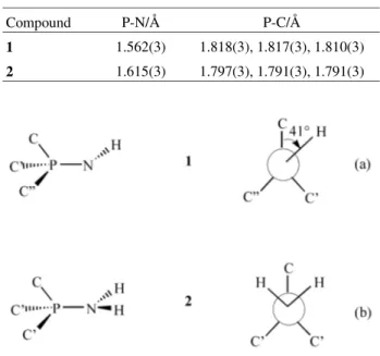
Table 1 summarises the bond distances involving phospho-rus. The P-N bond is significantly shorter for the parent iminophosphorane than for the bromide salt. Thus a reso-nance structure Ph3P+-NH2 is important for the latter, im-plying the existence of a lone pair on nitrogen. The local molecular symmetries about P for (1) and (2) found from crystallography have implications for the 13C NMR spectra discussed below. In (1), each molecule has local C1 sym-metry about P [i.e. all three ipso carbon atoms of the phenyl groups are different, Scheme 1(a)], whereas in (2) local Cs symmetry results from protonation of the imine N atom [i.e. two ipso carbon atoms of the phenyl groups (C’) are equiva-lent, Scheme 1(b)].
NMR Results, GIAO Calculations and
Discussion
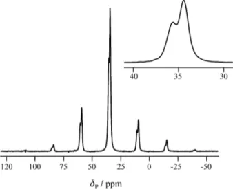
The 31P CPMAS spectrum of the bromide is shown in Fig. 1. There is a single centreband at a chemical shift δP = 34.8 ppm, close to that observed for a solution in deuterated chloroform (δP = 36.2 ppm). There is, however, a 1:2 splitting caused by residual dipolar coupling to the quadru-polar 14N nucleus11 (see below). Simulation of the spin-ning-sideband manifold gave shielding tensor information listed in Table 2, as are the corresponding data for the parent iminophosphorane. For the latter, solution-state measure-ments gave δP = 17.5 and 22 ppm when toluene and THF were used as solvents, respectively. It is clear that there are no substantial molecular structural differences between the solution and solid states of these compounds,although for 1 weak intermolecular C - H...N interactions were inferred from the neutron data6, which are unlikely to be maintained in solution. Gaussian 94 GIAO calculations were
per-Scheme 1.
Table 1. Bond lengths from X-ray crystallographic data at 150 K (1)6 and 153 K (2)5.
Compound P-N/Å P-C/Å
1 1.562(3) 1.818(3), 1.817(3), 1.810(3)
2 1.615(3) 1.797(3), 1.791(3), 1.791(3)
formed at the hf/6-31g* level and are also reported in Table 2. The agreement between the observed and calculated values of the shielding anisotropies and asymmetries for both 1 and 2 is remarkably good. Such calculations gave absolute isotropic shielding constants of 392.9 and 402.5 ppm for the bromide salt and parent iminophosphorane respectively. To relate these to the chemical shift scale, the absolute shielding of the reference compound, H3PO4 was taken to be 328.4 ppm12, giving shifts of δiso = -64.5 and -74.1 ppm respectively (i.e. considerably in error). When the basis set was increased to the 6-311g(2d,p) level, these values improved to -1 and -9 ppm respectively. Use of the B3LYP hybrid density functional method gave even better calculated values (+61 and +47 ppm respectively) for the isotropic shifts, though they are deshielded in both cases compared to the experimental values. It may be seen that the errors in the simpler treatment (hf/6-31g*) are substan-tially lower for the shielding anisotropy and asymmetry,
which use internal differences, than for the isotropic pa-rameters.
The static 31P spectrum of the bromide salt is illustrated in Fig. 2. This was acquired with CP, a Hahn echo, and proton decoupling during acquisition. The general shape makes it clear that the asymmetry is non-zero and that there is some broadening from homonuclear (P,P) dipolar cou-pling, making the turning points indistinct. As the crystal structure is known, the (31P,14N) dipolar coupling constant can be calculated at D = 835 Hz. We assume the indirect coupling tensor is negligible (Jiso = 32.9 Hz for N -phenyl(triphenyl)iminophosphorane13). The spectrum was iteratively fitted, with variation of the shielding anisotropy and asymmetry, the intrinsic linewidth and the Euler angles (αS and βS) relating the shielding and dipolar tensors. These parameters are equivalent to the polar angles of the P-N internuclear vector in the principal axis system of the shielding tensor, with αS being defined relative to σ22. The result is shown in Fig. 2. It is found that ζ = 45 ppm and
η = 0.78, which are in reasonable agreement with the re-sults from the spinning-sideband analysis. The result for η Table 2. Chemical shift and shielding data from 31P CPMAS NMR and Gaussian 94 calculations (at the hf/6-31g* level).
Compound δiso/ppm σ iso/ppmd ζ/ppm η
Expt.c Calc. Expt.c Calc.
1a 21.8e 402.5 107(1.3) 98 0.19(0.03) 0.24
2b 34.8f 392.9 42(0.5) 32 0.52(0.01) 0.56
a Imino(triphenyl)phosphorane.
b Amino(triphenyl)phosphonium bromide.
c From fitting of spinning sideband intensities. Estimated statistical errors in brackets. d Calculated absolute shielding (see the text).
e Individual components: -85.2(1.3), 65.1(1.8) and 85.5(1.9) ppm. f Individual components: -7.2(0.5), 44.9(0.3) and 66.7(0.4) ppm.
Fig ur e 1. Ph osp ho ru s-3 1 CPM AS NMR spectrum of amino(triphenyl)phosphonium bromide, obtained at ambient probe tem-perature with sample spinning at 2 kHz and high-power proton decoupling. The inset shows the sum of all the sidebands (plus the centreband) and illustrates the splitting arising from residual dipolar coupling. The acqui-sition parameters were: pulse duration 4.5 µs, contact time 1 ms, recycle delay 30 s, and number of transients 32.
is likely to have a substantial uncertainty. The shielding and dipolar tensors are found to be mutually oriented with angles βS = 30° and αS = 70°. Changing these angles by more than 10° makes a noticeable difference to the fit, though the angle αS is less sensitive to changes than βS. The hf/6-31g* calculations give βS = 30° and αS = 2°, indicating that there is effective (local) CS symmetry for the P-N bond. From the fitted values for these angles and the ab initio calculated orientation in the molecular frame, it is clear that the largest component of the shielding tensor lies in the plane of the N-P-C(unique) atoms and approximately per-pendicular to the unique P-C bond. The component σ22 also lies in this N-P-C plane within experimental error (accord-ing to the ab initio calculations), whilst σ11 is perpendicular to the plane. For the parent iminophosphorane βS was found to be zero within experimental error (so that experimentally
αS becomes meaningless), σ33 being essentially along the P-N bond. The ab initio calculations suggest that, from a viewpoint down the N-P bond, σ11 lies at a dihedral angle of 12° to P-C (see Scheme 1(a)) and 30° to N-H.
The splittings, s, in the 31P spectrum arising from resid-ual dipolar coupling to 14N may be calculated using pertur-bation theory by the equation11:
s = (9Dχ/20vN)(3 cos2βD - 1 + ηsin2βD cos2αD) where D is the dipolar coupling constant, χ is the quadrupolar coupling constant, and η is the quadrupolar asymmetry, while αDand βD are the Euler angles of the P-N vector in the quadrupolar principal axes. There is some mutual compensation between changes in χ a nd βD. Measured values of s are 101 and 75 Hz, with estimated errors of ±20 Hz (the intrinsic linewidths of the 31P peaks be i ng ca. 60 H z ), fo r t he bromi de and pare nt iminophosphorane respectively. Both splittings are found to be positive, i.e. the smaller component lies at the higher frequency.
Calculations with the 6-31g* basis set were used to estimate the electric field gradients at the nitrogen nuclei of the bromide salt and parent iminophosphorane and were found to be 0.92 and -0.71 atomic units respectively. The value of the quadrupole moment of 14N has been taken14 as 1.61 x 10⁻³⁰ m2, giving the quadrupole coupling constant data (including orientation information) quoted in Table 3. The calculated values of s are in reasonable agreement with the experimental data, given the errors involved in both
experiment and ab initio calculations. For the bromide salt, the calculated value of χ is negative, in agreement with the experimental measurement. This situation occurs because the electric field gradient arises mainly from a lone pair of electrons (in the resonance form with a P-N single bond); the orientation revealed by the calculations, βD = 95°, is in the direction where a lone pair would be expected. In this case χ (i.e.s) has been underestimated, indicating that the lone pair contribution may be larger than the calculations suggest. For the parent iminophosphorane the largest com-ponent of the EFG is aligned at an angle of 27° to the P-N bond, which is approximately where the lone pair of elec-trons (or the average of the two lone pairs for the resonance form Ph3P+-N-H) is expected.
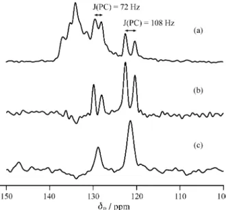
Figure 3 shows carbon-13 CPMAS spectra for the bro-mide under different conditions. The dipolar-dephased spectrum (Fig. 3b) shows clearly that one phenyl group is substantially different from the other two, as expected from the diffraction evidence. The fact that the observed doublet splittings arise from (P,C) coupling is proved by triple-channel experiments, i.e. observation of the
dipolar-Table 3. Residual dipolar coupling informationa.
Compound D/Hzb χ/MHz η αD βD s/Hz Calc. s/Hz Expt.
1 923 2.71 0.67 -27 27° 114 75
2 835 -3.53 0.25 0 95° 66 101
a χ, η, αD, βD, and thence the experimental value of s, are computed using the hf/6-31g* level of theory. b Calculated from the crystal structure.
Figure 3. Carbon-13 CPMAS spectra of amino(triphenyl)phosphonium bromide, obtained at ambient probe temperature: (a) full spectrum, with 1H decoupling during acquisition; (b) dipolar-dephased spectrum, with 1H
decoupling during acquisition; (c) dipolar-dephased spectrum, with both 1H and 31P decoupling during acquisition. Acquisition parameters were:
pulse duration 5 µs, contact time 1 ms, dephasing time 50 µs, recycle delay 5 s, spin rate 4 kHz, and number of transients (a,b) 1500, (c) 1328.
dephased 13C spectrum with simultaneous decoupling of 1H and 31P (Fig. 3c). The ipso-carbon isotropic chemical shifts are δC = 121.4 and 128.7 ppm, with (P,C) isotropic coupling constants 108 and 72 Hz in magnitude respec-tively. The chemical shifts of the remaining carbon nuclei are not clear. For the parent iminophosphorane the 13C spectra have very poor signal-to-noise (S/N) ratios because a long recycle delay is required for relaxation, so definitive data are not readily available. However, the double-decou-pled dipolar-dephased spectrum shows three sharp peaks at
δC = 128.3, 130.6 and 135.6 ppm, in agreement with the existence of three non-equivalent phenyl groups.
The 15N CPMAS spectrum of the bromide has been measured using the Varian UnityPlus 300 spectrometer (contact time 4 ms, recycle delay 45 s). Accumulation of the signal over a full day was barely sufficient to measure the isotropic chemical shift as δN = -340 ppm, and no shielding tensor information was accessible - no spinning sidebands were visible, probably because of the low S/N. Ab initio calculations yield the data in Table 4. The values for the shielding anisotropies and asymmetries may be expected to be more reliable than the isotropic data.
Conclusions
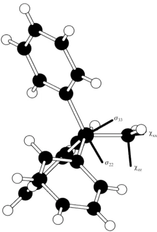
Insight into the molecular and electronic structure of imino(triphenyl)phosphorane and its hydrobromide salt in their solid states has been provided by detailed analyses of their CPMAS 13C and 31PNMR spectra. Molecular orbital calculations, based on known crystal structures, give good agreement with experimental data for 31P shielding anisot-ropy and asymmetry, and for 14N nuclear quadrupole cou-pling (accessed via residual dipolar coucou-pling effects in 31P spectra). The orientations of the tensors in the molecular frame for the bromide are shown in Fig. 4. Dipolar dephasing experiments on 13C spectra are in accord with the crystal structures, and (31P,13C) coupling is proved by triple-channel experiments.
Acknowledgements:
Two of us (JCC and RDP) thank the U.K. Engineering and Physical Sciences Research Council (EPSRC) for stu-dentships and to I.C.I. for scholarships. We are also grateful to EPSRC for access to the Solid-state NMR Service based at Durham.
References
1. (a) Armstrong, D.R.; Davidson, M.G.; Moncrieff, D. Angew. Chem. Int. Ed. Engl. 1995, 34, 478; (b) Bat-sanov, A.S.; Bolton, P.D.; Copley, R.C.B.; Davidson, M.G.; Howard, J.A.K.; Lustig, C.; Price, R.D. J.Or-ganomet. Chem. 1998, 550, 445; (c) Batsanov, A.S.; Davidson, M.G.; Howard, J.A.K.; Lamb, S.; Lustig, C.; Price, R.D. J.C.S. Chem. Comm. 1997, 1211. 2. Power, W.P.; Wasylishen, R.E.; Curtis, R.D. Can. J.
Chem. 1989, 67, 454.
3. Power, W.P. J. Am. Chem. Soc. 1995, 117, 1800. 4. Gudat, D.; Niecke, E.; Grossmann, G.; Krüger, K.
Magn. Reson. Chem. 1999, 37, 43.
5. Phol, E.; Gosink, H.J.; Herbst-Irmer, R.; Noltemeyer, M.; Roesky, H.W.; Sheldrick, G.M. Acta Crystallog. C1993, 49, 1280.
6. Davidson, M.G.; Goeta, A.E.; Howard, J.A.K.; Lehmann, C.W.; McIntyre, G.M.; Price, R.D. J. Or-ganomet. Chem. 1998, 550, 449.
7. Haeberlen, U. Adv. Magn. Reson. Suppl. No. 11976, 1.
Table 4. Shielding information for 15N, obtained from ab initio calcula-tions at the hf/6-31g* level.
Compound σiso/ppm δiso/ppma ζiso/ppm η
1 246.5 -362.2 57 0.30
2 283.1 -398.8 26 0.35
aBased on a shielding for NH
3 of 264.5 ppm (at a chemical shift of -380.2 ppm)15,16.
8. Gaussian 94, Revision E.2, Frisch, M.J.; Trucks, G.W.; Schlegel, H.B.; Gill, P.M.W.; Johnson, B.G.; Robb, M.A.; Cheeseman, J.R.; Keith, T.; Petersson, G.A.; Montgomery, J.A.; Raghavachari, K.; Al-La-ham, M.A.; Zakrzewski, V.G.; Ortiz, J.V.; Foresman, J.B.; Cioslowski, J.; Stefanov, B.B.; Nanayakkara, A.; Challacombe, M.; Peng, C.Y.; Ayala, P.Y.; Chen, W.; Wong, M.W.; Andres, J.L.; Replogle, E.S.; Gomperts, R.; Martin, R.L.; Fox, D.J.; Binkley, J.S.; Defrees, D.J.; Baker, J.; Stewart, J.P.; Head-Gordon, M.; Gon-zalez, C.; Pole, J.A. Gaussian Inc., Pittsburgh PA, 1995.
9. Ditchfield, R.; Mol. Phys.1974, 27, 789.
10. Wolinski, K.; Hinton, J.F.; Pulay, P. J. Am. Chem. Soc. 1990, 112, 8251.
11. Harris, R.K.; Olivieri, A.C. Prog. NMR Spectry. 1992, 24, 435.
12. Jameson, C.J.; De Dios, A.; Jameson, A.K. Chem. Phys. Lett. 1990, 167, 575.
13. Pomerantz, M.; Ziemnicka, B.T.; Merchant, Z.M.; Chou, W.; Perkins, W.B.; Bittner, S. J. Org. Chem. 1985, 50, 1757.
14. Brown, R.D.; Head-Gordon, M.P. Mol. Phys. 1987, 61, 1183.
15. Kukolich, S.G. J. Am. Chem. Soc. 1975, 97, 5704. 16. Jameson, C.J.; Jameson, A.K.; Oppusunggu, D.;
Wille, S.; Burrell, P.M.; Mason, J. J. Chem. Phys. 1981, 74, 81.