Quim. Nova, Vol. 29, No. 6, 1270-1278, 2006
Revisão
*e-mail: [email protected]
MYOGLOBINS – THE LINK BETWEEN DISCOLORATION AND LIPID OXIDATION IN MUSCLE AND MEAT
Jens K. S. Møller*
Instituto de Química, Universidade Estadual de Campinas, PO Box 6154, 13084-971 Campinas - SP, Brazil; Royal Veterinary and Agricultural University, Department of Food Science, Food Chemistry, Rolighedsvej 30, DK-1958 Frederiksberg C, Denmark Leif H. Skibsted
Royal Veterinary and Agricultural University, Department of Food Science, Food Chemistry, Rolighedsvej 30, DK-1958 Frederiksberg C, Denmark
Recebido em 9/1/05; aceito em 9/12/05; publicado na web em 6/7/06
Aerobic metabolism changes rapidly to glycolysis post-mortem resulting in a pH-decrease during the transformation of muscle in to meat affecting ligand binding and redox potential of the heme iron in myoglobin, the meat pigment. The “inorganic chemistry” of meat involves (i) redox-cycling between iron(II), iron(III), and iron(IV)/protein radicals; (ii) ligand exchange processes; and (iii) spin-equilibra with a change in coordination number for the heme iron. In addition to the function of myoglobin for oxygen storage, new physiological roles of myoglobin are currently being discovered, which notably find close parallels in the processes in fresh meat and nitrite-cured meat products. Myoglobin may be characterized as a bioreactor for small molecules like O2, NO,
CO, CO2, H2O, and HNO with importance in bio-regulation and in protection against oxidative stress in vivo otherwise affecting
lipids in membranes. Many of these processes may be recognised as colour changes in fresh meat and cured meat products under different atmospheric conditions, and could also be instructive for teaching purposes.
Keywords: myoglobin complexes; heme iron; oxidative processes.
INTRODUCTION
The primary physiological role of the small iron-containing globular protein, myoglobin (Mb), in mammals has been considered to maintain oxygen supply in skeletal muscles and other muscle tissue, where Mb acts in oxygen storage and the facilitated diffusion of molecular oxygen.1 New studies, however, strongly support other
physiological roles of Mb in mammals.2-4 Mb has served as a model
molecule for other larger and more complex metalloproteins over the years, and experimental findings for Mb have been useful for generalization in relation to molecular structure,5 biophysics and
mechanism of enzyme activation.6;7 Apart from the physiological
function of Mb and its role as a model for other macromolecules, the chemistry of Mb is important in food technology as the colour of both fresh meat and meat products depends on the redox status of the heme iron center.8;9
Mb is a small globular protein with about 150 amino acid residues and molecular weight of approx. 17 kD.10 A porphyrin ring,
protoporphyrin IX, is partly buried in the interior of Mb and a central iron atom is coordinated to this prosthetic group forming a heme moiety, which constitutes the active site of the molecule. The protein backbone and the prosthetic group are attached by a single coordinative bond between the His94 residue and the heme iron, while hydrophobic interactions between vinyl side chains of the prosthetic group and hydrophobic amino acid residues in the interior of Mb also helps to stabilize the association.
This present review covers mainly the chemistry behind the colour and colour changes of fresh meat and meat products during processing and storage. The “inorganic chemistry” of meat will be discussed in relation to the coordination properties of the central heme iron atom in Mb and its redox chemistry. The redox chemistry of Mb and other heme proteins will also be discussed in relation to oxidative
pro-cesses occurring in muscle during transformation to meat and during meat curing, and these processes may cause deterioration of freshness and initiate lipid oxidation leading to rancidity.
CHEMISTRY OF HEME IRON CENTRE IN MYOGLOBINS Under most physiological conditions, the iron atom in Mb and other heme proteins exists in either the ferrous(II) or the ferric(III) state with six or five electrons in the 3d orbital available for bonding (d6 or d5 electron configuration).9
The relatively high numbers of electrons present in the 3d orbital makes several different electronic configurations possible for the iron complex with small differences in electronic energy.8 Low spin FeII
complexes are diamagnetic, while all other spin states of both FeII and
FeIII contain at least one unpaired electron.
Ligands are ordered according to increasing “field strength” in what is known as the spectrochemical series depending on their effect on the splitting (Δ) between t2g and eg orbitals:
11
I- < Br- < Cl- < SCN- < F- < OH- < H
2O < NCS - < NO
2
- < CN- ≈ CO
Thus, weak field ligands such as the halides favour high spin states as these ligands only participate in σ-bonding, while strong field ligands like O2, NO and CN- favour low spin state complexes with
electron pairing of the d orbitals of iron.9;12 Thus, the bond formation
between the central metal and a strong field ligand may be considered to consist of two components: i) donation of σ-type electron density from the ligand to the central metal, and ii) donation of electron density from the d orbitals of the metal into the π*-antibonding of the ligand referred to as “back-bonding”.13 Regarding to physiologically relevant
METAL LIGAND ELECTRONIC DISTRIBUTION
Myoglobin heme iron has strong affinity for π-accepting ligands such as NO.13 Diatomic ligands like NO and O
2 exist in different
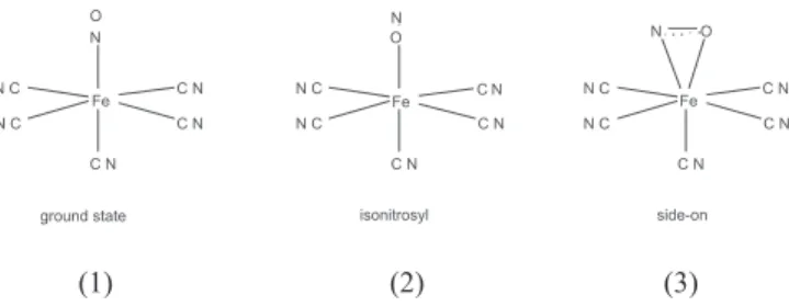
resonance forms, These ligands form complexes with transition metals that exhibit different electronic distributions, which besides the number of electrons in the complex are also affected by a varying degree of electronegativity of the diatomic ligands. Figure 1shows examples of bonding geometry and electronic resonance forms for complexes of FeII heme and three diatomic ligands of physiological
importance. The different interactions between heme iron and ligands are due to differences in the dipole moment and other electronic properties. Thus, distribution of charge along the ligand or ionic character of the ligand greatly affects the ability to participate in hydrogen bonding with the amino acid residue, His64, in the distal heme pocket of Mb, which accordingly also influences the stabilization of the metal ligand complex.
The notation for nitrosyl complexes incorporating bonding, bond geometry and reactivity developed by Enemark and Feltham has generally been accepted to predict the bonding geometry.12 The d
orbital electrons of the metal and the π* orbital electrons from the ligand together are counted as the overall number of electrons in the complex, and the nitrosylated complexes of FeIII or FeII Mb can
be written as {Fe(NO)}6 [Fe (d6-1) + 1 π* electron] and {Fe(NO)}7 [Fe (d6) + 1 π* electron], respectively, and used to predict the Fe-N-O bond angel, as linear for the FeIII complex and bent for the FeII
complex, respectively.
LIGAND BINDING AND DISCRIMINATION BY MYOGLOBIN
A wide range of ligands form complexes with either ferrous or ferric Mb in muscle and meat. The small diatomic molecules O2, NO and CO all bind reversibly as axial ligand in the sixth position of ferrous Mb, although their affinity for Mb varies significantly despite their similarities regarding size, charge and hydrophobicity. 14-16 Mb complexes with NO or O
2 differ from the complex formed
with CO regarding the ability to attain ionic character of the ligand due to variation in dipole moment, and also with respect to iron ligand bonding geometry of the complexes, Figure 1. The complexes
MbFeIINO and MbFeIIO
2 have a bent ligand geometry (112-147°), 17;18
whereas MbFeIICO has been found (somewhat depending on the
technique applied) to exhibit virtually a linear ligand geometry (155-180°).19 Several non-heme iron nitrosyl complexes exist and
especially nitroprusside, [Fe(CN)5NO]3-, has been studied as the
reactions of nitroprusside mimic the reactivity of metallonitrosyl in physiological environments.20 The geometry of the ground-state
(structure I) has a typical low-spin character with a Fe-NO+ core
and stronger π-back bonding to NO relative to the trans CN- resulting
in a slight displacement of the iron towards the NO ligand, but still the short Fe-NO bond is linear rather than bent. Following photoexcitation of nitroprusside linkage isomers have been found, e.g. a linear oxygen bound isonitrosyl (structure II) and a side-on complex with NO bound sideways (structure III).21 Such isomers
may also exist in nitrosylated heme protein and alter the reactivity of the heme Fe-NO complex, e.g. during photo-induced degradation of the nitrosylmyoglobin in meat products.
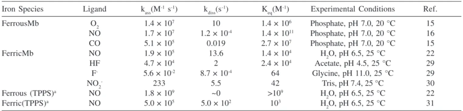
The differences in ligand geometry have been used to explain variation in ligand affinity for Mb, and the observed bond geometries can be assigned either to variations in electrostatic interaction between ligand and amino acid residues or to steric hindrance in the heme pocket. In Table 1, rate constants for ligand association, ligand dissociation and equilibrium constants for binding of various ligands to Mb or heme models are summarized. NO forms the most stable complexes with ferrous Mb, while CO has a significantly lower affinity for Mb closely followed by O2. However, studies with site-directed mutants of Mb suggest that the most important factor for overall stability of complexes of Mb is the ability to interact with hydrogen in His64, whereas steric hindrance of this residue is of minor significance.16 Moreover, the variation in stability observed for
MbFeIINO and MbFeIIO
2 can be partly assigned to differences in the
rate of bond formation between different ligands and heme iron. NO exhibits practically no energy barrier for bond formation, whereas the bond formation with O2 involves an energy barrier roughly equal to the energy for ligand escape from the heme pocket.14
Not surprisingly, the dynamic of recombination between heme iron and diatomic ligands, e.g. NO, CO, O2, depends on the presence or absence of the globin protein.22 This is further reflected in the
differences in the binding constants observed for heme-containing proteins and various modified heme model compounds (see Table 1). Several studies indicate that coordination of NO to heme iron weakens the fifth coordination position with proximal His93.23 In
addition, lowering of pH will further induce weakening or breaking of the proximal base coordinated to iron. Thus, Duprat et al. have found that when coordination between proximal His93 and heme iron is absent due to low pH or site-directed mutagenesis, the recombination rate of NO and heme iron is very fast resulting in a penta-coordinated state that is 3-fold more stable compared to NO bound in hexa-coordinated Mb.24;25 This transformation from a hexa- to
pentaco-ordinated NO complex is of biological relevance in heme proteins such as soluble guanylate cyclase,26;27 and flavo-heme enzymes, like
Nitric Oxide Synthases (NOS).28
Figure 1. Fe-ligand bonding geometry and simplified electronic distribution
COLOUR OF FRESH MEAT
The content of Mb in skeletal muscle tissue varies within different animal species, e.g. whale > beef > pork > poultry. Moreover, Mb concentration differs in muscles of the same animal and increases with age, e.g. beef > veal. The fact that whales along with other sea mammals all have relatively high Mb content in their skeletal muscle tissue is supportive of a function of Mb as an oxygen storage protein during prolonged periods of diving. Mb has recently also been isolated from human smooth muscle,32 and new findings
with respect to its role in cardiovascular biology suggest that Mb may also play other roles in the cells.33
In freshly cut meat, the primary pigment is an oxygenated form of ferrous Mb, MbFeIIO
2 (d
6), while a thin layer of the oxidized
form metmyoglobin, MbFeIIIOH 2 (d
5), will exist at a certain depth in
the meat followed by the reduced form deoxymyoglobin, MbFeII
(d6).9 The localisation of these three forms of Mb in raw meat can
be visualised as in Figure 2, and the depth of the MbFeIIO 2 (x in
mm) will be determined by the partial pressure of oxygen (pO2) in the air or headspace of a package surrounding the meat surface and may be calculated from Equation (1):
(1)
where Co is total oxygen concentration as determined by pO2, D is the diffusion coefficient of O2 and Ao is the O2 consumption rate due to activity of endogenous enzymes.
The bond formation between ferrous heme iron and O2 involves the p-orbital electrons of the O-atom, and as this ligand has strong electron withdrawing ability the resulting complex has partial ionic
character, often described as a partial ferric heme iron with superoxide anion coordinated.8 Moreover, this electronic structure
of the complex also yields a bent geometry relative to the heme plane and allows the O2 molecule to interact with an amino acid residue (His64) in the protein back-bone present in the distal heme pocket as shown in Figure 3.
The reaction known as autoxidation of MbFeIIO
2 in which the
red Mb derivative is spontaneously oxidized in the presence of molecular oxygen (eq. 2), is quite complex and has received much attention as this process besides being important for meat quality is also crucial in physiology.34 Site-directed mutagenesis of Mb
amino acid residues in the heme crevice reveals a pivotal role of these residues in determining the rate of autoxidation.35 Particular
the His64 has been found important for the robustness as native MbFeIIO
2 has an observed rate constant for autoxidation of 1.5 ×
10-5 s-1, while a mutant in which His64 was replaced by Ala exhibits
a rate constant of 0.016 s-1. The rate of autoxidation of MbFeIIO 2
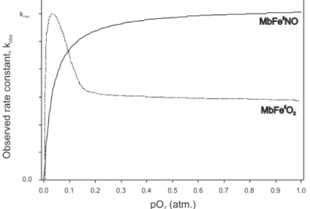
depends on the oxygen pressure initially showing a sharp linear increase with increasing pO2, passing through a maximal rate at a temperature-dependent pO2, and then leading to a significant decrease in the rate as the pO2 increases further,34 as is schematically
illustrated in Figure 4. This dependency of the autoxidation rate is consistent with a bimolecular mechanism in which the fraction of MbFeII reacts with unbound O
2 by an outer-sphere electron transfer
yielding MbFeIII and O 2
•- as seen in Equation 2.
The logarithmic transformed rate constant for autoxidation of MbFeIIO
2 depends linearly on pH under acidic conditions in support of Figure 2. Schematic illustration of myoglobin forms in layers of fresh meat in
equilibrium with atmospheric oxygen at the surface showing ligand coordinated and iron atom spin state
Table 1. Rate constants for ligand association (kass) and ligand dissociation (kdiss) together with equilibrium constant (Keq) for ligand binding to ferrous and ferric heme proteins and model compounds
Iron Species Ligand kass(M-1 s-1) k
diss(s
-1) K
eq(M
-1) Experimental Conditions Ref.
FerrousMb O2 1.4 × 10⁷ 10 1.4 × 10⁶ Phosphate, pH 7.0, 20 °C 15
NO 1.7 × 10⁷ 1.2 × 10⁻⁴ 1.4 × 10¹¹ Phosphate, pH 7.0, 20 °C 16
CO 5.1 × 10⁵ 0.019 2.7 × 10⁷ Phosphate, pH 7.0, 20 °C 15
FerricMb NO 1.9 × 10⁵ 13.6 1.4 × 10⁴ H
2O, pH 6.5, 25 °C 22
HF 4.7 × 10⁴ 2 2.4 × 10⁴ Acetate, pH 4.5, 25 °C 29
F- 5.6 × 10⁻² 8.7 × 10⁻⁴ 64 Glycine, pH 11.0, 25 °C 29
NO2- 233 5.5 42 Tris, pH 7.4, 25 °C 30
Ferrous (TPPS)a NO 1.8 × 10⁹ ~0 >109 H
2O, pH 6.5, 25 °C 22
Ferric(TPPS)a NO 5.0 × 10⁵ 5.0 × 10² 103 H
2O, pH 6.5, 25 °C 31
aTetra(4-sulfonatophenyl)porphine
Figure 3. Electronic distribution and metal-ligand geometry of Fe-O2 in
specific-acid catalysis with the stoechiometry shown in eq. 3.36 The
detailed mechanism is described by a two-state model with a single protolytic group, namely the distal His64 residue that when protonated forms H-bond with coordinated dioxygen and hereby assists in transferring a proton from the solvent to the bound, polarized dioxygen. A positive kinetic salt effect on the specific acid-catalyzed autoxidation of MbFeIIO
2 has been demonstrated, as the rate of proton-assisted
nucleophilic replacement of O2•- is found to increase with increasing
ionic strength.36 Porcine MbFeIIO
2 reacts with a significant lower rate
compared to the Mb complex isolated from bovine, corvine and ovine species and with different temperature dependence.37 For the
acid-catalyzed autoxidation of bovine MbFeIIO
2 the reported activation
enthalpy ΔH‡ = 117 kJ mol-1 and activation entropy ΔS‡ = 172 J mol-1 K -1 demonstrate a very high activation barrier and a dissociative activation,
respectively.36;37 Accordingly, the initial dissociation of oxygen as shown
in Equation 2 seems to be rate-determining rather than the subsequent electron transfer to create the superoxide anion radical. The observed kinetic salt effect for the rate of MbFeIIO
2 autoxidation is very similar to
that observed for autoreduction of certain hypervalent and prooxidative forms of Mb, as discussed in more detail below.
MbFeIIO
2 MbFe
II + O
2 MbFe
III + O 2
•– (2)
4MbFeIIO 2 + 4H
+ 4MbFeIII + 3O
2 + 2H2O (3)
At high hydrostatic pressures, two opposing factors were found to affect the observed rate for autoxidation of MbFeIIO
2: expansion in
the transition state (activation volume ΔV‡ of +12.7 ml mol-1), is in
agreement with the positive value of ΔS‡ that decreases the rate and a
pressure-induced decrease in solution pH increases the rate.38
The electronic structure of the FeII-O
2 heme moiety corresponds
to a structure with ionic character as shown in Figure 3, in effect resulting in a positive kinetic salt effect at reduced pH as in meat. During the transformation of muscle tissue to meat, post-mortem anaerobic glycolysis forms lactic acid, which helps to prevent microbial spoilage, but also increases autoxidation and decrease the colour stability. From a meat quality viewpoint, the desirable bright red pigment, MbFeIIO
2, should be protected and preserved during storage,
which may be achieved using modified atmosphere packaging with gas mixtures having high pO2 tensions (60-80%) along with CO2 (20-30%) to inhibit microbial growth.39 Moreover, MbFeIIO
2 is very
sensitive to light exposure and photooxidation of MbFeIIO
2 is strongly
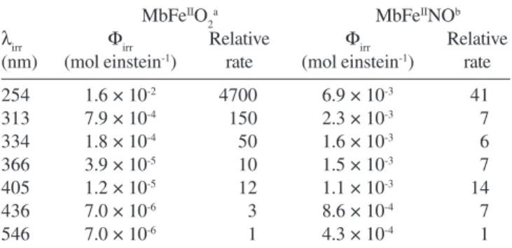
wavelength dependent with quantum yields, as determined using continuous wave photolysis with monochromatic light, having an exponential increase with increasing energy of the irradiation.40 Hence,
UV-light with λ = 254 nm was found to give a 4,700-fold increase
in relative rate of photooxidation in comparison to green light (λ = 546 nm). Therefore, the wavelength distribution is critical for colour stability as was shown for minced raw beef during frozen storage for which surface discoloration is often encountered (see Table 2).41
In raw meat from freshly slaughtered animals the enzyme system metmyoglobin reductase (MMR) is still active and reduces MbFeIII
to the physiological active MbFeII, which coordinates O
2 and reforms
the bright red colour.42;43 In Figure 5 the so-called colour cycle is shown
for fresh meat with various forms of Mb. The colour stability of meat during slaughter, de-boning, storage and retail display depends on post-mortem pH, de-boning/storage temperature, O2 tension, possible addition of salt, packaging and light exposure.
The uncatalyzed autoxidation of MbFeIIO
2 yields O2
•- as initial
reaction product, which will dismutate under the influence of superoxide dismutase (SOD) activity or as a result of acid catalysis generating H2O2 and O2. H2O2 is crucial for formation of prooxidative heme species in meat,44 as will be discussed in more detail below.
COLOUR OF CURED MEAT PRODUCTS
A general review of the chemistry of Mb and NO has recently been published,45 in which the complex reactions occurring in cured meat
products are discussed. The formation of the dominating heme pigment, nitrosylmyoglobin (MbFeIINO), in nitrite-cured meats involves a
complex series of reactions between added nitrate/nitrite and either endogenous or added reductants forming NO that readily associates to heme FeII in Mb.46 The exact reaction pathway from nitrite to NO in the
meat matrix is not fully established, but nitrite initially oxidizes the fresh meat pigment MbFeIIO
2 to MbFe
III, as shown in Equation 4. This
reaction is observed as a transient discoloration of the meat surface immediately after the addition of nitrite. The principal nitrosylating agent in cured meat has not been identified yet but several species have been suggested including the acid anhydride of nitrous acid, which, however, is only present in a minute fraction at the normal pH of meat with a value well above the pKa of HNO2. Nitrosylchloride, NOCl, which is known to act as a strong nitrosylating agent,47 is more likely
the reactant also in the meat matrix where it may be formed following the simultaneous addition of NaCl and NaNO2.48
4MbFeIIO 2 + 4NO2
- + 2H
2O 4MbFe
IIIOH + 4NO 3
- + O
2 (4)
Figure 4. Schematic curves of effect of oxygen partial pressure (pO2) on pseudo first order rate constants for autoxidation of MbFeIIO
2 and MbFe IINO
Figure 5. Colour cycle showing interchange of various myoglobin derivatives
in fresh meat during storage. The attractive, cherry-red oxymyoglobin (MbFeIIO
2) can be oxidised in an uncatalysed reaction into grey/brown metmyoglobin (MbFeIII) and superoxide anion (O
2
• –), which assisted by the
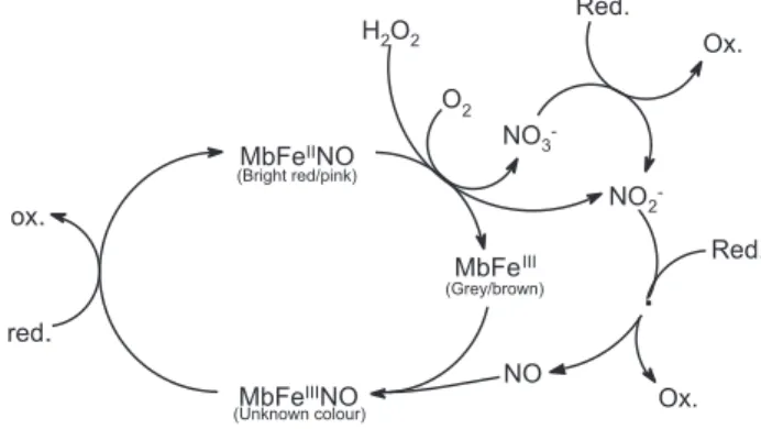
NO is unique as ligand as it binds to both the ferric and ferrous heme iron in Mb with equilibrium constants of 1.4 ×10⁴ M-1 and ≥1011
M-1, respectively.22 At higher pH values nitrosylmetmyoglobin
(MbFeIIINO) can via nucleophile attack of OH- undergo reductive
nitrosylation yielding MbFeIINO,49 although the significance of this
pathway is uncertain in the meat matrix. The meat pigment formed during nitrite-curing has been described as a dinitrosyl complex of myoglobin with a NO coordinated both at the distal and proximal side of the heme.46;50. The final proof of a pentacoordinate mononitrosyl
myoglobin as the principal pigment was presented by Bonnett and co-workers employing electron spin resonance (ESR) spectroscopy.51
There is several studies concerned about the chemical reactions of the pigment MbFeIINO in model systems including oxidative
degradation under conditions relevant to cured meat products during storage. In contrast to what was found for MbFeIIO
2, autoxidation of
MbFeIINO is not sensitive to pH and to changes in ionic strength as
caused by salt addition. The reaction of MbFeIINO with molecular
oxygen, known as autoxidation, is a relatively slow reaction, and the pseudo-first rate constant is very similar to the first-order rate constant found for dissociation of NO from MbFeIINO. Accordingly, it was
suggested that autoxidation should be described by two consecutive reaction steps,52 i.e. initial association of O
2 to coordinated NO forming
an intermediate peroxynitrite complex followed by an electron transfer and dissociation of the reaction products. More recent results, however, show that the initial step is a ligand exchange between coordinated NO and O2 present in solution.53 Thus, the rate constant of the initial
and rate-determining step exhibits a sharp linear increase in the rate, when pO2 is increased from low to atmospheric pressure, in which saturation is observed (Figure 4). This indicates the ligand exchange to exhibit saturation behaviour with respect to the incoming O2 ligand. The second reaction step has been assigned to a fast bimolecular oxidation of MbFeIIO
2 induced by NO (k2 ≈ 10
7 M-1 s-1). This reaction
is now believed to be important as a clearing pathway for excess NO in mammalian skeletal and cardiac muscle tissue under conditions of nitrosative stress.54 The NO molecule initially coordinated to MbFeIINO
seems to remain in the vicinity of the heme iron complex of O2 formed as intermediate in the exchange reaction. It further seems possible that NO is situated within the protein structure in which several cavities have been identified.55 It should be noted that NO is not released
from the Mb complex into the solvent according to this more detailed mechanism, which was based on additional experiments including bimolecular reactions between MbFeIINO and MbFeIIO
2. Mb
accordingly seems to serve as a chemical reactor for small molecules like O2 and NO as is depicted in Figure 6. The more detailed mechanism for autoxidation of MbFeIINO is accordingly suggested to be initiated
by an O2 substitution of NO at the heme iron centre (Figure 6.I). NO is subsequently located in one or more protein cavities,6 as has been
identified by Xe-binding (Figure 6.II). NO attacks coordinated O2 from
the cavities to form a transient, coordinated ONOO- (Figure 6.III),
which subsequently dissociates following isomerisation to nitrate, NO3-, that is expelled from the heme cleft (Figure 6.IV).
Figure 7shows the involvement of Mb in the colour cycle of cured meat products, and it includes the possible reformation of the pink form after oxidation to brown MbFeIII. Such colour re-establishment
has been observed in studies of colour stability of cooked cured ham during storage,56 although the initial colour intensity was never fully
restored.56;57 The pigment of nitrite-cured meat is much more
susceptible to light-induced degradation than the pigment of fresh meat. However, the quantum yield for MbFeIINO shows only moderate
wavelength dependence in contrast to MbFeIIO
2, but the quantum yields
are several magnitudes higher for MbFeIINO compared to MbFeIIO 2
for wavelength of relevance to food storage (Table 2).40;58 Furthermore,
the quantum yield for photooxidation of MbFeIINO increases linearly
with pO2,58 even at very low oxygen tension as demonstrated for
the pressure interval 0.0010 <pO2< 0.0150 atmospheres.59
Interestingly, a comparison of the stoechiometries for thermal and photo-induced oxidation of MbFeIINO shows as expected a 1:1
stoechiometry for O2 and MbFeIINO during thermal oxidation, while
photooxidation results in higher pigment degradation relative to the O2 present. This corresponds to a stoechiometry with approximately 20% excess degradation of MbFeIINO relative to
the O2 present, probably due to involvement of radical intermediates and chain reactions.
Evidence of damage to apomyoglobin (apoMb) during thermal
Figure 6. Reaction mechanism suggested for the autoxidation of nitrosylmyoglobin involving two consecutive steps. Schematic drawing of myoglobin molecule
was adapted from ref. 54, while the reaction mechanism is depicted according to ref. 53
Figure 7. Colour cycle of myoglobin derivatives in cured meat during storage. The attractive pink nitrosylmyoglobin (MbFeIINO) can be oxidised both by H
oxidation of MbFeIINO has also been presented as repeated
oxidation and nitrosylation of MbFeIINO after several cycles has
been found to yield hemichrome, a denatured ferric Mb form with two His residues bound to the central iron atom,60 which is detectable
by ESR spectroscopy.
MYOGLOBIN AND OXIDATIVE PROCESSES IN BIOLOGICAL SYSTEMS
The redox-activities involved in the discoloration of both fresh meat and nitrite-cured meat products affect the oxidative stability of the lipid fraction of the product, as iron redox cycling seems to initiate peroxidation and formation of low-molecular weight compounds responsible for off-flavours and rancidity. The interaction between meat discoloration and lipid oxidation in meat involves the brown MbFeIII, which has been found to have
pseudo-peroxidase activity and which forms several prooxidative Mb species during the catalytic cycle following reaction with H2O2 and other peroxides. MbFeIII also takes part in the propagation of lipid
peroxidation by cleavage of lipid hydroperoxides.44 H
2O2 is central
in triggering of processes leading to oxidative rancidity in fresh meat, as O2•- formed during autoxidation of MbFeIIO
2 yields H2O2
either spontaneously or mediated by SOD. In addition, H2O2 may also be produced in significant quantities as a result of growth of the catalase-negative lactic acid bacteria on the meat surface.
Transition metal redox couples such as FeIII/FeII or CuII/CuI have
standard reduction potentials allowing catalytic decomposition of lipid hydroperoxides (LOOH) to form LO• radicals (Equation 5), and can
thereby initiate the chain reaction characteristic for lipid autoxidation. Heme proteins or free heme groups released from proteins also have the ability to enhance lipid peroxidation processes,60 and Mb has been
active in this respect especially under acidic conditions. Mb reacts with H2O2 in a two-electron process in which a hypervalent perferrylmyoglobin, •MbFeIV=O, is formed with a tyrosyl radical at
Tyr103 (Equation 6). The initially formed hypervalent Mb then undergoes the so-called autoreduction to form ferrylmyoglobin, MbFeIV=O.61 Both of these hypervalent Mb species are known to
initiate lipid peroxidation by hydrogen abstraction from fatty acids.62
However, it should be noted that free heme per se has a higher pseudo-peroxidase activity compared to protein bound heme.63
LOOH + Fe2+ LO• + OH- + Fe3+ (5)
MbFeIII + H 2O2
•MbFeIV=O + H 3O
+ MbFeIV=O (6)
Not surprisingly, it has been shown that phenolic acids, known as
efficient antioxidants, inhibit the MbFeIII/H
2O2 induced oxidation of
low-density lipoproteins.64 More intriguing is the finding that free fatty
acids seem to prevent formation of activated hypervalent forms of Mb65 and hemoglobin (Hb)66 favouring formation of the inactive
hemichromes, as shown in Figure 8 for Mb. This inhibitory effect of free fatty acids on the formation of hypervalent Mb species has been investigated for conditions of excess of saturated fatty acids. Stearic acid and palmitic acid were both found to reduce •MbFeIV=O formation
from MbFeIII and H
2O2, while similar quantities of the monounsaturated
oleic acid did not affect the •MbFeIV=O formation.67 However, it is not
known whether this antioxidative mechanism is important in biological systems or in muscle-based food products, but free fatty acids have been found to increase in concentration during ischemia reperfusion,68
and extensive lipolysis also occurs during long maturation of dry-cured meat products, which attain an exceptional good oxidative stability.69;70
In this context, it should be noted that C8 – C14 saturated fatty acids recently have been found to inhibit the activity of cyclooxygenase enzymes, which are responsible for initiating inflammation processes in the body.71
The rate of MbFeIIO
2 autoxidation increases at reduced oxygen
tension,36;37 which again increases the oxidative stress of membrane
lipids, as O2•- is a product of MbFeIIO
2 autoxidation. The effect on
lipid oxidation of the post-mortem pH decrease in muscle-based foods60
has a parallel in the acidification and reduced oxygen tension occurring in tissue exposed to ischemia reperfusion during surgery,72 which also
increases autoxidation of oxygenated heme proteins. However, the rate for autoreduction of the prooxidative species, MbFeIV=O, has also
been found to be subject to specific acid catalysis and is accordingly pH dependent with a modest temperature dependence corresponding to the activation parameters: ΔH‡ = 58.5 kJ mol-1, ΔS‡ = 2.7 J mol-1 K -1.73 A positive kinetic salt effect was also observed for the autoreduction
of MbFeIV=O to yield MbFeIII, and any MbFeIV=O formed in either
fresh meat or cured meat products will be deactivated due to the low pH conditions and the presence of salt in competition with oxidation of the membrane phospholipids.
The effects of glycation of proteins in relation to various physiological dysfunctions is an emerging field within the medical sciences, and the covalent binding of blood glucose to circulating Hb is involved in several diabetes related pathological conditions.74-76
Glycation of Mb was recently demonstrated to enhance the prooxidative activity of Mb and to increase release of free iron ion Table 2. Quantum yields and relative rates for photooxidation of
oxymyoglobin and nitrosylmyoglobin exposed to monochromatic irradiation at 15 °C
MbFeIIO 2
a MbFeIINOb
λirr Φirr Relative Φirr Relative
(nm) (mol einstein-1) rate (mol einstein-1) rate
254 1.6 × 10⁻² 4700 6.9 × 10⁻³ 41
313 7.9 × 10⁻⁴ 150 2.3 × 10⁻³ 7
334 1.8 × 10⁻⁴ 50 1.6 × 10⁻³ 6
366 3.9 × 10⁻⁵ 10 1.5 × 10⁻³ 7
405 1.2 × 10⁻⁵ 12 1.1 × 10⁻³ 14
436 7.0 × 10⁻⁶ 3 8.6 × 10⁻⁴ 7
546 7.0 × 10⁻⁶ 1 4.3 × 10⁻⁴ 1
aResults from ref. 41; bresults from ref. 59
Figure 8. Prooxidative cycle of Mb activated by H2O2 and proposed
from this heme protein.77 Similar effects should be studied for meat
products with sugar added as flavouring agent or as substrate for microbial starter cultures.
HEME PROTEINS AND NO• IN OXIDATIVE PROCESSES Once initiated, lipid peroxidation, a chain reaction, will be further enhanced by radicals or redox active metals or will be inhibited by free radical scavenging compounds. Both heme proteins and NO belong to a class of chemical compounds that can be active as promoters or inhibitors of lipid peroxidation,78 and their role in
lipid peroxidation will be discussed with special focus on the reactivity of NO towards reaction intermediate in lipid oxidation. NO can act as a prooxidant or an antioxidant depending on the concentration of NO and the absence or presence of O2•-.79 NO reacts
extremely fast with O2•- (k 2 = 10
9 M-1 s-1) and the reaction product is
the strong prooxidant peroxynitrite, ONOO-.80 Equimolar fluxes of
O2•-, H
2O2 and NO have been found to enhance the oxidation of a
substrate (dihydrorhodamine) in a model system containing MbFeIII,
while further increases in NO concentration significantly reduced oxidation via inhibition of •MbFeIV=O/MbFeIV=O formation.81 Another
study showed that NO protects cardiomyocytes against tert-butyl hydroperoxide-induced formation of non-protein and protein-centered free radical species and concomitant peroxidation of membrane phospholipids, and NO was concluded to be an important antioxidant in heart tissue.82 In contrast, NO formation during
ischemia reperfusion of hearts isolated from wild-type or NOS knock-out mice showed a contribution of NO to oxidative injury, most likely due to interaction with O2•-.83 Recent findings with
transgenic mice with an over-expression of NOS, which lack Mb in their cardiac tissue, indicate that excessive formation of NO leads to heart insufficiencies, thus suggesting that Mb under normal circumstances can be an important factor in protecting the heart from nitrosative stress.84 Thus, in light of these conflicting results,
the role of NO in heart tissue under oxidative stress still remains to be clarified.
NO and its interaction with heme iron/Mb has been suggested to prevent oxidative rancidity in nitrite-cured meat.85;86 Oxidative rancidity
is seldom encountered in nitrite-cured meat due to the antioxidative capacity of MbFeIINO and possibly also other nitrite-derived
compounds.46 So far, only few studies have investigated the isolated
effect of MbFeIINO in oxidative processes, but in a carotene-linoleate
model system, it has been found that 2-10 µM of MbFeIII or MbFeIIO 2
acted as a prooxidative species. In contrast, MbFeIINO at all investigated
concentrations did not act as a prooxidant, and MbFeIINO even inhibits
the prooxidative effect of 2 µM MbFeIII.87 MbFeIINO, in presence of
excess MbFeIII, has further been found to significantly inhibit oxygen
consumption in a lipid peroxidating model system with methyl linoleate as substrate.88 NO has been found to inactivate the highly oxidizing
ferryl Mb species involved in oxidative stress.89 The reaction mechanism
includes two steps with a rapid initial formation of a reaction intermediate, nitritometmyoglobin (MbFeIIIONO), which subsequently decays on a
longer time scale (Equations 7-8). The second order rate constant for MbFeIV=O and NO is found to be 1.8 ×10⁷ M-1 s-1 at pH 7.5 and 20°C,
while the first order rate constant for decay of the reaction intermediate is 3.4 s-1. Thus, it seems that free NO in solution may either inhibit or in
the presence of O2•- promote lipid peroxidation. However, when NO is
bound to Mb, it may act as a bioactive reservoir that can be released to scavenge OH• or lipid derived radicals.
[MbFeIV=O MbFeIII=O •] + NO MbFeIIIONO (7)
MbFeIIIONO MbFeIII + NO 2
- (8)
The ability of nitrite (the oxidation product of NO) to inactivate MbFeIV=O has been found to occur at a markedly lower rate with a
second order rate constant between 13-16 M-1 s-1 determined at
ambient temperatures.89;90 Activation parameters of ΔH‡ = 30 kJ mol-1
and ΔS‡ = -123 J mol-1 K-1 are indicative of an associative activation
with a rate-determining intra-molecular electron transfer to yield a cation radical +•MbFeIII-O-.
The strong prooxidant ONOO- has been found to cause rapid
conversion of MbFeIIO
2 to MbFe
III under the conditions expected in
muscle foods, and the presence of CO2 and lowering of pH seem to reduce MbFeIIO
2 degradation slightly.
91 In the reaction between
MbFeIIO
2 and ONOO
-, it has been found that ONOO- oxidizes the
small fraction of deoxygenated Mb (in equilibrium with MbFeIIO 2)
to hypervalent ferryl Mb (Equation 9), while a subsequent reduction by ONOO- yields MbFeIII and peroxynitrite radical (eq. 10).92 The
second order rate constants (20°C and pH 7.3) are found to be very similar and to have values 5.4 × 10⁴ and 2.2 × 10⁴ M-1 s-1, respectively.
MbFeII + ONOOH MbFeIV=O + HNO
2 (9)
MbFeIV=O + HOONO MbFeIII + OONO• + OH- (10)
The effect of CO2 is interesting, since in the presence of 1.2 mM CO2 the rate of the initial reactions step involving MbFeII and ONOO
-(eq. 9) increases significantly (4.1 × 10⁵ M-1 s-1), while the rate of the
second reaction step remains practically unaltered.93 The possible
formation of traces of ONOO- during the autoxidation of MbFeIINO
should also be considered, and a recent study of this autoxidation reaction demonstrates the initial rate-determining reaction step to be unaffected by varying levels of CO2, while the second reaction step was affected by elevated levels of CO2.94
Analysis of nitrated amino acid residues in Mb or haemoglobin (Hb) following exposure to variable amounts of ONOO- shows that
only low quantities of 3-nitrotyrosine can be detected after the reaction with the intact heme proteins.95 However, when apoMb or a cyano
complex of ferric Mb are submitted to similar treatment, significantly larger yields for 3-nitrotyrosine and even lower quantities of nitrated tryptophan is observed in apoMb indicating that the Mb heme iron may act as an efficient scavenger of ONOO-, thereby protecting not
only its globin part, but also other proteins such as the cytochromes from nitration. This further supports the theory of Mb as a protector of cellular respiration in addition to the function of Mb in facilitated diffusion of O2 in muscle tissue. Likewise, in a study of the reaction between Hb and ONOO- nitrated amino acid residues were not
detected, unless large excess of oxidant compounds was added.96 In
contrast, when Hb was incubated with nitrite and H2O2, nitration of other proteins and apoHb were observed,97 and a similar reaction
pattern has also been observed for Mb.98 However, blocking of the
central iron atom via formation of a cyano Hb complex again showed that it is the pseudo-peroxidase activity of Hb and other heme proteins that accounts for this nitration mechanism.
The mechanism of the early processes involved in the formation of back-bone protein radicals may also have negative effects on the oxidative status of protein and lipids in meats during maturation and storage. In fact, the above-mentioned findings may have implications for meat products packaged in modified atmosphere (MA) where CO2 is often used to inhibit microbial growth. So far, these mechanisms are quite speculative, as studies have been conducted only in model systems, and their importance in meats or meat products should be investigated in order to fully clarify the role of reactive nitrogen species in oxidative processes taking place for the post-mortem conditions found in muscle foods.
result of remaining activity of NOS in the muscle tissue,99 and the
concomitant formation of O2•- may generate ONOO-, which will
initiate oxidative processes degrading both lipids and pigments of fresh meat during the initial handling and storage.91;100 However,
further studies are needed to fully assess the potential damaging effect of such reactions, also since hypervalent Mb species so far have not been detected in muscle tissue intended for consumption. Protein oxidation receives increasing attention in relation to oxidative stress in biological system as it seems linked to certain diseases. For meat and meat products modifications of functional properties due to radical damage of proteins also need to be considered. For model systems containing the free amino acid tyrosine, it has been shown that MbFeIV=O accelerates the formation of dityrosine
possibly via formation of tyrosyl radicals followed by dimerization.101 Likewise, the strong prooxidant, ONOO-, has been
shown to modify specific side chains of amino acid residues in Hb with long-lived tyrosyl radicals being formed upon exposure of erythrocytes to ONOO-.102 In addition, the Cys110 amino acid
residue unique to human Mb has been found to form an initial thiyl radical upon reaction with ONOO- as shown by ESR spin trapping.
The final product is, however, either found to be a Mb dimer formed as Tyr103 radicals subsequently form an intermolecular crosslink to another Tyr103 or a 3-nitrotyrosine also in the 103-position.103
CONCLUSION
Colour is an important quality parameter for both fresh meat and nitrite-cured meat products and depends on the redox status and ligand bound to heme iron in Mb. A basic understanding of Mb chemistry is accordingly crucial, and the “inorganic chemistry” of meat should include a quantitative description of Mb complex formation with small ligands, such as O2, NO, H2O and CO, and the kinetics of transformations of these complexes under varying conditions of temperature, oxygen pressure, pH, ionic strength and light exposure. The practical aspect of colour stability of meat and meat products for the meat industry and retail trade has initiated numerous investigations over the last 50 years and an increased understanding of the complex chemistry. The discovery of the physiological importance of NO and the possible role of hypervalent Mb and Hb during oxidative stress have added new perspectives to the dynamic description of electron transfer and ligand exchange reactions of these heme pigments. Other functions of Mb than oxygen transport and storage seem to be important and these in vivo functions include activity as a pseudo-enzyme in specific muscle tissue similar to NO dioxygenases known from microorganisms. In effect, Mb acts as a cellular protector against nitrosative stress during excess production of NO. Future research should focus on the role of Mb as a mediator of reactions between small molecules important as bio-regulators and include investigations of reaction dynamics possibly occurring within the protein structure using time-resolved spectroscopy.
ACKNOWLEDGEMENT
The continuing support from LMC-Centre of Advanced Food Studies is greatly acknowledged. J. K. S. Møller wishes to thank the Norma and Frode S. Jacobsen Foundation for a travel grant to work V in Brazil.
REFERENCES
1. Kagen, L. J.; Myoglobin. Biochemical, Physiological, and Clinical Aspects,
Columbia University Press: New York, 1973. 2. Brunori, M.; Trends Biochem. Sci.2001, 26, 209. 3. Brunori, M.; Trends Biochem. Sci.2001, 26, 21.
4. Wittenberg, J. B.; Wittenberg, B. A.; J. Exp. Biol.2003, 206, 2011. 5. Frauenfelder, H.; Fenimore, P. W.; McMahon, B. H.; Biophys. Chem.2002,
98, 35.
6. Brunori, M.; Biophys. Chem.2000, 86, 221. 7. Sharma, V. S.; Magde, D.; Methods1999, 19, 494. 8. Griddings, G. G.; Crit. Rev. Food Nutr.1977, 9, 81.
9. Livingston, D. J.; Brown, W. D.; Food Technol.1981, May, 244. 10. Antonini, E.; Brunori, M.; Hemoglobin and Myoglobin in their Reactions
with Ligands, North-Holland Publishing Company: Amsterdam, 1971.
11. Schläfer, H. L.; Gliemann, G.; Basic Principles of Ligand Field Theory,
John Wiley & Sons Ltd.: London, 1969.
12. Enemark, J. H.; Feltham, R. D.; Coord. Chem. Rev.1974, 13, 339. 13. Richter-Addo, G. B.; Legzdins, P.; Metal Nitrosyls, Oxford University
Press: New York, 1992.
14. Olson, J. S.; Phillips, G. N.; J. Biol. Chem.1996, 271, 17593.
15. Springer, B. A.; Sligar, S. G.; Olson, J. S.; Phillips, G. N.; Chem. Rev.1994,
94, 699.
16. Olson, J. S.; Phillips, G. N.; J. Biol. Ing. Chem.1997, 2, 544.
17. Copeland, D. M.; West, A. H.; Richter-Addo, G. B.; Proteins: Struct.,
Funct., Genet.2003, 53, 182.
18. Brucker, E. A.; Olson, J. S.; Ikeda-Saito, M.; Phillips, G. N.; Proteins:
Struct., Funct., Genet.1998, 30, 352.
19. Spiro, T. G.; Kozlowski, P. M.; Acc. Chem. Res.2001, 34, 137.
20. Clarke, M. J.; Gaul, J. B. In Structure and Bonding; Clarke, M. J.; Goodenough, J. B.; Ibers, J. A.; Jørgensen, C. K.; Mingos, D. M. P.; Neilands, J. B.; Palmer, G. A.; Reinen, D.; Sadler, P. J.; Weiss, R.; Williams, R. J. P., eds.; Springer-Verlag: Berlin, 1993.
21. Carducci, M. D.; Pressprich, M. R.; Coppens, P.; J. Am. Chem. Soc.1997,
119, 2669.
22. Hoshino, M.; Laverman, L. E.; Ford, P. C.; Coord. Chem. Rev.1999, 187, 75. 23. Andersen, H. J.; Johansen, H. S.; Shek, C. K.; Skibsted, L. H.; Z. Lebensm.
Unters. For.1990, 191, 293.
24. Duprat, A. F.; Traylor, T. G.; Wu, G. Z.; Coletta, M.; Sharma, V. S.; Walda, K. N.; Magde, D.; Biochemistry1995, 34, 2634.
25. Decatur, S. M.; Franzen, S.; DePillis, G. D.; Dyer, R. B.; Woodruff, W. H.; Boxer, S. G.; Biochemistry1996, 35, 4939.
26. Dierks, E. A.; Hu, S. Z.; Vogel, K. M.; Yu, A. E.; Spiro, T. G.; Burstyn, J.
N.; J. Am. Chem. Soc.1997, 119, 7316.
27. Zhao, Y. D.; Hoganson, C.; Babcock, G. T.; Marletta, M. A.; Biochemistry
1998, 37, 12458.
28. Migita, C. T.; Salerno, J. C.; Masters, B. S.; Martasek, P.; McMillan, K.; Ikeda-Saito, M.; Biochemistry1997, 36, 10987.
29. Merryweather, J.; Summers, F.; Vitello, L. B.; Erman, J. E.; Arch. Biochem.
Biophys.1998, 358, 359.
30. Wanat, A.; Gdula-Argasinska, J.; Rutkowska-Zbik, D.; Witko, M.; Stochel, G.; van Eldik, R.; J. Biol. Ing. Chem.2002, 7, 165.
31. Laverman, L. E.; Hoshino, M.; Ford, P. C.; J. Am. Chem. Soc.1997, 119, 12663.
32. Qiu, Y.; Sutton, L.; Riggs, A. F.; J. Biol. Chem.1998, 273, 23426. 33. Garry, D. J.; Kanatous, S. B.; Mammen, P. P. A.; Trends Cardiovas. Med.
2003, 13, 111.
34. Shikama, K.; Chem. Rev.1998, 98, 1357.
35. Brantley, R. E., Jr.; Smerdon, S. J.; Wilkinson, A. J.; Singleton, E. W.; Olson, J. S.; J. Biol. Chem.1993, 268, 6995.
36. Andersen, H. J.; Bertelsen, G.; Skibsted, L. H.; Acta Chem. Scand. A1988,
42, 226.
37. Gutzke, D.; Trout, G. R.; J. Agric. Food Chem.2002, 50, 2673. 38. Bruun-Jensen, L.; Skibsted, L. H.; Meat Sci.1996, 44, 145. 39. Jakobsen, M.; Bertelsen, G.; J. Muscle Foods2002, 13, 143. 40. Bertelsen, G.; Skibsted, L. H.; Meat Sci.1987, 19, 243.
41. Andersen, H. J.; Bertelsen, G.; Skibsted, L. H.; Meat Sci.1990, 28, 87. 42. Mikkelsen, A.; Juncher, D.; Skibsted, L. H.; Meat Sci.1999, 51, 155. 43. Mikkelsen, A.; Skibsted, L. H.; Z. Lebensm. Unters. For.1992, 194, 9. 44. Skibsted, L. H.; Mikkelsen, A.; Bertelsen, G. In Flavors of Meat, Meat
Products and Seafoods; Shahidi, F., ed.; Blackie Academic & Professional:
London, 1994.
45. Møller, J. K. S.; Skibsted, L. H.; Chem. Rev.2002, 102, 1167.
46. Skibsted, L. H. In The Chemistry of Muscle-based Foods; Johnston, D. E.; Knight, M. K.; Ledward, D. A., eds.; The Royal Society of Chemistry: Cambridge, UK, 1992.
47. Koppenol, W. H.; FEBS Lett.1994, 347, 5.
48. Sebranek, J. G.; Fox, J. B. J.; J. Sci. Food. Agric.1985, 36, 1169. 49. Hoshino, M.; Maeda, M.; Konishi, M.; Seki, H.; Ford, P. C.; J. Am. Chem.
Soc.1996, 118, 5702.
50. Pegg, R. B.; Shahidi, F.; Nitrite Curing of Meat. The N-Nitrosamine
Problem and Nitrite Alternatives, Food & Nutrition Press, Inc.:
51. Bonnett, R.; Chandra, S.; Charalambides, A. A.; Sales, K. D.; Scourides, P. A.; J. Chem. Soc., Perkin Trans 11980, 8, 1706.
52. Arnold, E. V.; Bohle, D. S.; Methods Enzymol.1996, 269, 41. 53. Møller, J. K. S.; Skibsted, L. H.; Chem. Eur .J.2004, 10, 2291. 54. Frauenfelder, H.; McMahon, B. H.; Austin, R. H.; Chu, K.; Groves, J. T.;
Proc. Natl. Acad. Sci. U.S.A.2001, 98, 2370.
55. Brunori, M.; Gibson, Q. H.; Embo Reports2001, 2, 674.
56. Møller, J. K. S.; Jensen, J. S.; Olsen, M. B.; Skibsted, L. H.; Bertelsen,
G.; Meat Sci.2000, 54, 399.
57. Andersen, H. J.; Rasmussen, M. A.; Int. J. Food Sci. Technol.1992, 27, 1. 58. Andersen, H. J.; Skibsted, L. H.; J. Agric. Food Chem.1992, 40, 1741. 59. Møller, J. K. S.; Bertelsen, G.; Skibsted, L. H.; Meat Sci.2002, 60, 421. 60. Baron, C. P.; Andersen, H. J.; J. Agric. Food Chem.2002, 50, 3887. 61. Irwin, J. A.; Ostdal, H.; Davies, M. J.; Arch. Biochem. Biophys.1999, 362, 94. 62. Giulivi, C.; Cadenas, E.; Methods Enzymol.1994, 233, 189.
63. Grinberg, L. N.; O’Brien, P. J.; Hrkal, Z.; Free Radical Biol. Med.1999,
27, 214.
64. Laranjinha, J.; Vieira, O.; Almeida, L.; Madeira, V.; Biochem. Pharmacol.
1996, 51, 395.
65. Baron, C. P.; Skibsted, L. H.; Andersen, H. J.; J. Agric. Food Chem.2002,
50, 883.
66. Harrington, J. P.; Newton, P.; Crumpton, T.; Keaton, L.; Int. J. Biochem.
1993, 25, 665.
67. Saifutdinov, R. G.; Larina, L. I.; Vakul’skaya, T. I.; Voronkov, M. G.;
Electron Paramagnetic Resonance in Biochemistry and Medicine, Kluwer
Academic/Plenum Publishers: New York, 2001.
68. Hendrickson, S. C.; St. Louis, J. D.; Lowe, J. E.; Abdelaleem, S.; Mol. Cell.
Biochem.1997, 166, 85.
69. Coutron-Gambotti, C.; Gandemer, G.; Food Chem.1999, 64, 95. 70. Vestergaard, C. S.; Schivazappa, C.; Virgili, R.; Meat Sci.2000, 55, 1. 71. Henry, G. E.; Momin, R. A.; Nair, M. G.; Dewitt, D. L.; J. Agric. Food
Chem.2002, 50, 2231.
72. Gunther, M. R.; Sampath, V.; Caughey, W. S.; Free Radical Biol. Med.
1999, 26, 1388.
73. Mikkelsen, A.; Skibsted, L. H.; Z. Lebensm. Unters. For. 1995, 200, 171. 74. Inouye, M.; Mio, T.; Sumino, K.; Clinica Chimica Acta1999, 285, 35. 75. Inouye, M.; Mio, T.; Sumino, K.; Metabolism1999, 48, 205.
76. Warren, J. D.; Blumbergs, P. C.; Thompson, P. D.; Muscle & Nerve2002,
25, 332.
77. Roy, A.; Sen, S.; Chakraborti, A. S.; Free Radical Res.2004, 38, 139.
78. Eiserich, J. P.; Patel, R. P.; O’Donnell, V. B.; Mol. Aspects Med.1998, 19, 221. 79. Hogg, N.; Kalyanaraman, B.; Biochim. Biophys. Acta1999, 1411, 378. 80. Koppenol, W. H.; Redox Report2001, 6, 339.
81. Jourd´heuil, D.; Mills, L.; Miles, A. M.; Grisham, M. B.; Nitric Oxide Biol.
Chem.1998, 2, 37.
82. Gorbunov, N. V.; Tyurina, Y. Y.; Salama, G.; Day, B. W.; Claycamp, H. G.; Argyros, G.; Elsayed, N. M.; Kagan, V. E.; Biochem. Biophys. Res. Commun.
1998, 244, 647.
83. Flogel, U.; Decking, U. K. M.; Godecke, A.; Schrader, J.; J. Mol. Cell.
Cardiol.1999, 31, 827.
84. Godecke, A.; Molojavyi, A.; Heger, J.; Flogel, U.; Ding, Z. P.; Jacoby, C.; Schrader, J. R.; J. Biol. Chem.2003, 278, 21761.
85. Kanner, J.; Harel, S.; Shagalovich, J.; Berman, S.; J. Agric. Food Chem.
1984, 32, 512.
86. Morrissey, P. A.; Tichivangana, J. Z.; Meat Sci.1985, 14, 175. 87. Kanner, J.; Methods Enzymol.1996, 269, 218.
88. Møller, J. K. S.; Sosniecki, L.; Skibsted, L. H.; Biochim. Biophys. Acta
2002, 1570, 129.
89. Herold, S.; Rehmann, F. J. K.; J. Biol. Ing. Chem.2001, 6, 543. 90. Kroger-Ohlsen, M. V.; Skibsted, L. H.; Food Chem.2000, 70, 209. 91. Connolly, B. J.; Brannan, R. G.; Decker, E. A.; J. Agric. Food Chem.2002,
50, 5220.
92. Exner, M.; Herold, S.; Chem. Res. Toxicol.2000, 13, 287.
93. Herold, S.; Exner, M.; Boccini, F.; Chem. Res. Toxicol.2003, 16, 390. 94. Møller, J. K. S.; Nannerup, L.; Skibsted, L. H.; Meat Sci.2005, 69, 71. 95. Herold, S.; Shivashankar, K.; Mehl, M.; Biochemistry2002, 41, 13460. 96. Romero, N.; Radi, R.; Linares, E.; Augusto, O.; Detweiler, C. D.; Mason,
R. P.; Denicola, A.; J. Biol. Chem.2003, 278, 44049.
97. Grzelak, A.; Balcerczyk, A.; Mateja, A.; Bartosz, G.; Biochim. Biophys. Acta2001, 1528, 97.
98. Bourassa, J. L.; Ives, E. P.; Marqueling, A. L.; Shimanovich, R.; Groves, J. T.; J. Am. Chem. Soc.2001, 123, 5142.
99. Brannan, R. G.; Decker, E. A.; Meat Sci.2002, 62, 229.
100. Brannan, R. G.; Decker, E. A.; J. Agric. Food Chem.2001, 49, 3074. 101. Ostdal, H.; Sorensen, G.; Daneshvar, B.; Skibsted, L. H.; Eur. Food Res.
Technol.2003, 216, 23.
102. Minetti, M.; Scorza, G.; Pietraforte, D.; Biochemistry1999, 38, 2078. 103. Witting, P. K.; Mauk, A. G.; Douglas, D. J.; Stocker, R.; Biochem. Biophys.