O
RIGINALA
RTICLE Revista Brasileira de FisioterapiaHeart rate responses during isometric
exercises in patients undergoing a phase III
cardiac rehabilitation program
Resposta da frequência cardíaca durante o exercício isométrico de pacientes
submetidos à reabilitação cardíaca fase III
Poliana H. Leite1, Ruth C. Melo2, Marcelo F. Mello1, Ester da Silva3, Audrey Borghi-Silva1, Aparecida M. Catai1
Abstract
Background: The magnitude of cardiovascular responses is dependent on the static and dynamic components as well as the duration
and intensity of the contraction performed. Objective: To evaluate the heart rate responses to different percentages of isometric contractions in 12 patients (63±11.6 years) with coronary artery disease and/or risk factors for coronary artery disease that were participating in a phase III cardiac rehabilitation program. Methods: Heart rate variation (ΔHR) was evaluated during maximum (MVC, five and ten seconds in duration) and submaximal (SMVC, 30 and 60% of MVC-5, until muscle exhaustion) voluntary contraction, using a handgrip dynamometer. Additionally, the representative index of cardiac vagal modulation (RMSSD index) was calculated at rest (pre-contraction), at the final 30 seconds of SMVC and during recovery (post-contraction). Results:ΔHR showed higher values in MVC-10 versus MVC-5 (17±5.5 vs 12±4.2 bpm, p<0.05) and the SMVC-60 vs SMVC-30 (19±5.8 vs 15±5.1 bpm, p<0.05). However, results for CVM-10 showed similar ΔHR compared to results for CVSM (p> 0.05). RICVM at rest decreased (p<0.05) during SMVC-30 (30% = 27.9±17.1 vs 12.9±8.5 ms) and SMVC-60 (60% =25.8±18.2 vs 9.96±4.2 ms), but returned to the baseline values when the contraction was interrupted. Conclusions: In patients with coronary artery disease and/or risk factors for coronary heart disease, low intensity isometric contraction, maintained over long periods of time, presents the same effect on the responses of HR, compared to a high intensity or maximal isometric contraction of briefly duration.
Key words: isometric contraction; heart rate; autonomic nervous system; cardiovascular diseases.
Resumo
Contextualização: A magnitude das respostas cardiovasculares depende dos componentes estático e dinâmico bem como da duração
e intensidade da contração realizada. Objetivo: Avaliar as respostas da frequência cardíaca (FC) frente a diferentes percentuais de contração isométrica em 12 pacientes (63±11,6 anos; média±dp) com doença da artéria coronária e/ou fatores de risco para ela, participantes de um programa de reabilitação cardíaca fase III. Métodos: A variação da frequência cardíaca (∆FC) foi avaliada durante as contrações voluntárias máximas (CVM; 5” e 10” de duração) e submáximas (CVSM; 30 e 60% da CVM-5, até exaustão muscular) de preensão palmar, utilizando-se um dinamômetro (hand grip). Adicionalmente, o RMSSD dos iR-R em ms (índice representante da modulação vagal cardíaca) foi calculado em repouso (pré-contração) nos últimos 30 segundos da CVSM e na recuperação (pós-contração). Resultados: A ∆FC apresentou maiores valores em CVM-10 vs CVM-5 (17±5,5 vs 12±4,2 bpm, p<0,05) e no CVSM-60 vs CVSM-30 (19±5,8 vs 15±5,1 bpm, p<0,05). No entanto, os resultados para CVM-10 mostraram ∆FC similar quando comparados aos resultados obtidos para CVSM (p>0,05). RMSSD de repouso reduziu-se (p<0,05) durante a CVSM-30 (30%=29,9±17,1 vs 12,9±8,5ms) e CVSM-60 (60%=25,8±18,2 vs 9,96±4,2 ms), mas retornou aos valores basais quando a contração foi interrompida. Conclusões: Em pacientes com doença da artéria coronária e/ou fatores de risco para ela, a contração isométrica de baixa intensidade mantida por longos períodos de tempo apresenta os mesmos efeitos sobre as respostas da FC, quando comparada à contração isométrica de alta ou máxima intensidade, porém de breve duração.
Palavras-chave: contração isométrica; frequência cardíaca; sistema nervoso autonômico; doenças cardiovasculares.
Received: 05/05/2009 – Revised: 23/11/2009 – Accepted: 21/12/2009
1 Physical Therapy Department, Laboratory of Cardiovascular Physical Therapy, Nucleus of Research in Physical Exercise (NUPEF), Universidade Federal de São Carlos (UFSCar), São Carlos (SP), Brazil
2 School of Arts, Sciences and Humanities (EACH), Universidade de São Paulo (USP), São Paulo (SP), Brazil 3 Faculty of Health Sciences, Universidade Metodista de Piracicaba (UNIMEP), Piracicaba (SP), Brazil
Correspondence to: Aparecida Maria Catai, Departamento de Fisioterapia, Universidade Federal de São Carlos (UFSCar), Rodovia Washington Luís, Km 235, Bairro Monjolinho, CEP 13565-905, São Carlos (SP), Brasil, e-mail: [email protected]
Introduction
Every physical activity requires quick adjustments on cardio-vascular system (CVS) to maintain circulatory homeostasis1,2.
Heart rate (HR) regulation and the modulation of its oscilla-tions are very dependent on the autonomic nervous system (ANS), through the stimulation or inhibition of its eferent, the parasympathetic nervous system (PNS), through the vagus nerve, and the sympathetic nervous system (SNS)2.
It is well documented that isometric contractions increase HR. It is characterized by a quick initial response, attributed to the inhibition of vagus modulation on sinus node (SN). his occurs in the irst 10 seconds of exercise2. Depending on the
intensity and duration of the isometric contraction performed, the HR increases gradually, especially due to the sympathetic modulation of the ANS2-7.
Some studies have reported that the improvements on muscle strength and the resistance training are safe for low-risk patients8-10. However, it is important to note that this type
of training mentioned includes both isotonic and isometric exercises, and the magnitude of cardiovascular responses de-pends on the static and dynamic components as well as the duration and intensity of the exercise10.
Based on the fact that isometric contraction causes a high overload and increases sympathetic activity7,11, the prescription
of the isometric exercises alone must be avoided in programs of cardiovascular rehabilitation9. However, some current studies
have reported that the isometric training with handgrip modi-ies some cardiovascular risk factors (that is, reduces blood pressure (BP), improves endothelial function and increases HR variability) in patients with arterial hypertension12,13.
herefore, studies considering the cardiovascular responses during isometric contractions in diferent intensities and dura-tions are needed to generate evidences regarding the adequate prescription of this modality of exercise for patients enrolled in programs of cardiovascular rehabilitation.
he hypothesis of the this study is that the isometric con-traction in 100% of the maximal voluntary concon-traction (MVC) (5” and 10”) would lead to lower responses of the HR than the sub-maximal percentage of the MVC with longer duration.
he objective of the this study was to evaluate the responses of HR during three isometric contractions of diferent intensi-ties in patients with coronary artery disease and/or risk factor for coronary artery disease.
Methods
Twelve men (63±11.6 years) with coronary artery dis-ease and/or risk factors for coronary artery disdis-ease (Table 1)
participated in this study. All this patients were enrolled, for at least six months, in a phase III cardiovascular rehabilitation pro-gram. his rehabilitation program (60’/session, 3 sessions/week) was predominantly aerobic, including exercises in treadmill and cycloergometer, at an intensity of 70 to 75% of the HR observed during the clinical ergometric test. Patient were submitted to clinical exams (general examination, conventional electrocar-diogram (ECG), physical exams (maximal ergometric test and/or limited by symptom, performed by a cardiologist) and laboratory tests (total cholesterol and fractions, triglycerides, fasting blood glucose, complete blood count, type 1 urine and urea).
Smokers and patients with musculo skeletal diseases were excluded. All patients were informed about the experimen-tal proceedings and, after agreeing with it, they signed the informed consent, which had been approved by the Ethics Committee of Research in Humans from the Universidade Fed-eral de São Carlos (UFSCar), São Carlos (SP), Brazil (protocol 071/2005). All participants were evaluated at the same time of the day considering the inluence of circadian cicle.
he data collection was performed in an acclimatized room. he temperature and relative humidity were maintained at 22-23°C and 50-60%, respectively. he tests were performed in non-consecutive days, separated by a ive-day interval. Pa-tients were previously familiarized with the proceedings and equipments to be used in the study. hey were oriented to avoid cafeine, alcohol and physical exercise and to maintain the usual medication in the day before and in the experiment day. Before the starting of the test, the participants were asked about the occurrence of a regular sleep at night. hey were also examined to certiicate that the basal conditions were within the normality limits (BP≤130/85mmHg; HR: 60-80bpm)14.
During the experiments, the HR and the intervals between two waves R of the ECG (R-R interval) were collected at each heart beat from the CM5 derivation by a cardiac monitor of one channel (TC–500, ECAFIX, São Paulo, SP, Brazil) coupled to an analogue-digital converter Lab – PC + (National Instruments, Co, Austin, TX, USA), which is an interface between the cardiac monitor and the computer. hen, the analogical sign of the ECG was converted in binary values to a computer input at a sam-pling rate of 500Hz which, through a speciic software15, allowed
the data processing and analysis to be performed later.
Prior to the measure of the HR subjects remained at rest (20’) to obtain its stabilization. hen, HR and R-R interval were obtained during the rest pre-contraction (60”); the isometric contractions (5” and 10” to the maximal and until muscle exhaustion to sub-maximal) and recovery post-con-traction (120”) were performed with the patient seated. Fur-thermore, blood pressure (BP) was measured with a mercury sphygmomanometer before and immediately after muscle contraction.
he isometric contractions were performed using an ana-logue hand grip dynamometer (Jamar®
- Sammons Preston, INC Bolingbrook, IL, USA) with the patient sitting in a chair with back support and adjustable arm support, so that the lex-ion forearm angle was maintained at 90°.
On the irst day of the study, the participants performed two sets of three MVC, which were randomly sustained for 5 (MVC-5) and 10 (MVC-10) seconds. On the second day, patients were instructed to maintain a 30% (SMVC-30) and 60% (SMVC-60) contraction of the MVC-5, in a randomized order by draw, until muscle exhaustion. On both test days, patients rested for a three minutes period or until the return of HR to the basal levels. In ad-dition, they were instructed to maintain normal breathing and to avoid the Valsalva maneuver during exercise.
he cardiovascular responses to isometric contraction were evaluated by the diference between HR peak (highest value observed before the end of contraction) and HR rest (pre-con-traction). he duration of voluntary sub-maximal contraction (SMVC) was not predetermined that is, the subjects maintained the muscle contraction until exhaustion. he RMSSD index (square root of the sum of the square of the diferences between the R-R interval in ms in the record divided by the number of R-R interval in ms at a given time minus one) was also calculated dur-ing the SMVC-30 bedur-ing the period of analysis was chosen based in the individual time to exhaustion observed for the SMVC-60. Moreover the autonomic modulation of HR was assessed at rest (pre-contraction), in the 30 seconds preceding the end of the contraction and in the irst seconds of the recovery period.
For data analysis, the SMVC at 30% (30% of MVC, consider-ing 5”) was divided in SMVC-30%A=value calculated based in
time to exhaustion at 60% SMVC and SMVC-30%B= value cal-culated based in time to exhaustion at SMCV at 30% of SMCV.
Statistical analysis
Data are presented as mean ± SD. he diferences of HR and RICVM among all contractions were compared through one-way analysis of variance for repeated measures (one-way ANOVA). Blood pressure values pre and post contraction were compared by Student’s t test. he level of signiicance was set at p<0.05.
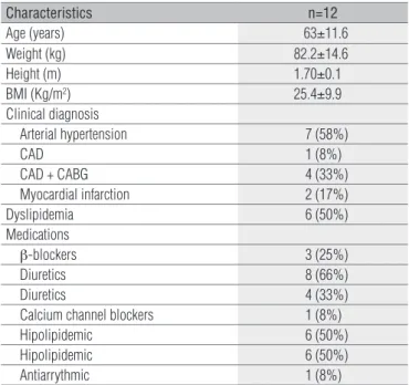
Table 1. Patients’ characteristics.
Characteristics n=12
Age (years) 63±11.6
Weight (kg) 82.2±14.6
Height (m) 1.70±0.1
BMI (Kg/m2) 25.4±9.9
Clinical diagnosis
Arterial hypertension 7 (58%)
CAD 1 (8%)
CAD + CABG 4 (33%)
Myocardial infarction 2 (17%)
Dyslipidemia 6 (50%)
Medications
β-blockers 3 (25%)
Diuretics 8 (66%)
Diuretics 4 (33%)
Calcium channel blockers 1 (8%)
Hipolipidemic 6 (50%)
Hipolipidemic 6 (50%)
Antiarrythmic 1 (8%)
BMI= body mass index; CAD= coronary artery disease; CABG= coronary artery bypass graft.
Table 2. Isometric contractions.
MVC SMVC
5 10 30%A 30%B 60%
Time (sec) 5 10 69±13.8 198±58.0 69±13.8
Intensity (%) 100 100 30 30 60
HRrest (bpm) 61±9.1 60±9.7 61±8.7 61±9.0 61±8.7
HRpeak (bpm) 73±10.2 77±12.7 67±10.6* 76±11.1 79±12.4†
∆HR (bpm) 12±4.2 17±5.5† 6±3.9* 15±5.1 19±5.8†§
RICVM (ms)
Rest - - 27.9±17.1 27.9±17.1 25.8±18.2
Exercise - - 16.8±11.5 12.9±8.5+ 9.96±4.2+‡
Recovery - - 27.6±19.1 27.6±19.1 28.9±10.6
Strength (Kgf) 37.9±7.1 35.4±5.3 12.7±2.3 12.7±2.3 23.2±4.1
SBP (mmHg)
Rest 120.4±6.3 120.4±6.3 120.0±12.9 120.0±12.9 120.0±12.9
Recovery 129.2±11.3+ 128.3±12.9+ - 127.9±12.3+ 129.2±10.6+
DBP (mmHg)
Rest 80.4±4.8 80.4±4.8 79.6±9.3 79.6±9.3 79.6±9.3
Recovery 85.0±6.8+ 86.7±7.2+ - 80.8±9.3 82.1±10.0
Values shown as mean±SD. HR=heart rate; SBP=systolic blood pressure; DBP=dyastolic blood pressure; MVC=maximal voluntary contraction; SMVC=submaximal voluntary contraction; 30=intensity at 30% of MVC (5”); A=value calculated based on time to exhaustion of SMVC at 60%; B= value calculated based on time to exhaustion of SMVC at 30%; 60=intensity at 60% of MVC (5”); *p<0.05 vs. all the contractions studied; †p<0.05 vs. MVC-5; §p<0.05 vs. SMVC-30%B; +p<0.05 vs. rest and recovery conditions (when applied); ‡p<0.05 vs. SMVC-30%A.
Figure 1. Variation of heart rate during maximal (MVC) and sub-maximal (SMVC) voluntary contractions.
A=value calculated based on time to exhaustion of SMVC at 60%; B= value calcula-ted based on time to exhaustion of SMVC at 30%; *p<0.05 vs. MVC-5 †p<0.05 vs.
all the contractions studied; §p<0.05 vs. SMVC-30%B.
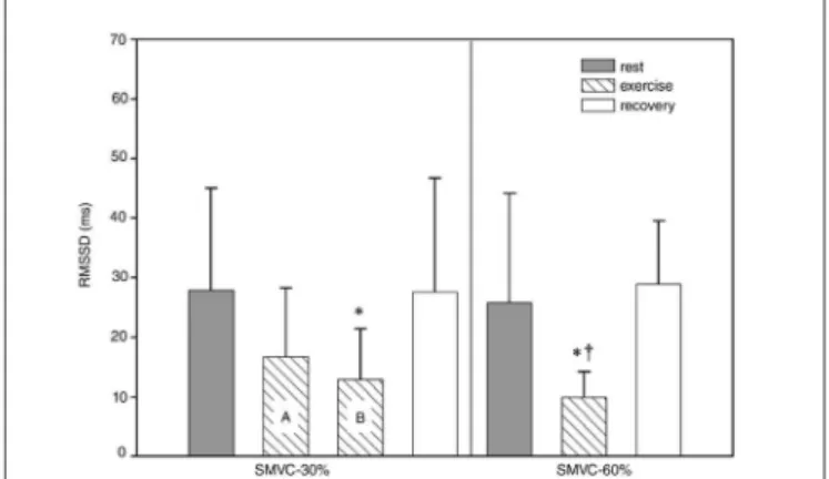
A=value calculated based on time to exhaustion of SMVC at 60%; B= value calcu-lated based on time to exhaustion of SMVC at 30%; *p<0.05 vs. rest and recovery conditions; †p<0.05 vs. SMVC-30%A.
Figure 2. Autonomic modulation of heart rate through the RMSSD index of the R-R intervals in ms, assessed at rest, during sub-maximal voluntary contractions (SMVC) and recovery.
Results
Table 1 presents the patients’ characteristics, the clinical di-agnosis and the medications in use. No diferences were found for resting HR for all contractions analyzed (Table 2). For the HR peak, the SMVC-30%A, determined based on time to exhaustion of SMVC-60, showed the lowest value compared to the other contractions (p<0.05) (Table 2). Moreover, the MVC-5 had HR peak signiicantly lower than the SMVC-60 (p<0.05) (Table 2).
Considering the ∆HR, the SMVC-30%A produced the low-est cardiovascular response (p<0.05) in comparison with to the remaining contractions, while the SMVC-60 presented the highest ∆HR among the contractions measured (except for MVC-10). For the SMVC-30%B, in which ∆HR was calculated
considering muscle exhaustion; it was observed a ∆HR similar to the MVC-5 and MVC-10 (Table 2 and Figure 1).
At rest, there were no statistical diferences for the RMSSD index of the SMVC-30 and SMVC-60. As expected, the RMSSD index reduced during isometric contraction, reaching statisti-cal signiicance (p<0.05), except for SMVC-30%A. Moreover, when this index was calculated considering time to exhaus-tion of SMVC-60, a lower value of RMSSD index was observed for CVSM-60 when compared to SMVC-30%A (p<0.05). In the recovery period after contraction, the values of RMSSD index were similar for both intensities tested (p>0.05) (Table 2 and Figure 2).he values of systolic blood pressure (SBP) and dia-stolic blood pressure (DBP), measured in the irst seconds of the recovery period were higher in comparison to the resting values (p<0.05), except the SMVC, which presented similar values of DBP (p>0.05) between the rest and recovery periods (Table 2).
Discussion
his study investigated the response of the HR during iso-metric contractions, with diferent intensities and durations, in patients with cardiovascular disease and/or risk factors for cardiovascular disease. he magnitude of cardiovascular responses, evaluated through ∆HR and index RMSSD of R-R interval in ms, showed to be dependent of the intensity and duration of the isometric contractions. hus, for an isometric contraction of low intensity, maintained for long period of time, there were observed efects on HR responses similar to those of a high or maximal intensity contraction, maintained for a short period of time.
Cardiovascular system has an important role on homeo-stasis maintenance. During physical exercise, hemodynamic adjustments occur to allow the appropriate distribution of blood to supply the demands of muscles in activity. Moreover, the magnitude of these adjustments seems to depend on the exercise’s characteristics2,16.
he isometric exercise promotes a signiicant increase on HR, BP and peripheral vascular resistance11 being the
mechanisms responsible for these responses are central and peripheral1,11,16,17. he central mechanism activates neuronal
pathways from central nervous system to modify the activities of sympathetic and parasympathetic systems, consequently determining some cardiovascular responses1. In addition,
evidences from electromyography records show that the acti-vation of more motor units of muscle ibers recruited during a contraction is related to the neural mechanism of central command, which determines immediate changes in the level of eferent activity from SNS and PNS, acting on heart, and of SNS, acting on blood vessels17,18.
Moreover, the relex neural mechanism, related to mechan-ical and metabolic activities from the muscle in contraction, also determines the level of autonomic activity on cardiovascu-lar system. Neural impulses related to the mechanical activity are transmitted initially by muscle receptors through aferent ibers from groups III and IV and reach areas of cardiovascular control almost simultaneously to the neural impulses from central command4,19,20. he neural impulses related to muscle
metabolic activity are transmitted primarily by aferent muscle ibers from group IV and reach the area of vascular control with a delay of some seconds17,21,22. he aferent muscle
recep-tors from groups III and IV are divided in ergoreceprecep-tors (group III), which are activated by muscle contraction, and nocicep-tors (group IV), activated by stimuli responsible for muscle pain sensation20,23.
hus, the reduction on oxygen supply of active muscles, which is caused by a mechanical obstruction of blood vessels during isometric contraction of high intensity, causes an in-crease on metabolites on the muscle and, consequently, stimu-lates pressor relex of exercise19.
The elevation of HR occurs suddenly in the beggining of the isometric exercise, being its magnitude seems to be directly related to the levels of muscle tension2,24. This
initial elevation of HR that occurs within the first seconds of contraction (5” to 30”) is also associated to the intensity of the exercise and is attributed to the withdrawal of vagal modulation on sinus node. However, if the exercise is main-tained until exhaustion, HR will increase gradually due to the increased sympathetic modulation acting on heart2,7,21.
In this study, it was possible to observe that the fast in-crease of HR, evaluated through ∆HR, for the same tension (MVC), is dependent on the period in which the contraction is maintained. Therefore, our results are in agreement with the authors mentioned above, since MVC maintained for 5 seconds may not have been long enough to generate maxi-mal vagal withdrawal.
Iellamo et al.7 evaluated the autonomic control of HR
in young subjects through the analysis of rate domain dur-ing 4 minutes of isometric contraction of knee extension (30% of MVC). The authors observed a reduction on vagal modulation and an increase on cardiac sympathetic modu-lation, which suggests the participation of the sympathetic component on HR regulation during low intensity and long duration exercises. Although Stewart etal.25 had shown a
reduction on vagal modulation during hand grip exercises (35% MVC) in young subjects, they were not able to repro-duce the same results of Iellamo et al.7. However, the authors
observed a reduction on sympathetic modulation in the first minute of exercise and its return for pre-exercise basal levels. Since Stewart et al.25 used only periods of 1 minute
to analyze HR variability, it is possible that sympathetic modulation has been underestimated, which could explain partially the divergence between the results found on the two studies discussed above.
In this study, patients were unable to maintain, at SMVC, the time required to perform the analysis of ΔHR in the rate domain (30%=3 minutes and 60%=1 minute, approximately) since it requires, at least, 4-5 minutes of data recording and, also, with the ECG signal remaining stable26. In this context,
the autonomic control of HR was assessed only through the RMSSD index of R-R interval in ms (time domain), which represents the fast oscillatory component, that is, the vagal modulation responsible by the variation between the car-diac cycles. Since the RMSSD index reduced during isomet-ric contraction for both sub-maximal intensities studied, our results agree partially with those of Iellamo et al.7 and
Stewart et al.25. However, nothing can be asserted on the
sympathetic modulation during isometric contraction from these results.
The literature has reported that the magnitude of HR re-sponse during isometric exercise is related to muscle mass, duration and tension developed during contraction17,27. In
this study, we sought to study the effect of different intensi-ties and times of contraction on HR response. Thus, the protocol used tested only one muscle group (palmar flex-ors) at a specific angle, that is, at the same muscle length since the wrist was in a neutral position. Under these con-ditions, HR has shown to be influenced by duration and intensity of isometric contraction, since maximal efforts of short duration produced similar responses to sustained sub-maximal efforts. Furthermore, the effect of time was shown when the ΔHR was compared at the same intensity (SMVC-30% A versus SMVC-30% B) but with a different duration (69” versus 198”).
In this study, patients had adequate cardiovascular re-sponses to isometric exercise and none had signs or symp-toms that required exercise interruption. It is noteworthy that, prior to the start of the experiment, all patients under-went a clinical ergometric test and, besides this, they already participated of an aerobic physical training for at least six months, so they presented adequate aerobic capacity and, also, were using speciic medication. hus, the prescription of isometric exercises seems to be promising and safe for low risk patients. Low risk patients are those with good func-tional capacity, controlled hypertension, with no evidence of myocardial ischemia at rest or induced by efort, without severe left ventricular dysfunction or complex ventricular ar-rhythmia, which are common characteristics of the patients from the this study, whom are enrolled in programs of car-diac rehabilitation (phase III). For this, they shall be correctly
1. Williamson JW, Fadel PJ, Mitchell JH. New insights into central cardiovascular control during exercise in humans: a central command update. Exp Physiol. 2006;91(1):51-8.
2. Freeman JV, Dewey FE, Hadley DM, Myers J, Froelicher VF. Autonomic nervous system interaction with the cardiovascular system during exercise. Prog Cardiovasc Dis. 2006;48(5):342-62.
3. Galvez JM, Alonso JP, Sangrador LA, Navarro G. Effect of muscle mass and intensity of isometric contraction on heart rate. J Appl Physiol. 2000;88(2):487-92.
4. Stebbins CL, Walser B, Jafarzadeh M. Cardiovascular responses to static and dynamic contraction during comparable workloads in humans. Am J Physiol Regul Integr Comp Physiol. 2002;283(3):R568-75.
5. Koba S, Hayashi N, Miura A, Endo M, Fukuba Y, Yoshida T. Pressor response to static and dynamic knee extensions at equivalent workload in humans. Jpn J Physiol. 2004;54(5):471-81.
6. Iellamo F, Massaro M, Raimondi G, Peruzzi G, Legramante JM. Role of muscular factors in cardiorespiratory responses to static exercise: contribution of reflex mechanisms. J Appl Physiol. 1999;86(1):174-80.
7. Iellamo F, Pizzinelli P, Massaro M, Raimondi G, Peruzzi G, Legramante JM. Muscle metaboreflex contribution to sinus node regulation during static exercise: insights from spectral analysis of heart rate variability. Circulation. 1999;100(1):27-32.
8. Pollock ML, Franklin BA, Balady GJ, Chaitman BL, Fleg JL, Fletcher B, et al. AHA Science Advisory. Resistance exercise in individuals with and without cardiovascular disease: benefits, rationale, safety, and prescription: an advisory from the Committee on Exercise, Rehabilitation, and Prevention, Council on Clinical Cardiology, American Heart Association; Position paper endorsed by the American College of Sports Medicine. Circulation. 2000;101(7):828-33.
9. Bjarnason-Wehrens B, Mayer-Berger W, Meister ER, Baum K, Hambrecht R, Gielen S, et al. Recommendations for resistance exercise in cardiac rehabilitation. Recommendations of the German Federation for Cardiovascular Prevention and Rehabilitation. Eur J Cardiovasc Prev Rehabil. 2004;11(4):352-61.
10. Williams MA, Haskell WL, Ades PA, Amsterdam EA, Bittner V, Franklin BA, et al. Resistance exercise in individuals with and without cardiovascular disease: 2007 update: a scientific statement from the American Heart Association Council on Clinical Cardiology and Council on Nutrition, Physical Activity, and Metabolism. Circulation. 2007;116(5):572-84.
11. Rowland T, Fernhall B. Cardiovascular responses to static exercise: a re-appraisal. Int J Sports Med. 2007;28(11):905-8.
12. Taylor AC, McCartney N, Kamath MV, Wiley RL. Isometric training lowers resting blood pressure and modulates autonomic control. Med Sci Sports Exerc. 2003;35(2):251-6.
13. McGowan CL, Visocchi A, Faulkner M, Verduyn R, Rakobowchuk M, Levy AS, et al. Isometric handgrip training improves local flow-mediated dilation in medicated hypertensives. Eur J Appl Physiol. 2007;99(3):227-34.
14. Mion Jr D, Kohlmann Jr O, Machado CA, Amodeo C, Gomes MAM, Praxedes JN, et al. V Diretrizes brasileiras de hipertensão arterial. Arq Bras Cardiol. 2007;89(3):e24-79.
15. Silva E, Catai AM, Trevelin LC, Guimaraes JO, Silva Jr LP, Silva LMP, et al. Design of computerized system to evaluate the cardiac function during dynamic exercise. Phys Med Biol. 1984;33:409.
16. Iellamo F. Neural mechanisms of cardiovascular regulation during exercise. Auton Neurosci. 2001;90(1-2):66-75.
examined and guided during the performance of this type of exercise.
he responses of the BP are also directly related to the duration of isometric contraction. However, the assessment of BP during MVC could not be performed due to the limita-tion in contraclimita-tion duralimita-tion and the absence of a non-invasive equipment for checking the BP during exercise continuously; with regards to the SMVC, the variability of the duration of contraction of the subjects did not allow a standardized data collection, reason for why they are not presented in this manu-script. Considering this, we decided to assess BP responses immediately after the interruption of the contractions (MVC and SMVC), and the values of SBP showed to be higher in rela-tion to rest values. For future studies, it would be interesting to examine BP during isometric contractions that could lead to additional contributions.
Overall, in patients with cardiovascular diseases and/or risk factors for cardiovascular diseases, HR response and its autonomic control seem to be dependent on the intensity and duration of isometric contractions. In addition, all pa-tients had adequate HR responses during exercise, suggest-ing its prescription in cardiovascular rehabilitation for low risk patients, since they have characteristics similar of those from this study, and the exercises are carefully selected and guided.
It is also noteworthy that some participants (n=3) were using drugs that directly afect the responses of HR (e.g. beta blockers). As beta blockers are often used in the treatment
of patients with stable coronary artery disease, hypertension and congestive heart failure28,29 mainly due to its positive
ef-fect on their prognosis, in clinical practice is very common to ind patients in Phase III of the cardiovascular rehabilitation using beta blockers in combination to other drugs. herefore, the results of this study should be interpreted with caution and should not be transferred to all types of people and / or patients.
In conclusion, in the patients studied, the results showed that isometric contraction of low intensity sustained for long periods of time has the same efects on HR responses than an isometric contraction of high or maximal intensity with short duration.
Considering that the response of PA is directly related to the isometric exercise, its evaluation would bring relevant and complementary data to this study. For future studies it would be interesting: a) to assess BP continuously during isometric contraction, with a non invasive method; b) to evaluate the chronic efects of isometric training in low risk patients and c) to work with a control group.
Acknowledgments
Research supported by Conselho Nacional de Desenvol-vimento Cientíico e Tecnológico (CNPq - 309312/2009-4) and Fundação de Amparo à Pesquisa do Estado de São Paulo (FAPESP - 05/54838-9).
388
17. Mitchell JH. J.B. Wolffe memorial lecture. Neural control of the circulation during exercise. Med Sci Sports Exerc. 1990;22(2):141-54.
18. Gallo L Jr, Maciel BC, Marin-Neto JA, Martins LE, Lima-Filho EC, Manco JC. The use of isometric exercise as a means of evaluating the parasympathetic contribution to the tachycardia induced by dynamic exercise in normal man. Pflugers Arch. 1988;412(1-2):128-32.
19. Daniels JW, Stebbins CL, Longhurst JC. Hemodynamic responses to static and dynamic muscle contractions at equivalent workloads. Am J Physiol Regul Integr Comp Physiol. 2000;279(5):R1849-55.
20. Ichinose M, Saito M, Kondo N, Nishiyasu T. Baroreflex and muscle metaboreflex: control of muscle sympathetic nerve activity. Med Sci Sports Exerc. 2008;40(12):2037-45.
21. Williamson JW, Nobrega AC, Winchester PK, Zim S, Mitchell JH. Instantaneous heart rate increase with dynamic exercise: central command and muscle-heart reflex contributions. J Appl Physiol. 1995;78(4):1273-9.
22. O’Leary DS. Heart rate control during exercise by baroreceptors and skeletal muscle afferents. Med Sci Sports Exerc. 1996;28(2):210-7.
23. Ray CA, Carter JR. Central modulation of exercise-induced muscle pain in humans. J Physiol. 2007;585(Pt 1):287-94.
24. Silva E, Oliveira L, Catai AM, Ferreira Filho P, Berzin F, Gallo Junior L. Evaluation of electromyographic activity and heart rate responses to isometric exercise. The role played by muscular mass and type. Braz J Med Biol Res. 1999;32(1):115-20.
25. Stewart JM, Montgomery LD, Glover JL, Medow MS. Changes in regional blood volume and blood flow during static handgrip. Am J Physiol Heart Circ Physiol. 2007;292(1):H215-23.
26. Heart rate variability: standards of measurement, physiological interpretation and clinical use. Task Force of the European Society of Cardiology and the North American Society of Pacing and Electrophysiology. Circulation. 1996;93(5):1043-65.
27. Seals DR, Washburn RA, Hanson PG, Painter PL, Nagle FJ. Increased cardiovascular response to static contraction of larger muscle groups. J Appl Physiol. 1983;54(2):434-7.
28. Rosendorff C. Hypertension and coronary artery disease: a summary of the American Heart Association scientific statement. J Clin Hypertens (Greenwich). 2007;9(10):790-5.
29. Jessup M, Abraham WT, Casey DE, Feldman AM, Francis GS, Ganiats TG, et al. 2009 focused update: ACCF/AHA Guidelines for the diagnosis and management of heart failure in adults: a report of the American College of Cardiology Foundation/American Heart Association Task Force on Practice Guidelines: developed in collaboration with the International Society for Heart and Lung Transplantation. Circulation. 2009;119(14):1977-2016.