Hyperhomocystinemia in patients
with coronary artery disease
1Instituto do Coração, 2Laboratório de Hematologia Molecular (LIM-31),
Disciplina de Hematologia e Hemoterapia, Faculdade de Medicina, Universidade de São Paulo, São Paulo, SP, Brasil
3Pontifícia Universidade Católica do Paraná, Curitiba, PR, Brasil
J.R. Faria-Neto1,3,
A.C.P. Chagas1,
S.P. Bydlowski2,
P.A. Lemos Neto1,
D.A. Chamone2,
J.A.F. Ramirez1
and P.L. da Luz1
Abstract
Hyperhomocystinemia has been related to an increased risk of cardio-vascular disease in several studies. The C677T polymorphism for the gene that encodes the methylenetetrahydrofolate reductase enzyme (MTHFR) and low plasma folate levels are common causes of hyper-homocystinemia. Due to differences in nutritional patterns and ge-netic background among different countries, we evaluated the role of hyperhomocystinemia as a coronary artery disease (CAD) risk factor in a Brazilian population. The relation between homocysteine (Hcy) and the extent of CAD, measured by an angiographic score, was determined. A total of 236 patients referred for coronary angiography for clinical reasons were included. CAD was found in 148 (62.7%) patients and 88 subjects had normal or near normal arteries. Patients with CAD had higher Hcy levels [mean (SD)] than those without disease (14 (6.8) vs 12.5 (4.0) µM; P = 0.04). Hyperhomocystinemia
(Hcy >17.8 µM) prevalence was higher in the CAD group: 31.1 vs
12.2% (P = 0.01). After adjustment for major risk factors, we found an independent association between hyperhomocystinemia and CAD (OR = 2.48; 95% CI = 1.02-6.14). Patients with a more advanced coronary score had a higher frequency of hyperhomocystinemia and tended to have higher mean Hcy levels. An inverse relation between plasma folate and Hcy levels was found (r = -0.14; P = 0.04). Individuals with the MTHFR C677T polymorphism had a higher prevalence of hyperhomocystinemia than those without the mutated allele. We conclude that hyperhomocystinemia is independently asso-ciated with CAD, with a positive association between Hcy level and disease severity.
Correspondence
A.C.P. Chagas
Unidade Clínica de Aterosclerose Instituto do Coração, FM, USP Av. Dr. Enéas C. Aguiar, 44 05403-000 São Paulo, SP Brasil
Fax: +55-11-3069-5447
E-mail: [email protected]
Research supported by FAPESP (Nos. 98/3168-8 and 98/03167-1).
Received June 21, 2004 Accepted November 28, 2005
Key words
•Hyperhomocystinemia •Homocysteine
•Methylenetetrahydrofolate
reductase
•Atherosclerosis •Folic acid deficiency
Introduction
Atherosclerosis is the leading cause of mortality in the Western world. In most pa-tients, traditional risk factors such as dyslipi-demia, hypertension, smoking, and diabetes can be identified. The reduction of
could explain the disease in these patients; hyperhomocystinemia is one of them.
Homocysteine (Hcy) is a sulfhydryl amino acid involved in methionine metabo-lism. The detrimental effect of severe hyper-homocystinemia on the cardiovascular sys-tem was first described by McCully (1) in 1969. He reported diffuse atherosclerotic lesions in a post-mortem study of two chil-dren with homocystinuria, an inborn error of Hcy metabolism in which extremely high plasma Hcy levels are found. Fatal events before the age of 30 occur in 25% of these patients (2).
The harmful effect of hyperhomocystine-mia on the vascular system has been con-firmed by experimental studies in animals (3). After the introduction of reliable routine methods for Hcy determination, clinical stud-ies showed the association of mild to moder-ate hyperhomocystinemia not only with cor-onary disease (4-6), but also with stroke (7) and peripheral artery disease (8).
Plasma Hcy concentration is dependent on some nutritional and genetic factors. A low plasma folate level is a common cause of hyperhomocystinemia, but vitamin B12 deficiency may also play a role. Folate is a substrate for the remethylation cycle of Hcy, and vitamin B12 is a co-factor in its metabo-lism. Folate supplementation is an effective therapy for Hcy normalization (9).
A common polymorphism for the en-zyme 5,10-methylenetetrahydrofolate reduc-tase (MTHFR) is related to mild to moderate elevation of plasma Hcy levels. This enzyme converts 5,10-methylenetetrahydrofolate to 5-methyltetrahydrofolate, required for the conversion of Hcy to methionine. The C677T polymorphism results in an amino acid change from alanine to valine. This substitu-tion causes thermolability and reducsubstitu-tion of enzyme activity (10).
Since genetic and nutritional differences are present among different countries, it is reasonable to assume that Hcy may play a different role as a coronary risk factor
ac-cording to the population studied. To the best of our knowledge, no study assessed the relation between Hcy and coronary disease in any Latin American country, a develop-ing region susceptible to nutritional defi-ciencies (11). Although the prevalence of malnutrition is decreasing in Brazil, some diseases like anemia, highly dependent on iron, folate and vitamin B12 intake are now reaching epidemic levels (12).
The aim of the present cross-sectional study was to evaluate the role of hyperho-mocystinemia as a coronary risk factor in a Brazilian population. Moreover, using an angiographic index, we evaluated the asso-ciation between plasma Hcy level and the extent of coronary disease.
Patients and Methods
Patients
On the assumption that up to 25% of patients would have normal or near normal arteries upon an angiogram, sample size was calculated with α = 0.05 and a power of 90%
to detect a 16% difference in hyperho-mocystinemia prevalence. So, 236 patients referred to the Heart Institute for a selective coronary angiogram were enrolled. All pa-tients had been previously seen by their at-tending physician, and the angiography was requested based on the clinical judgment of the latter. Patients whose coronary angio-gram was requested for a reason other than suspected coronary disease were also in-cluded. We excluded patients younger than 18 years, with acute coronary syndrome in the last 30 days, those who had taken any medication containing vitamins or folic acid in the 6 weeks before enrollment, and pa-tients with creatinine above 1.5 mg/dL.
the same technique and always the same calibrated equipment. The study was ap-proved by the institutional Ethics Commit-tee and written informed consent was ob-tained from all patients.
Coronary angiography
Angiograms were evaluated by an expe-rienced angiographer blind to the clinical data. CAD was considered to be present when any stenotic lesion ≥40% in any of major epicardial coronaries and their branches was observed. Subjects with normal or near-normal arteries (no lesion greater than 40%) formed a control group.
All angiograms were scored according to the Friesinger index (13). This index ranges from 0 (completely normal arteries) to 15. Each of the 3 arteries (right coronary, cir-cumflex and anterior descending artery), with their major branches, was analyzed inde-pendently and scored from 0 to 5. The final score was the sum of the results for each artery. An artery with no wall irregularity was scored as 0. Score 1 was determined by parietal irregularities, less than 30%. If the artery had a single stenotic lesion causing a narrowing of less than 70%, the score was 2. The same degree of obstruction, but at more than one specific site of the artery, was scored as 3. An artery with any lesion greater than 70% was scored as 4. A score of 5 was assigned when complete occlusion of the proximal right coronary, circumflex or ante-rior descending artery was found. Lesions in the left main coronary were assessed using the same scoring system, but doubled (the lesion was considered in two arteries).
Measurement of plasma homocysteine
Blood samples were collected before an-giography, after a 12-h fast. The samples were kept on ice and immediately centri-fuged at 0ºC to obtain plasma. Plasma Hcy levels were quantified by high-performance
liquid chromatography using a Shimadzu Class-Vp System. The technique described by Fiskerstrand et al. (14) was used.
Genetic analysis
Genotyping for the C677T polymorphism of MTHFR was performed by PCR amplifi-cation. The reaction primers have been de-scribed elsewhere (15). The resulting frag-ments were separated by 1.5% agarose gel electrophoresis.
Definitions of risk factors
Age was evaluated as a continuous and categorical variable. Hypertension was de-fined as a history of systolic blood pressure >140 mmHg and/or diastolic pressure >90 mmHg, confirmed by measurement at en-rollment, or antihypertensive therapy. Pa-tients were considered to be smokers when regularly using tobacco for the last 6 months. Diabetes was defined as fasting glucose above 126 mg/dL or use of hypoglycemic drug therapy. Hyperlipidemia was present if total cholesterol >240 mg/dL, or LDL-cho-lesterol >160 mg/dL or if the patient was on lipid-lowering therapy.
Hyperhomocystinemia was defined ac-cording to values found in patients with normal or near-normal coronary arteries. We defined hyperhomocystinemia as any value above the 90th percentile for this group.
Statistical analysis
Continuous variables are reported as means ± SD. Categorical variables are pre-sented as percentages. Log transformation was used for analysis of skewed data (Hcy). Univariate (2-sided t-tests and chi-square
and hyperhomocystinemia was modulated by any other variable, we used multiple lo-gistic regression analysis.
Results
A total of 236 patients had undergone angiography. Most were referred for a coro-nary angiogram for corocoro-nary disease inves-tigation (83.9%). Heart failure was the rea-son for the angiographic study in 6.8%, valve disease in 6.3% and other reasons in 3% of cases. Only 13 patients (5.5%) had no risk factor for CAD. One risk factor was present in 72 patients (30.5%), while 86 (36.5%) had two risk factors, 56 (23.7%) had three, and 9 (9.8%) had all of them.
Coronary angiograms showed 88 sub-jects with no stenotic lesion >40%, a result considered to indicate normal or
near-nor-mal arteries. One hundred and forty-eight patients (62.7%) had coronary disease (at least one stenotic lesion >40% in a major epicardic coronary).
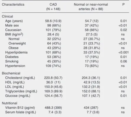
Characteristics of the patients
Patients with CAD were older than those without significant coronary abnormalities (58.6 vs 54.7 years; P = 0.01), and were
predominantly males. Body mass index was similar in both groups, despite a higher preva-lence of overweight subjects among CAD patients. The prevalence of traditional risk factors was different in the two groups; hy-perlipidemia was found in 68% of patients with CAD and in 37.5% of those without CAD (P < 0.001). There was also a signifi-cant difference regarding diabetes (36 vs
19%; P = 0.01) and a trend toward a higher frequency of smoking among patients with CAD. No difference was observed regard-ing hypertension.
In relation to biochemical characteris-tics, plasma levels of total cholesterol and LDL-cholesterol were higher in patients with CAD, whereas the inverse relation was seen with HDL-cholesterol levels. Plasma glu-cose levels were also higher in patients with CAD. No statistically significant difference was found in vitamin B12 or plasma folate levels between groups. Clinical and bio-chemical features are listed in Table 1.
Homocysteine
Mean plasma Hcy level and prevalence of hyperhomocystinemia were higher in pa-tients with CAD compared to those with normal or near-normal arteries: Hcy level [mean (SD)] was 14.4 (6.8) vs 12.5 (4.0) µM
(P = 0.04) and hyperhomocystinemia was found in 31.1 vs 12.2% (P = 0.02).
By univariate analysis, the OR for coro-nary disease was 2.55 (95% CI = 1.15-5.67) in patients with hyperhomocystinemia rela-tive to subjects with plasma Hcy level below Table 1. Clinical characteristics of 236 patients with and without coronary artery
disease (CAD).
Characteristics CAD Normal or near-normal P
(N = 148) arteries (N = 88)
Clinical
Age (years) 58.6 (10.9) 54.7 (12) 0.01
Male sex 98 (66%) 37 (42%) <0.01
Caucasian 101 (79%) 58 (66%) 0.02
BMI (kg/m2) 28.4 (5) 27.5 (5) ns
Normal 32 (22%) 27 (30.7%) ns
Overweight 64 (43%) 21 (23.7%) <0.01
Obese 43 (29%) 28 (31.8%) ns
Hyperlipidemia 101 (68%) 33 (37.5%) <0.001
Diabetes 53 (36%) 17 (19%) 0.01
Smoking 45 (30%) 17 (19%) 0.06
Hypertension 109 (74%) 73 (83%) ns
Biochemical
Cholesterol (mg/dL) 220.8 (50.7) 204.3 (36.1) 0.01
HDL (mg/dL) 36.0 (11) 42.9 (13.5) <0.01
LDL (mg/dL) 150.9 (45.6) 132.2 (31.9) <0.01 Triglycerides (mg/dL) 169.3 (89.9) 152.0 (88.1) ns Glucose (mg/dL) 124.4 (56.7) 107.1 (42.7) 0.01
Nutritional
Vitamin B12 (pg/ml) 488.3 (399) 434 (287) ns Serum folate (ng/dL) 7.4 (3.3) 7.7 (3.6) ns
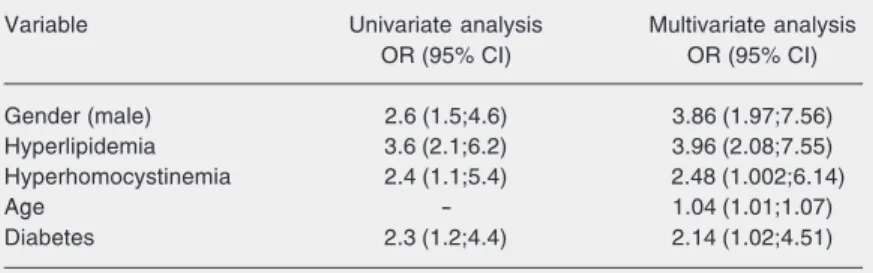
17.3 µM. In a stepwise multivariate regres-sion analysis, where age, creatinine, gender, hyperlipidemia, diabetes, and tobacco use were included, an independent association between hyperhomocystinemia and CAD was found (OR = 2.48; 95% CI = 1.002-6.14). Age, male sex, hyperlipidemia, and diabetes were also independently associated with CAD (Table 2).
When plasma Hcy level was evaluated according to Friesinger score, we found a trend toward more advanced CAD in pa-tients with hyperhomocystinemia. Papa-tients with no arterial wall abnormalities (score 0) had the lowest mean Hcy level: 12 ± 3.6 µM. Those with a score between 1 and 5 had 13.8 ± 5.3 µM, and those with a score of 6 to 10 had 14.1 ± 7.4 µM. The highest mean plasma Hcy level was found in patients with the more severe CAD (scores 11 to 15): 14.5 ± 7.1 µM (P = ns by ANOVA). The difference between the two extreme groups was statis-tically significant (P = 0.04). The same trend was found regarding the prevalence of hy-perhomocystinemia. Among patients with normal arteries (score 0), only 6% had hy-perhomocystinemia. This proportion in-creased in patients with more extensive dis-ease: 18% in patients with a score of 1 to 5, 21% in patients with a score of 6 to 10, and 23.5% in patients with a score of 11 to 15 (P = 0.19 between all groups; P = 0.03 between patients with score 0 and score 11-15 ).
Homocysteine, MTHFR polymorphism and folate status
The C677T MTHFR polymorphism was found in the heterozygous form (CT geno-type) in 30.9% of the study population, while 9.1% had the homozygous form (TT geno-type). The same proportion was found de-spite gender and race. We found no differ-ence in TT genotype prevaldiffer-ence between patients with CAD and those without dis-ease (data not shown).
Hcy level was similar regardless of
MTHFR genotype. Mean concentration was 12.3 (4.6) µM in patients with the CC geno-type, 15.6 (8.8) µM in patients with the CT genotype, and 12.8 (4.7) µM in patients with the TT genotype (P = ns). Nevertheless, patients with the mutant allele had a higher prevalence of hyperhomocystinemia: Hcy above 17.3 µM was present in 29% of ho-mozygous individuals (TT), while this preva-lence was 26% in patients with genotype CT and only 11% in the group with the wild genotype (genotype CC); P = 0.03.
Regarding nutritional factors, we found opposite results regarding vitamin B12 and folate. There was no significant correlation between Hcy and plasma vitamin B12 levels (r = -0.035; P = ns), but we found a negative and significant correlation with folate (r = -0.14; P = 0.04). Comparing patients accord-ing to folate quartiles, we found that those with the lowest folate levels (1st quartile, folate <4.9 ng/dL) had a higher prevalence of hyperhomocystinemia than those in the highest quartile (folate >9.4 ng/dL) (30.3 vs
9.2%; P < 0.01).
The correlation between Hcy and folate status differed according to MTHFR geno-type (Table 3). In patients without the mu-tant allele, this correlation was not statisti-cally significant (r = -0.11; P = ns). How-ever, in patients with genotypes CT and TT combined, a statistically significant correla-tion was demonstrable (r = -0.25; P < 0.05). The wild genotype seems to exert a
protec-Table 2. Variables independently associated with coronary artery disease (CAD) after stepwise multiple regression analysis.
Variable Univariate analysis Multivariate analysis
OR (95% CI) OR (95% CI)
Gender (male) 2.6 (1.5;4.6) 3.86 (1.97;7.56)
Hyperlipidemia 3.6 (2.1;6.2) 3.96 (2.08;7.55) Hyperhomocystinemia 2.4 (1.1;5.4) 2.48 (1.002;6.14)
Age - 1.04 (1.01;1.07)
Diabetes 2.3 (1.2;4.4) 2.14 (1.02;4.51)
tive effect against hyperhomocystinemia even in patients in the lowest folate quartile. In the same way, patients in the top folate quartile had low Hcy levels even when they carried the mutant allele.
Additional factors related to homocysteine
Men have had higher Hcy levels than women (14.9 (6.6) vs 12.4 (5.6) µM; P <
0.001). Age was analyzed in two distinct ways, i.e., as a continuous and categorical variable (≤60 or >60 years old). There was no relation between Hcy level and age in neither way, although patients with hyper-homocystinemia were older than those with Hcy <17.3 µM (60 vs 56 years; P = 0.04).
Smoking, frequently related to high-plasma Hcy levels, was not a cause of hyper-homocystinemia in our population. Ho-mocysteine was 13.7 (5.0) µM in smokers and 13.8 (6.7) µM in non-smokers. Despite the fact that no patient with creatinine above 1.5 mg/dL was included in the study, we found a consistent and positive correlation between creatinine and Hcy (r = 0.42; P < 0.001).
Discussion
This is the first study from a Latin Ameri-can country to show an independent associa-tion between hyperhomocystinemia and CAD. Our results are in agreement with the meta-analysis by Christen et al. (16), in which 43 studies were analyzed. In another meta-analysis, Boushley et al. (17) evaluated 27 studies and showed that Hcy is an independ-ent and gradual risk factor for CAD. The OR for CAD ranged from 1.6 for men to 1.8 for women, for each Hcy level elevation of 5 µM. The increase in coronary risk resulting from this increase is similar to an increase of 20 mg/dL in total cholesterol levels. The authors considered that 10% of the risk of the general population for the development of CAD can be attributed to Hcy.
Prospective studies reported conflicting results. Some identified Hcy as an independ-ent risk factor (5,18), but others did not (19,20). Recently, Wald et al. (21) published a comprehensive meta-analysis of 16 pro-spective studies. Data from 144,936 patients with 3144 combined events were analyzed. The OR for a 5 µM increase in Hcy level was
Table 3. Comparison between patients with poor (bottom quartile: ≤4.9 ng/dL) and high (top quartile) folate status in terms of mean plasma homocysteine (Mean Hcy) levels and frequency of hyperhomocystinemia (Hyper Hcy) according to MTHFR genotype.
Folate quartiles P
1st (N = 56) 2nd (N = 58) 3rd (N = 55) 4th (N = 54)
All patients
Mean Hcy (µM) 15.7 ± 7.5 14.2 ± 8.0 12.5 ± 3.7 12.5 ± 4.3 0.05
Hyper Hcy 30.3% 20.0% 9.1% 9.2% <0.01
CC genotype (60% patients)
Mean Hcy (µM) 13.4 ± 5.1 13.0 ± 4.7 11.2 ± 4.4 12.3 ± 4.1 ns
Hyper Hcy 18.5% 15.9% 14.8% 10.7% ns
CT (30.9% patients) + TT (9.1% patients) genotype
Mean Hcy (µM) 16.7 ± 7.7 17.4 ± 12.2 13.4 ± 3.1 11.8 ± 4.3 0.04
Hyper Hcy 35.7% 33.3% 10.5% 7.1% ns
1.23 (95% CI = 1.14-1.32). Zylberstein et al. (22) published the results of a 24-year fol-low-up evaluating Hcy as a risk factor for coronary morbidity and mortality in 1368 women. For the fifth Hcy quintile, relative risk was 1.86 (95% CI = 1.06 to 3.26) for acute myocardial infarction and 5.14 (95% CI = 2.22 to 11.92) for death due to acute myocardial infarction.
Toole et al. (23) analyzed the effect of homocysteine-lowering therapy upon recur-rent stroke. Despite persistent association between baseline Hcy levels and outcome, folate, vitamins B12 and B6 alone or in com-bination did not prevent new vascular event after 2 years. However, this and other stud-ies had their statistical power questioned (24). Also, the mandatory fortification of food with folate in certain countries has masked the effect of treatment on stroke risk (25).
Only a few studies have assessed the relationship between plasma Hcy levels and the extent of coronary disease. In 70 pa-tients, Tsai et al. (26) demonstrated a posi-tive correlation between Hcy levels and the extent of coronary disease, but clearly only in patients with a low CAD risk profile (less than 3 risk factors). Evaluating only the number of injured vessels, Chao et al. (27) showed that patients with progressively higher Hcy levels have more arteries in-jured. The Friesinger score that we used is not only accurate for evaluation of extension and severity of coronary disease, but also as a prognostic indicator. In the CASS study (28), its prognostic value was better than scores that considered only the number of injured vessels or coronaries with proximal stenotic lesions.
The mechanism(s) by which Hcy is atherogenic is still not completely under-stood. Endothelial dysfunction seems to play a major role. Hcy is a highly reactiveamino acid that produces endothelial injury in both experimental animals and cell cultures (30). Woo et al. (31) demonstrated impaired
flow-mediated dilatation of the brachial ar-tery in 17 healthy individuals who had no other risk factor except hyperhomocystine-mia; furthermore, Hcy levels and endotheli-al function were corrected with folic acid supplementation. Since toxicity is depend-ent on the degree of hyperhomocystinemia, this could explain, at least in part, the graded effect we found between Hcy concentration and the extent of CAD.
The inverse and significant correlation of Hcy with folate, but not with vitamin B12, are compatible with the metabolic pathway of Hcy. Folate is a substrate in the remethyl-ation cycle, where Hcy is converted to me-thionine, in which it donates the methyl group for this reaction. On the other hand, vitamins B12, B2 and B6 function only as enzyme co-factors. Although median folate level was similar and within the normal range in both patients and controls, a considerable propor-tion of patients had low-plasma folate levels. The lower limit proposed by the WHO is 6 ng/mL. Almost 30% of patients had folate below this limit, and their Hcy levels were higher than those of patients with folate within the normal range. Larger studies have previously shown this relation. In the Framinghan study (32), patients in the low-est decile of folate had mean Hcy 15.6 µM, while those in the superior decile had 11 µM. The data regarding the prevalence of C677T MTHFR polymorphism showed that the TT genotype occurs in our population at a similar frequency as that observed in Cau-casian populations (33). Arruda et al. (34), evaluated the C677T polymorphism in three distinct ethnic groups in Brazil. Genotype TT was found in a similar prevalence among Caucasian descent (10%). The prevalence was lower among Black (1.45%) and Indian populations (1.2%).
or heterozygosis (30.9%). This group is par-ticularly susceptible to hyperhomocystine-mia when exposed to low folate levels. Ac-cordingly, countries such as the United States and Canada already enrich their grains with folic acid in an effort to reduce folate-neural tube defects. If lowering Hcy levels is proved to reduce cardiovascular risk, a high folate intake should be recommended for every-one. On the other hand, patients with the wild genotype (CC) seem to be less prone to increased Hcy levels, even in the presence of low folate levels.
Our findings are relevant since several studies have shown that Hcy levels differ amongcountries with similar nutritional hab-its. Thus, it may be useful to know the data for our population. Alfthan et al. (35) have demonstrated these inter-country differences among several European countries, Japan and Israel. Moreover, these investigators demonstrated a directrelationship between plasma Hcy concentration and mortality from all cardiovascular diseases according to WHO data.
References
1. McCully KS (1969). Vascular pathology of homocysteinemia: impli-cations for the pathogenesis of arteriosclerosis. American Journal of Pathology, 56: 111-128.
2. Bolander-Gouaille C (2002). Focus on Homocysteine and the Vita-mins Involved in its Metabolism. Springer Verlag, Paris, France, 15. 3. Harker CA, Slichter SF, Scott CR et al. (1974). Homocysteinemia: vascular injury and arterial thrombosis. New England Journal of Medicine, 291: 537-543.
4. Clarke R, Daly L, Robinson K et al. (1991). Hyperhomocysteinemia: an independent risk factor for vascular disease. New England Jour-nal of Medicine, 324: 1149-1155.
5. Stampfer MJ, Malinow MR, Willett WC et al. (1992). A prospective study of plasma homocyst(e)-ine and risk of myocardial infarction in US physicians. Journal of the American Medical Association, 268: 877-881.
6. Shai I, Stampfer MJ, Ma J et al. (2004). Homocysteine as a risk factor for coronary heart diseases and its association with inflamma-tory biomarkers, lipids and dietary factors. Atherosclerosis, 177: 375-381.
7. Perry IJ, Refsum H & Morris RW (1995). Prospective study of serum total homocysteine concentration and risk of stroke in middle-aged British men. Lancet, 346: 1395-1398.
8. Boers GH, Smals AG, Trijbels FJ et al. (1985). Heterozygosity for homocystinuria in premature peripheral and cerebral occlusive arte-rial disease. New England Journal of Medicine, 313: 709-715. 9. Homocysteine Lowering Trialists’ Collaboration (1998). Lowering
blood homocysteine with folic acid based supplements: meta-analy-sis of randomized trials. British Medical Journal, 316: 894-898. 10. Rozen R (1977). Genetic predisposition to hyperhomocystinemia:
deficiency of methylenetetrahydrofolate reductase (MTHFR). Jour-nal of Thrombosis and Haemostasis, 78: 523-526.
11. United Nations/Statistics Division: World and regional trends: data for years around 1990 and 2000 http:/unstats.un.org/unsd/mi/ mi_wrtrends1.asp.
12. Malaquias Filho M & Rissin A (2003). Nutritional transition in Brazil: geographic and temporal trends. Cadernos de Saúde Pública, 19 (Suppl I): S181-S191.
13. Friesinger GC, Page EE & Ross RS (1970). Prognostic significance of coronary arteriography. Transactions of the Association of Ameri-can Physicians, 83: 78-92.
14. Fiskerstrand T, Refsum H, Kvalheim G et al. (1993). Homocysteine and other thiols in plasma and urine: automated determination and sample stability. Clinical Chemistry, 39: 263-271.
15. Bravo-Osorio M & BydLowski SP (2000). Detection of methylenetet-rahydrofolate reductase (MTHFR) C677T and prothrombin G20210A mutations: second restriction site for digestion control of PCR prod-ucts. Clinica Chimica Acta, 301: 219-223.
16. Christen WG, Ajani UA, Glynn RJ et al. (2000). Blood levels of homocysteine and increased risks of cardiovascular disease: causal or casual? Archives of Internal Medicine, 160: 422-434.
17. Boushley CJ, Beresford SAA, Omenn GS et al. (1995). A quantita-tive assessment of plasma homocysteine as a risk factor for vascu-lar disease: probable benefits of increasing folic acid intakes. Jour-nal of the American Medical Association, 274: 1049-1057. 18. Arnesen E, Refsum H & Bonaa KH (1995). Serum total
homocys-teine and coronary heart disease. International Journal of Epidemi-ology, 24: 704-709.
19. Evans RW, Shaten BJ, Hempel JD et al. (1997). Homocyst(e)ine and risk of cardiovascular disease in the Multiple Risk Factor Inter-vention Trial. Arteriosclerosis, Thrombosis, and Vascular Biology, 17: 1947-1953.
20. Folsom AR, Nieto FJ, McGovern PG et al. (1998). Prospective study of coronary heart disease incidence in relation to fasting total ho-mocysteine, related genetic polymorphisms, and B vitamins. The Atherosclerosis Risk in Communities (ARIC) study. Circulation, 98: 204-210.
21. Wald DS, Law M & Morris JK (2002). Homocysteine and cardiovas-cular disease: evidence on causality from a meta-analysis. British Medical Journal, 325: 1202.
22. Zylberstein DE, Bengtsson C, Bjorkelund C et al. (2004). Serum homocysteine in relation to mortality and morbidity from coronary heart disease: a 24-year follow-up of the population study of women in Gothenburg. Circulation, 109: 601-606.
ho-mocysteine in patients with ischemic stroke to prevent recurrent stroke, myocardial infarction, and death. The Vitamin Intervention for Stroke Prevention (VISP) randomized controlled trial. Journal of the American Medical Association, 291: 565-575.
24. Bostom AG, Selhub J, Jacques PF et al. (2001). Power shortage: clinical trials testing the “Homocysteine Hypothesis” against a back-ground of folic acid-fortified cereal grain flour. Annals of Internal Medicine, 135: 133-137.
25. Schwammenthal Y & Tanne D (2004). Homocysteine, B-vitamin supplementation, and stroke prevention: from observational to interventional trials. Lancet Neurology, 3: 493-495.
26. Tsai WC, Li YH, Tsai LM et al. (2000). Correlation of homocysteine levels with the extent of coronary atherosclerosis in patients with low cardiovascular risk profiles. American Journal of Cardiology, 85: 49-52.
27. Chao CL, Tsai HH, Lee CM et al. (1999). The graded effect of hyperhomocystinemia on the severity and extent of coronary athero-sclerosis. Atherosclerosis, 147: 379-386.
28. Ringqvist I, Fisher LD, Mock M et al. (1983). Prognostic value of angiographic indices of coronary artery disease from the Coronary Artery Surgery Study (CASS). Journal of Clinical Investigation, 71: 1854-1866.
29. Ray JG (1998). Meta-analysis of hyperhomocystinemia as a risk factor for venous thromboembolic disease. Archives of Internal Medi-cine, 158: 2101-2106.
30. Wall RT, Harlan JM, Harker LA et al. (1980). Homocysteine-induced endothelial cell injury in vitro: a model for the study of vascular injury. Thrombosis Research, 18: 113-121.
31. Woo KS, Chook P & Lolin YI (1999). Folic acid improves arterial endothelial function in adults with hyperhomocystinemia. Journal of the American College of Cardiology, 34: 2002-2006.
32. Selhub J, Jacques PF, Wilson PW et al. (1993). Vitamin status and intake as primary determinants of homocysteinemia in an elderly population. Journal of the American Medical Association, 270: 2693-2698.
33. Brattstrom L, Zhang Y, Hurtig M et al. (1998). A common methylene-tetrahydrofolate reductase gene mutation and longevity. Atheroscle-rosis, 141: 315-319.
34. Arruda VR, Siqueira LH, Goncalves MS et al. (1998). Prevalence of the mutation C677 → T in the methylenetetrahydrofolate reductase gene among distinct ethnic groups in Brazil. American Journal of Medical Genetics, 78: 332-335.