Changes in lower incisor inclination and the
occurrence of gingival recession
Luciane Q. Closs*, Betina Grehs**, Dirceu Barnabé Raveli***, Cassiano Kuchenbecker Rösing****
Abstract
Objectives: The aim of this study was to investigate whether altering the labial-lingual posi-tion of lower incisors in adolescents might predispose to the development of gingival
reces-sion. Methods: Records from 189 Caucasian adolescents (107 female and 81 male) pre and
post orthodontic treatment were selected. Patients yielded mean ± SD values of initial records age 11.2 ± 1.9 years and final records age 14.7 ± 1.8 years. The presence of gingival recession was evaluated in models and photographs. The inclination of lower incisors to the mandibular plane angle (IMPA) was measured on lateral cephalograms, pre and post treatment. Results: No significant association was observed between changes in tooth inclination and the pres-ence of gingival recessions, based on chi-square analysis (p = 0.277). Data demonstrated that in 107 patients (56.6%) incisors were proclined, in 64 patients (33.9%) incisors were retro-inclined and 18 patients (9.5%) did not show any changes in tooth inclination. In the cases where new gingival recessions occurred, 64.9% had been moved buccally, 26.3% had been moved lingually and 8.8% did not change inclination. In the group of patients that displayed coronal migration of the gingival margin, 60% were moved lingually, 30% were moved buc-cally and 10% did not change inclination. Conclusions: Even though the percentage of cases where teeth were proclined showed a larger number of new gingival recessions, it was not statistically significant.
Keywords: Lower incisors. Inclination. Gingival recession. Orthodontic tooth movement.
* Doctor in Orthodontics from the University of Araraquara (Unesp). Professor and coordinator of Ulbra’s (Canoas/RS) School of Orthodontics. ** Master’s candidate at the Araraquara (Unesp) School of Orthodontics.
*** Associate Professor of the Department of Children Care and Coordinator of the postgraduate Orthodontics course – Araraquara (Unesp) School of Dentistry.
**** Associate Periodontics Professor of the Rio Grande do Sul Federal University (UFRGS) and the Lutheran University of Brazil (Ulbra). Doctor in Periodontics – Araraquara (Unesp) School of Dentistry.
INTRODUCTION
Determining the ideal position of lower inci-sors has hitherto posed a challenge in Orthodon-tics both in terms of stability13,15 and periodontal
conditions4,18. When teeth move across alveolar
bone, hard and soft tissues undergo remodeling, which involves a variety of cells and tissue reac-tions in tandem with periodontal fibers19.
Some studies have reported that tooth move-ments outside the alveolar bone resulting from excessive inclination predispose to a loss of gin-gival attachment buccally, which leads to gingin-gival recession3,11,27,28. Others report that there is no
ev-idence associating tooth movement with gingival recession development1,2,14,21.
dogs12 have demonstrated that fenestrations can
be produced in alveolar bone when lower incisors are moved buccally3. Nevertheless, the bone
re-modeling which occurs following this movement does not seem to entail any attachment tissue loss since this tooth movement occurs inside the den-tal arch and the supracrisden-tal supporting tissue is kept inflammation-free12.
Engelking and Zachrisson9, repositioning
mon-key incisors to their original location after these teeth had been orthodontically protruded showed that there occurred buccal bone reapposition in the coronal orientation. This observation dem-onstrated that there could be an improvement in bone fenestration and gingival recession9,12.
The position of the tooth upon eruption has also been regarded as a local factor which may lead to gingival recession. However, the presence of trauma caused by brushing and plaque-related gingival lesions should be considered a key caus-ative factor in orthodontic patient recession25.
At the time orthodontic planning is made pa-tients very often present with a retrusive mandible and teeth which are already excessively inclined. In order to enhance aesthetics and avoid surgical procedures teeth are kept at or moved to an even more proclined position. Additionally, most stud-ies investigating the impact of changes in tooth inclination and changes in the gingival margin were performed on non-specific age brackets6 or
on adult patients10,11,21.
The purpose of this study was to investigate whether altering the labial-lingual position of lower incisors in adolescents might predispose to the development of gingival recession.
MATERIALS AND METHODS Sample
Intraoral photographs, preliminary casts and cephalograms of 189 Caucasian adolescents (107 female and 81 male) were selected, prior to and following orthodontic treatment. These cases were selected from records of 209 patients
who had finished orthodontic treatment in pri-vate clinics using fixed appliances. Patients were treated with Standard Edgewise and Straight wire appliances. The mean starting age was 11.2 ± 1.9 years standard deviation and a final mean age of 14.7± 1.8 years. The active treatment mean was 1.99 ± 0.89 years.
Inclusion criteria
To take part in this study, patients had to pres-ent with either Angle’s Class I or Class II, sectional or vertical problems; the presence of diastema or maximum 4 mm lower incisor crowding; treated with no extractions; with permanent lower inci-sors and cuspids erupted; apparent periodontal health; final records issued no earlier than 28 days following appliance removal; and cephalograms should be clear enough as to allow scanning.
All patients were given oral hygiene instruc-tions immediately following orthodontic appli-ance placement and during treatment.
Exclusion criteria
Excluded patients comprised those whose in-cisors were missing or not yet erupted, with an Angle Class III malocclusion, who reported pre-existing systemic diseases or who were on medi-cation due to gingival alterations. Follow-up of periodontal conditions was performed based on individual needs. About 20% of the records were disregarded on account of inaccurate casts or pho-tographs, which compromised assessment.
Main variable
The independent variable was gingival reces-sion, which was assessed by means of visually evaluating the preliminary casts and with the aid of pre and post treatment intraoral photographs. Teeth were considered to present with recession when exposure of the amelocemental junction was detected. The amount of recession was quan-tified using a digital gauge (Mitutoyo Digimatic®,
the nearest tenth of a millimeter. (Fig. 1). Cases were assessed prior to and following orthodon-tic treatment, whereas teeth were classified as in: (a) unaltered gingival margin position; (b) coronal migration; or (c) apical migration of the gingival margin.
Prior to starting the study, the position of the gingival margins was gauged by evaluating the photographs and casts within a time interval of one week, without any knowledge of the previous evaluation. The Kappa test was used to evaluate intra-observer concordance. Perfect reproducibil-ity was achieved once (Kappa = 1) after extensive practice.
After the data had been collected, a multiplica-tion factor was set to calculate the actual amount of gingival recession since the measurements were performed on photographs, which did not reflect the actual size of the variable being measured. To correct photo magnification, the size of the upper right incisor crown was compared – on the pho-tograph – with the dimensions of the same tooth on the cast. The following equation was used: Ac-tual recession equals the recession measured on the photograph times the size of the cast crown, divided by the crown length – measured on the photograph – as suggested by Djeu, Hayes and Zawaideh7.
Actual recession = recession measured on the photograph x size of cast crown
crown length on the photograph
Independent variable
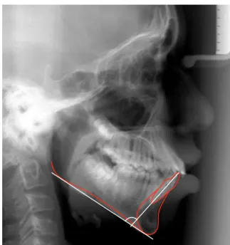
The lateral cephalograms were scanned and cephalometric points marked and scanned with Radioceph software (Radiomemory, Belo Hori-zonte, Brazil) run by a well-trained observer who was not informed about the gingival recession measurements. Lower incisor inclination was as-sessed by altering the IMPA angle (Fig. 2). Ini-tial and final measurements were assessed and divided into 3 groups: Patients presenting with proclined teeth, patients with retroinclined teeth
and patients with no buccolingual changes in the lower incisors.
Evaluation of method error
In order to evaluate the method error in lo-cating the points and the radiographic overlay, 10 cephalograms were randomly selected after being traced and superimposed twice. The following equation was used to calculate the methodologi-cal error Sx = Σd2/2n, where “d” is the difference
between the duplicated measurements and “n” are the duplicated records5. The radiographic variable
FIGURE 1 - Recession measurement.
In the group of patients who experienced coronal migration of gingival margin, 60% were moved to-wards lingual, 30% were moved toto-wards buccal and 10% did not show any inclination changes. In the cases where the gingival margin was altered, 54.9% were moved towards buccal, 45.2% were moved towards lingual and 9.8% had no inclina-tion changes at all.
A Chi-square analysis failed to demonstrate a significant association (p = 0.277) between the changes in tooth inclination and the presence of new gingival recessions (Table 1).
In patients where recession occurred, the mean inclination was +2º, whereas the first and third quartiles were -1.54 and +5.68º, respective-ly. When the gingival margin migrated coronally, the mean value of lingual-oriented movement was -1.13º. Individuals whose tooth inclinations were changed yielded a mean value of +0.89º, error did not exceed 0.58º. The cephalometric
measurements were rounded to the nearest 0.5º.
Statistical analysis
The relative and absolute frequencies depen-dent and independepen-dent variables were obtained. Chi-square analysis was used to investigate the association between gingival recession and tooth inclination. Tooth inclination variation (Δ incli-nation) was calculated to assess the differences between the variables, and the non-parametric Kruskal-Wallis16 test was applied to the patients,
who came to the analysis unit. An alpha level of 0.05 was defined.
RESULTS
The cases were divided according to the changes which took place in the lower incisor gin-gival margin after treatment. The collected data showed that the incisors of 107 patients (56.6%) were moved towards buccal, the incisors of 64 patients (33.9%) were moved towards lingual, whereas 18 patients (9.5%) did not display any changes in tooth inclination. The cases were fur-ther subdivided into 3 groups: (a) occurrence of gingival recession, (b) coronal migration of gingi-val margin and (c) unchanged position of gingigingi-val margin. In those cases where gingival recession occurred, 64.9% were moved towards buccal, 26.3% were moved towards lingual and 8.8% did not show any inclination changes (Graph 1).
TABLE 1 - Association between changes in the gingival margin following orthodontic treatment and orthodontic inclination movement.
Gingival margin position classification
Inclination Coronal gingival migration
Unchanged gingival margin
Gingival
recession total
n % n % n % n %
retroinclined 6 60,0 43 35,2 15 26,3 64 33,99
unchanged 1 10,0 12 9,8 5 8,8 18 9,5
proclined 3 30,0 67 54,9 37 64,9 107 56,6
total 10 100 122 100 57 100 189 100
GRAPH 1 - Changes in tooth inclination and new recessions. 9,5%
33,9% 56,6% Teeth 8.8% Yielded new
recessions
Teeth 26.3% Yielded new
recessions Teeth 64.9% Yielded new recessions
X2 = 5,10; p = 0,2777.
Proclined teeth
Retroclined teeth
avoid methodological errors. The fact that the two observers were blinded when analyzing the other variables ensured an unbiased evaluation.
Although adults often seek orthodontic treat-ment, this population is not the prevailing patient base at orthodontic clinics. The population of the present study fell within an age bracket analogous to the majority of patients who seek orthodontic treatment and who still have growth potential23,24.
The few studies which evaluated the effects of lower incisor inclination and the gingival margin position exclusively were either conducted on non-specific age groups6 or on adults3,7,27. One
such study, performed by Ruf, Hansen and Pan-cherz21, which addressed the same issues as the
present study, analyzed orthodontic treatment us-ing a specific orthodontic appliance for a shorter period of time and with a smaller sample size.
Many studies have demonstrated that behav-ioral factors, particularly the pattern of oral hy-giene in children and adolescents during orth-odontic treatment, are, more often than not, inadequate8,20. Considering this observation, any
other controllable risk indicator should be known. The methodology employed in this investiga-tion to assess gingival recession has been reported in a number of other studies2,21,27. It is
unques-tionable that a clinical inspection of the gingival margin position directly in the subjects’ mouths would have been desirable. However, in view of the sample size and observation time – prior to and following orthodontic treatment completion – the likelihood of errors was minimized. In the present study, gingival recessions were assessed by visually inspecting intraoral photographs and preliminary casts. Other studies have evaluated the length of the clinical crown1,2,7. This, however, may not
con-stitute an accurate indicator of gingival recession. The simple mathematical analysis of crown length is error-prone since teeth can extrude without the occurrence of gingival margin apical migration and therefore result in crown lengthening. This may lead professionals to mistakenly classify such whereas the first and third quartiles were -2.08º
and +4.88º, respectively. The lowest mean value (77.70) was observed when the gingival migra-tion moved coronally, whereas the highest mean value (100.82) was observed in individuals who presented with new recessions.
When the group of patients was divided by gender, no significant difference was observed.
DISCUSSION
The goal of the present study was to investigate the possible association between changes in buc-colingual inclination of lower anterior teeth during orthodontic treatment and the development of gin-gival recessions. Although some authors12,21 assert
that any tooth that is moved outside the dentoal-veolar envelope is predisposed to bone fenestration followed by gingival recession, such phenomenon was not found to be true in this study.
In the group of patients whose teeth were moved towards buccal (56.6%), as well as those whose teeth were moved towards lingual (33.9%), no association with the development of gingival recession was noted.
Many methodological considerations could be made to better assess the present study. Firstly, this is a retrospective longitudinal study where only radiographs, photographs and models were analyzed. The limitations of this approach should be taken into account. Nevertheless, by calibrat-ing and traincalibrat-ing observers, the authors tried to
Δinclination
classification 1st Quartile 2nd Quartile 3rd Quartile Mean
Coronal gingival
migration -2,34 -1,13 3,60 77,7
Unchanged
gingival margin -2,08 0,89 4,88 93,70
Gingival
recession -1,54 2,00 5,68 100,82
Kruskal-Walllis: p = 0,424.
patients as gingival recession cases.
The changes resulting from tooth inclination were assessed with the aid of the National Insti-tute for Pure and Applied Mathematics (IMPA) standards. The mean value was reached based on a lateral cephalogram. This mean value does not reflect the position of each individual lower inci-sor with accuracy but rather the position of the most prominent tooth in that region. In fact, the use of cephalograms is a very common method described in the literature as a tool to determine lower incisor inclination3,7,11,24.
This is a retrospective-longitudinal study where individuals who met the inclusion crite-ria were selected irrespective of the directional movement of their lower incisor teeth. In order to evaluate the process through which recessions evolve, it would be necessary to perform interme-diate assessments in predetermined periods during the treatment as well as to secure control of hy-giene habits. The results achieved with this study, however, can mirror with greater clarity the actual impact of orthodontic treatment on gingival mar-gin position, free from the Hawthorne effect.
A retrospective study, which sought to evalu-ate the periodontal conditions found after man-dibular incisor proclination, was conducted with a sample of Class II patients treated during mixed dentition2. In this study, a group of Class II
pa-tients who had not presented with proclined teeth was used as control group. In terms of recession, the findings were similar to the present study. No difference was noted between the test and control groups in the number of lower incisors which de-veloped recession from the pre to the post-treat-ment periods.
Once it had been observed in other stud-ies that bone compensatory formation can oc-cur when teeth are moved to their original po-sitions12,19, it might be assumed that the gingival
margin would follow the bone closely and thus no recession would take place. The present study shows that although no strong correlations were
detected, a tendency exists towards the coronal migration of the gingival margin of teeth which are moved lingually, compared with teeth which are moved buccally.
On the other hand, in a study using an animal model26 where the upper incisors had been moved
buccally, a significant gingival margin apical mi-gration was observed after tooth movement. Such results may have been negatively impacted by the fact that the teeth involved presented with signs of gingival inflammation.
Another study1, which assessed gingival
reces-sion in adults following excessive incisor proclina-tion, showed that 10% of the patients developed new recessions and 5% displayed gingival margin coronal migration. Similarly to the present study, most subjects (85%) did not show any gingival margin alterations.
Previous studies yielded contradictory re-sults3,6. Geiger and Wasserman10 investigated the
relationship between occlusion and periodontal disease and postulated that lower incisor labial gingival recession would be connected to lin-guoversion (with a lower than 85º IMPA score). Yared, Zenobio and Pacheco27, however, noted
that lower central incisors with a 95º IMPA score and gingival margin thickness smaller that 0.5 mm yielded severe recessions. Based on the results of this study, the increased risk reported by some authors3,11,27,28 of lower incisor proclination is not
clearly supported by the evidence. In fact, 15 indi-viduals who experienced gingival recession in the present study had their incisors retroclined and 5 of them did not show any changes in tooth incli-nation. This could lead one to question the recom-mendation made by some authors10,18 to perform
a pretreatment gingival graft as a preventive mea-sure in those cases where teeth need to undergo extensive motion. It should be reported that none of the patients in this study were subjected to gin-gival graft either prior to or following orthodontic treatment.
move-ment executed, there was an increase in the num-ber of patients (57) who presented with new post-treatment gingival recessions. Other independent variables, such as buccal hygiene pattern, amount and thickness of gingival tissue and features of the alveolar process should be further investigated.
CONCLUSION
The results of the present study indicate no as-sociation whatsoever between lower incisor incli-nation and gingival recession.
1. ALLAIS, D.; MELSEN, B. Does labial movement of lower incisors influence the level of the gingival margin? A case-control study of adult orthodontic patients. Eur. J. Orthod., Oxford, v. 25, no. 4, p. 343-352, Aug. 2003.
2. ÅRTUN, J.; GROBÉTY, D. Periodontal status of mandibular incisors after pronounced orthodontic advancement during
adolescence: A follow–up evaluation. Am. J. Orthod.
Dentofacial Orthop., St. Louis, v. 119, no. 1, p. 2-10, Jan. 2001. 3. ARTUN, J.; KROGSTAD, O. Periodontal status of mandibular
incisors following excessive proclination: a study in adults with
surgically treated mandibular prognathism. Am. J. Orthod.
Dentofacial Orthop., St. Louis, v. 91, no. 3, p. 225-233, Mar. 1987.
4. COATOAM, G. W.; BEHRENTS, R. G.; BISSADA, N. F. The width of keratinized gingiva during orthodontic treatment: Its significance and impact on periodontal status. J. Periodontol., Chicago, v. 52, no. 6, p. 307-313, June 1981.
5. DAHLBERG, G. Statistical methods for medical and biological
students. London: George Allen & Unwin, 1940. p. 122-132. 6. DE LOOR, P.; DE SMIT, A. A.; ADRIAENS, P. A. Periodontal
changes induced by orthodontically changed incisor inclination. Eur. J. Orthod., Oxford, v. 17, no. 1, p. 57, Jan. 1995. Abstact.
7. DJEU, G.; HAYES, C.; ZAWAIDEH, S. Correlation between mandibular central incisor proclination and gingival recession
during fixed appliance therapy. Angle Orthod., Appleton,
v. 72, no. 3, p. 238-245, 2002.
8. DUBEY, R.; JALILI, V. P.; GARG, S. Oral hygiene and gingival status in orthodontic patients. J. Pierre Fauchard Acad., Indore, v. 7, no. 2, p. 43-54, June 1993.
9. ENGELKING, G.; ZACHRISSON, B. U. Effects of incisor repositioning on monkey periodontium after expansion through the cortical plate. Am. J. Orthod., St. Louis, v. 82, no. 1, p. 23-32, July 1982.
10. GEIGER, A. M.; WASSERMAN, B. H. Relationship of occlusion and periodontal disease: part IX - incisor inclination and periodontal status. Angle Orthod., Appleton, v. 46, no. 2, p. 99-110, Nov. 1976.
11. HOLLENDER, L.; RONNERMAN, A.; THILANDER, B. Root resorption, marginal bone support and clinical crown length in orthodontically treated patients. Eur. J. Orthod., Oxford, v. 2, no. 4, p. 197-205, Apr. 1980.
12. KARRING, T.; NYMAN, S.; THILANDER, B.; MAGNUSSON, I. Bone regeneration in orthodontically produced alveolar bone dehiscences. J. Periodontal Res., Copenhagen, v. 17, no. 3, p. 309-315, May 1982.
13. LITTLE, R. M. Stability and relapse of dental arch alignment.
Br. J. Orthod., Oxford, v. 17, no. 3, p. 235-241, Aug. 1990. 14. MELSEN, B.; ALLAIS, D. Factors of importance for the
development of dehiscences during labial movement of mandibular incisors: a retrospective study of adult patients.
Am. J. Orthod. Dentofacial Orthop., St. Louis, v. 127, no. 5, p. 555-560, May 2005.
15. MERRIFIELD, L. L. Dimensions of the denture: back to basics.
Am. J. Orthod. Dentofacial Orthop., St. Louis, v. 106, no. 5, p. 535-542, Nov. 1994.
16. MONTGOMERY, D. C. Design and analysis of experiments.
5th ed. New York: John Wiley, 2001.
17. NAHÁS, A. C. R.; FREITAS, M. R.; NAHÁS, D.; JANSON, G. P.; HENRIQUES, J. F. C. A inter-relação Ortodontia e Periodontia na prevenção e controle das recessões gengivais decorrentes
do tratamento ortodôntico. Rev. Dental Press Ortodon.
Ortop. Facial, Maringá, v. 5, n. 6, p. 51-56, nov./dez. 2000. 18. NGAN, P. W.; BURCH, J. G.; WEI, S. H. Grafted and ungrafted
labial gingival recession in pediatric orthodontic patients: effects of retraction and inflammation. Quintessence Int., Berlin, v. 22, no. 2 p. 103-111, Feb. 1991.
19. REITAN, K. Clinical and histologic observations on tooth
movement during and after orthodontic treatment. Am. J.
Orthod., St. Louis, v. 53, no. 10, p. 721-745, Oct. 1967. 20. RINCHUSE, D. J.; RINCHUSE, D. J.; ZULLO, T. G. Oral hygiene
compliance: a clinical investigation. J. Clin. Orthod., Boulder, v. 26, no. 1, p. 33-38, Jan. 1992.
21. RUF, S.; HANSEN, K.; PANCHERZ, H. Does orthodontic proclination of lower incisors in children and adolescents cause
gingival recession? Am. J. Orthod. Dentofacial Orthop.,
St. Louis, v. 114, no. 1, p. 100-106, July 1998.
22. STEINER, G. G.; PEARSON, J. K.; AINAMO, J. Changes of the marginal periodontium as a result of labial tooth movement in monkeys. J. Periodontol., Chicago, v. 52, no. 6, p. 314-320, June 1981.
23. TAUSCHE, E.; LUCK, O.; HARZER, W. Prevalence of malocclusions in the early mixed dentition and orthodontic treatment need. Eur. J. Orthod., Oxford, v. 26, no. 3, p. 237-244, June 2004.
24. VAKIPARTA, M. K.; KEROSUO, H. M.; NYSTROM, M. E.; HEIKINHEIMO, K. A. Orthodontic treatment need from eight to 12 years of age in an early treatment oriented public health
care system: a prospective study. Angle Orthod., Appleton,
v. 75, no. 3, p. 344-339, May 2005.
25. WENNSTRÖM, J. L. Mucogingival consideration in orthodontic treatment. Semin. Orthod., Philadelphia, v. 2, no. 1, p. 46-54, Mar. 1996.
26. WENNSTROM, J. L.; LINDHE, J.; SINCLAIR, F.; THILANDER, B. Some periodontal tissue reactions to orthodontic tooth
movement in monkeys. J. Clin. Periodontol., Copenhagen,
v. 14, no. 3, p. 121-129, Mar. 1987.
27. YARED, K. F. G.; ZENOBIO, E. G.; PACHECO, W. Periodontal status of mandibular central incisors after orthodontic proclination in adults. Am. J. Orthod. Dentofacial Orthop., St. Louis, v. 130, no. 1, p. 1-8, July 2006.
28. YARED, K. F. G.; ZENOBIO, E. G.; PACHECO, W. Projeção ortodôntica de incisivos inferiores: um risco à recessão
periodontal? Rev. Dental Press Ortodon. Ortop. Facial,
Maringá, v. 11, n. 5, p. 35-41, set./out. 2006.
Contact Luciane Q. Closs
Rua Marcelo Gama 1249 - 3º andar CEP: 90.540-41 - Porto Alegre/RS Email: [email protected] REFERENCES