460
Rev. Col. Bras. Cir. 2010; 37(6): 460-462
B a t i s t a B a t i s t aB a t i s t a B a t i s t aB a t i s t a Cecum volvulus complicated by septic jaundiceCase ReportCase ReportCase ReportCase ReportCase Report
Cecum volvulus complicated by septic jaundice
Cecum volvulus complicated by septic jaundice
Cecum volvulus complicated by septic jaundice
Cecum volvulus complicated by septic jaundice
Cecum volvulus complicated by septic jaundice
Volvo de ceco complicado por icterícia séptica
Volvo de ceco complicado por icterícia séptica
Volvo de ceco complicado por icterícia séptica
Volvo de ceco complicado por icterícia séptica
Volvo de ceco complicado por icterícia séptica
THALES PAULO BATISTA, ACBC-PE1; JÂNIO CIPRIANO ROLIM2 ; ALLYSSON ANTONIO RIBEIRO GOMES2;
WANESKA LUCENA NÓBREGADE CARVALHO3; RENATO JOSÉDOS SANTOS2
Work performed in the Surgey Service of The Hospital de Guarnição de João Pessoa (HGuJP).
1. Master’s Degree, Health Sciences, the Universidade de Pernambuco (UPE) – Pernambuco – Brazil; 2. General Surgeon, Hospital de Guarnição de João Pessoa (HGuJP) – João Pessoa – Paraíba – Brazil; 3. Intensivist, Hospital de Guarnição de João Pessoa (HGuJP) – João Pessoa – Paraíba – Brazil.
INTRODUCTION
INTRODUCTION
INTRODUCTION
INTRODUCTION
INTRODUCTION
T
he cecal volvulus is uncommon in western adults1 and results from a twist of this colonic segment over its mesentery, causing intestinal obstruction and strangulation. It presents with variable forms of intesti-nal obstruction and its treatment is often individualized, the patient’s condition and perioperative cecal viability being taken into account.We report a case of intestinal obstruction due to cecal volvulus treated with cecopexia and complicated with septic jaundice.
CASE REPORT
CASE REPORT
CASE REPORT
CASE REPORT
CASE REPORT
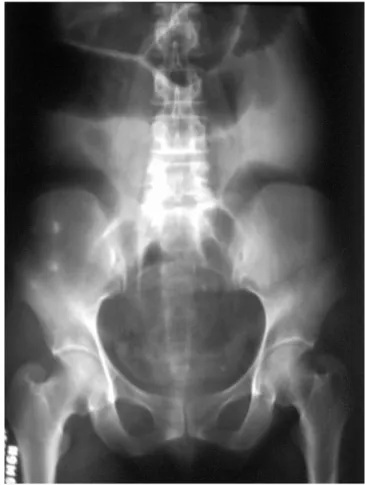
A female patient, age 55, was admitted to the emergency room of HGuJP with crampy abdominal pain of sudden onset and high intensity associated with nausea, vomiting, abdominal distension and stop of the elimination of intestinal gas with two days of evolution and progressive deterioration. She was previously healthy, had regular bowel habits and reported no other similar events. Among the tests carried out urgently, the white blood count showed leukocytosis with left shift (WBC = 13,200; metamyelocytes = 2%; bands = 51%; neutrophils = 43%; lymphocytes = 3%; monocytes = 1%) and the abdominal radiography showed signs of intestinal obstruction caused by intestinal dilatation and air-liquid levels at different segments (Figure 1).
With the diagnosis of obstructive acute abdomen, she was referred for surgical treatment by laparotomy under general anesthesia, in which the perioperative finding was a cecal volvulus without, however, gangrene or bowel perforation. The colonic segment showed marked mobility and poor fixation due to its long mesentery and the absence of caecal ligaments to the abdominal wall. The treatment performed then was manual reduction of the torsion and cecopexia with simple stitches of nonabsorbable monofilament suture, securing the cecum to the right parietal peritoneum and retroperitoneum.
The patient regained bowel movements and improved abdominal pain and distention in 48 hours. However, after 72 hours a rapidly and progressive jaundice ensued (Total Bilirubin = 18.2 mg/dl, Indirect Bilirubin = 7.5 mg/dl, Direct Bilirubin = 10.7 mg/dl, Alkaline Phosphatase = 135U/l, AST = 162U/ml, ALT = 61U/ml, Amylase = 141U/l), with persistent leukocytosis, tachycardia, tachypnea and the irregular fever highs. Ab-dominal CT scan was then performed and showed no evidence of abdominal abscesses, intrahepatic or pancreatic changes, showing only slight segmental distension of the cecum and proximal right colon. After
Figure 1 Figure 1Figure 1 Figure 1
B a t i s t a B a t i s t a B a t i s t a B a t i s t a B a t i s t a
Cecum volvulus complicated by septic jaundice 461
Rev. Col. Bras. Cir. 2010; 37(6): 460-462
drug-induced hepatitis was ruled out, blood and urine cultures for common germs were collected, as well as new laboratory exams, followed by the exchange of antimicrobial treatment for ciprofloxacin and metronidazole. The culture did not return any bacterial growth, the patient evolved with progressive clinical and laboratory improvement and was discharged on the 12th postoperative day.
DISCUSSION
DISCUSSION
DISCUSSION
DISCUSSION
DISCUSSION
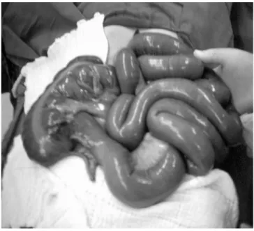
The cecal volvulus is an uncommon entity in Western countries, accounting for only 1% of cases of intestinal obstruction in adults1. As predisposing factors, there are chronic use of laxatives, residue-rich diet, Chagas’s disease, neurological and psychiatric comorbidities, female gender and advanced age. The occurrence of cecal torsion, however, probably depends on anatomical bases as poor intestinal rotation, dilation, poor fixation and hypermotility1-3. In this report, the last two mechanisms were involved (Figure 2), but there was no comorbidities or semi-occlusion antecedents. Its clinical picture is highly variable, appearing more often in the form of recurrent abdominal pain episodes and self-limited intestinal semi obstruction in women with a mean age of 50-60 years and associated comorbidities. Other times it can manifest suddenly with typical obstruction accompanied by intestinal strangulation and sepsis1,3-5.
Treatment should be individualized depending on clinical circumstances and surgical findings. Uncomplicated cases can be treated with manual reduction and cecopexia or reduction by barium enema or colonoscopy in selected patients. In the presence of gross dilatation, perforation or gangrene, surgical treatment is necessary, the ileo-colic resection with primary anastomosis being recommended1-5. In recent years cecostomy has been abandoned due to high complication rates, mortality and recurrence5, while the laparoscopic cecopexia tactics have been used with good results1.
Jaundice may occur after surgery due to the formation of liver abscess by extrinsic compression of the biliary tree and because of complications related to the direct manipulation of the biliary tract and pancreas. It may also be the result of hepatotoxicity by many drugs or arise in cases of sepsis of different origin through mechanisms that involve reduced uptake and biliary conjugation and/or intrahepatic bile stasis. In the case presented, the literature review of side effects from drug therapy ruled out the possibility of drug-induced hepatitis, since none of them had a significant hepatotoxic effect described. Likewise, the computerized tomography scans and laboratory helped to exclude other causes of postoperative jaundice and pointed sepsis by bacterial translocation as the likely cau-se. The analysis of cultures performed was impaired due to the ongoing systemic antimicrobial treatment started empirically at admission, which, however, did not prevent adequate control of the infectious process.
Figure 2 Figure 2 Figure 2 Figure 2
-Figure 2 - Cecum exteriorized from the abdominal cavity, demonstrating its hypermobility and inadequate fixation.
A B S T R A C T A B S T R A C TA B S T R A C T A B S T R A C TA B S T R A C T
Cecal volvulus is an uncommon cause of acute bowel obstruction in adults. The mechanism is torsion of the enlarged, poorly-fixed or hypermobile cecum. Patients with this condition may display highly variable clinical presentations, ranging from intermittent, self-limiting abdominal discomfort to acute abdominal pain associated with intestinal strangulation and sepsis. The treatment needs to be individualized for each case, but surgical management is required in almost every case. In the presence of gangrene or perforation of the cecum, resection and primary ileocolic anastomosis is recommended. However, in non-complicated cases detorsion and cecopexy are adequate. The authors report one case of cecal volvulus in a 55-year-old women treated with cecopexy that complicated with septic jaundice.
Key words: Key words: Key words:
462
Rev. Col. Bras. Cir. 2010; 37(6): 460-462
B a t i s t a B a t i s t aB a t i s t a B a t i s t aB a t i s t a Cecum volvulus complicated by septic jaundice
REFERENCES
REFERENCES
REFERENCES
REFERENCES
REFERENCES
1. Rodriguez-Hermosa JI, Martin A, Farres R, Pont J, Codina-Cazador A, Ruiz B, et al. [Intestinal occlusion due to cecal volvulus]. Cir Esp. 2005;78(6):385-7.
2. Jones IT, Fazio VW. Colonic volvulus: etiology and management. Dig Dis. 1989;7(4):203-9.
3. Abita T, Lachachi F, Durand-Fontanier S, Maisonnette F, Roudaut PY, Valleix D, et al. [Cecal volvulus]. J Chir (Paris). 2005;142(4):220-4.
4. Gupta S, Gupta SK. Acute caecal volvulus: report of 22 cases and review of literature. Ital J Gastroenterol. 1993;25(7):380-4. 5. Rabinovici R, Simansky DA, Kaplan O, Mavor E, Manny J. Cecal
volvulus. Dis Colon Rectum. 1990;33(9):765-9.
Received 10/10/2006
Accepted for publication 11/12/2006 Conflict of interest: none
Source of funding: none
How to cite this article: How to cite this article:How to cite this article: How to cite this article: How to cite this article:
Batista TP, Rolim JC, Gomes AAR, Carvalho WLN, Santos RJ. Cecum volvulus complicated by septic jaundice. Rev Col Bras Cir. [periódico na Internet] 2010; 37(6). Disponível em URL: http://www.scielo.br/rcbc