DOI: http://dx.doi.org/10.1590/2446-4740.00915
*e-mail: [email protected]
Received: 06 July 2015 / Accepted: 06 July 2016
Effects of vibration therapy in the musculoskeletal system in post‑surgical
breast cancer women: longitudinal controlled clinical study
Izabela dos Santos Mendes*, Fernanda Pupio Silva Lima, Sergio Takeshi Tatsukawa de Freitas, Tamires de Souza Moreira Prianti, Adriano Oliveira Andrade, Mario Oliveira Lima
Abstract Introduction: The biomechanical changes that arise after breast cancer increase the need for new rehabilitation programs. The aim of this study was to evaluate medium- and long-term effects of vibration therapy on pain intensity, range of motion, myoelectric activity, and muscle strength of post-surgical breast cancer women. Methods: This controlled longitudinal clinical study was composed of 14 breast cancer women, who underwent vibration therapy treatment (VTG), and 14 healthy women, who constituted the control group (CG). The VTG performed ten 15-minutes sessions of vibration therapy on their affected upper limb. The volunteers were evaluated before and after treatment protocol, and three months later. Results: We observed an attenuation of pain intensity after vibration therapy (p < 0.0001) and signiicant increase in range of motion during extension, abduction, and adduction movements of the horizontal shoulder. We noticed a trend in the reduction of compensatory movements, which activated the muscle contraction mechanism. The scapular dynamometer values for shoulder strength were signiicant. The VTG had less muscle strength than the CG in all situations: before treatment (p < 0.0001), after treatment (p = 0.0024), and 3 months later (p = 0.0008). The VTG increased muscle strength after treatment (p = 0.0005) and 3 months later (p = 0.0006). Conclusion: Vibration therapy attenuated pain symptoms, improved shoulder movements, activated muscle contraction mechanism, and increased shoulder strength, which may be beneits of the conducted physical therapy.
Keywords: Breast cancer, Vibration therapy, Pain, Range of motion, Muscle activation, Muscle strength.
Introduction
Breast cancer is considered a disease of great importance in health-care services, so prevention and control measures have been adopted to promote positive changes in its population scenario. Breast cancer is the leading cause of death due to cancer in the female population of developing countries. According to the Brazilian Instituto Nacional do Câncer (INCA), the survival of these patients has increased about 50% to 60% over the past 40 years (Brasil, 2014).
Currently there are two options of treatment, 1) conservative therapy that includes breast lumpectomy or local excision followed by dissection of the lymph nodes with or without performing radiotherapy; 2) or mastectomy, which consists of excision of all breast tissue, and is divided according to axillary and muscle dissection (Tovar et al., 2014).
Regardless of the surgical technique chosen, other therapeutic modalities are associated with both treatments, such as chemotherapy, radiotherapy, and endocrine therapy. These methods lead to the development of biomechanical changes in the shoulder joint, mainly due to intense pain, muscle weakness, restriction of
movement, among other physical disabilities that can occur from 12 months to 3 years later (Londen et al., 2014; Springer et al., 2010).
Pain is a high impact factor on the psychological, social, and physical health, added to the primary impairments related to cancer and surgical interventions (Walczyńska-Dragon and Baron, 2011). Chronic pain is a debilitating clinical symptom, which prevails in 25% to 60% of post-surgical breast cancer women and can last for many years after treatment. Nerve injury is considered an important factor for the development of chronic pain and is responsible for triggering neuropathic pain, local sensitization, and hyperalgesia, mainly in breast area of the ipsilateral limb and cervical-thoracic region (Hadi et al., 2012).
Disorders that involve the shoulder joint and
surgery ipsilateral upper limb affect about 70-80% of women and are common in clinical practice.
Movements of lexion and abduction are the most
affected, instigate reduction of function, and muscle activation mechanisms, probably because of pain, neurological, and muscular affections (Xu et al., 2014).
Muscles in the shoulder girdle are responsible for
providing stability and mobility to the scapula. Deltoid
and rotator cuff muscles provide compressive force to the glenohumeral joint, preventing the humeral head from improper movement, and scapulothoracic muscles that are involved in humerus abduction assist them. The imbalance between these muscles may induce movement disorders of the glenohumeral joint and cause joint instability and imbalance in muscle activity, which may affect other muscles (Jung and Moon, 2015).
Thus, rehabilitation programs should be carried out early to prevent functional limitations. Multiple therapeutic resources can be used for this purpose, such as manual lymphatic drainage (Martín et al., 2011), kinesiotherapy (Mutrie et al., 2012.), skin care (Rodrick et al., 2013.), pneumatic compression and compression bandages (Atalay et al., 2015; Stout et al., 2012), and the vibration therapy (Mendes et al., 2014; Souza et al., 2014).
Based on neurophysiological mechanisms, vibration therapy is responsible for promoting normal patterns of motor activity due to modulation of the excitability of motor neurons and corticospinal tract. The generated mechanical stimuli are transmitted through neural networks, which stimulate muscle spindles and sensory receptors in the muscle belly, which, in its turn, may
inluence the muscle contraction mechanism and
somatosensory stimulation (Mikhael et al., 2010). Recent studies recommend the use of vibration therapy to relieve manifestations of pain and functional impairment (Aman et al., 2015; Yang and Seo, 2015), attenuate of muscle tension and muscle soreness (Veqar and Imtiyaz, 2014), ix neurological disorders (Silva et al., 2011), improve balance and proprioception (Martínez et al., 2013) and improve cardiovascular conditions (Naghii et al., 2012).
Considering the beneits of vibration therapy
reported in previous studies, we believed that it could help to minimize pain symptoms, improve range of motion and strength, and promote muscle synergism in post-surgical breast cancer women. However, there are no studies concerning the use of vibratory stimulation in rehabilitation programs after breast cancer.
Therefore, the aim of this study was to evaluate the medium- and long-term effects of vibration therapy in pain intensity, range of motion, myoelectric activity, and muscle strength in post-surgical breast cancer women.
Methods
This is a longitudinal controlled clinical study conducted in the Laboratório de Engenharia de Reabilitação Sensório Motora at the Universidade
do Vale do Paraíba (Univap), after approval by the Research Ethics Committee of the Univap under Protocol CAAE 07694812.7.0000.5501 and the Clinical Trials registry (NCT01893944). All of the individuals signed the informed consent and were informed about the measures taken.
Twenty-eight women participated in this study and were divided into two distinct groups: vibration therapy group (VTG) consisting of 14 post-surgical breast cancer women (mean age = 56.3 ± 10.9 years); and control group (CG) consisting of 14 healthy women (mean age = 50.28 ± 7.40 years). The VTG underwent vibratory therapy and the CG only took part of evaluation protocol with electromyography and dynamometer, in order to give a reference of standard behavior and muscle strength in healthy women.
The calculation of sample size was performed
according to the 95% conidence interval and statistical
power 73.7%, which considered the 19 patients who met the criteria for inclusion and exclusion in the screening center. The initial population was 19 post-breast cancer women; however, 5 patients did not complete the total number of sessions and were excluded from the study; thus, the total diminished to 14 women by the end of the study.
Inclusion criteria (VTG): women who underwent conservative or not conservative surgery due to breast cancer and axillary lymphadenectomy, over a year prior; age range 40-70 years; who were not performing radiation therapy or chemotherapy and who had no other carcinomas. Inclusion criteria (CG): women who had no problems in the shoulder joint.
Initially, we evaluated all volunteers, considering the parameters of pain intensity, range of motion, myoelectric activity, and muscle strength.
All participants of the VTG received in writing some important treatment information about the committed upper limb and guidance on potential factors that could interfere in the study, such as not to perform any physical activity or take part in other treatment parallel to this. Then we applied the treatment protocol comprised of ten consecutive sessions of vibration therapy for 15-minutes. After completion of the treatment, at the eleventh session, we reevaluated all VTG volunteers following the same initial evaluation parameters. Three months after the end of the proposed treatment, the volunteers returned to undergo the same assessment protocol again, aiming to monitor the long-term effects of vibration therapy protocol.
Assessment tools
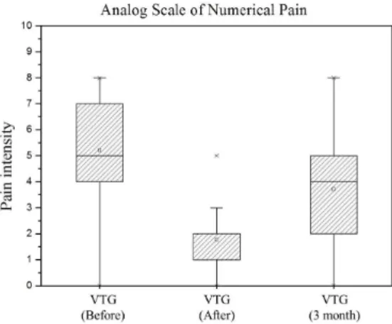
Analog Scale of Numerical Pain: It consisted of a numbered horizontal line, 0-10, with the ends indicating “no pain” and “worst possible pain” (Georgiou et al., 2015; Brasil, 2002). The volunteers were instructed to rate the pain within the existing scale value, according to the intensity of their sensation.
Surface electromyography (EMG): The acquisition of the muscle electrical signal was performed by an eight channel electromyography, EMG830 WF model (EMG System do Brasil Ltda, São José dos Campos/SP/Brasil), with the following technical
characteristics: analog-to-digital conversion board (A / D) with 16-bit resolution; bipolar pre-ampliied electrode with a differential gain of 20 times, totaling a inal ampliication of 1000 times; impedance > 10 MOhms, signal-to-noise ratio < 3 μVolts RMS; common mode rejection ratio > 120 dB; analog band-pass ilter of
20-500 Hz; sampling frequency set to 2 kHz per channel.
Signals were measured by 10 mm disc-shaped surface Ag/AgCl (silver/silver chloride) electrodes
and bipolar active (preampliier) electrodes that were
placed 20 mm apart from center to center. They were
placed with ixing disks and tape on the skin after
it had been shaved and cleaned with a 70% alcohol cotton swab (International…, 1999).
Surface electrodes were placed in pairs between the motor point and the distal tendon of the biceps,
triceps, deltoid (middle ibers), and trapezius (upper ibers), as speciied in the Surface- EMG protocol for
the Non Invasive Muscle Assessment (Seniam, 2015), following the longitudinal direction of the muscle
ibers. The reference electrode was anointed with
gel and placed on the styloid process of the ulna on the side contralateral to the member that underwent surgical intervention.
The EMG signals capture consisted of each volunteer standing upright, as they performed a
harmonious and comfortable movement of lexion
and abduction; they repeated both movements three times, totaling six repetitions for twenty seconds. All recordings were performed in upper limb ipsilateral to the surgical procedure.
Initially, these EMG signals were normalized regarding to amplitude, consisting of the adaptation of signal values in the desired range (min –1, and most 1) by means of the MATLAB 6.5.1 software (The Mathworks…, 2015), using routine and speciic functions.
After normalization, the data were processed by
EMGWorks Analysis of Delsys software (Delsys,
2015), using band pass ilter 4th order Butterworth, cutoff frequency set from 20 Hz to 400 Hz for noise elimination.
For data analysis, considering the total collection time of 20 seconds, only excerpts were selected related to muscle contraction, and relaxation periods were excluded, yielding the root mean square RMS - Root
Mean Square of each contraction. Data were grouped into a spreadsheet from Microsoft Ofice Excel 2013,
where rows represent individuals and columns the RMS values, with an average of 3 contractions obtained
for each muscle for both lexion and abduction kept
for later statistical analysis.
Dynamometry: The measurement of muscle strength was performed using the portable dynamometer scapular EMG System do Brasil Ltda São José dos Campos/SP/Brasil, dynamometer model scapular
DFE021115/200, electromyography, and this was
connected to the computer network. The calibration parameters followed were 2,000 Hz sampling frequency and Kgf unit.
The dynamometry test consisted of each volunteer standing upright with shoulders in abduction of
90°, with elbow lexion and both hands holding the
device in front of herself. The dynamometer reading was calibrated before each measurement and was triggered when the volunteer was oriented to pull the dynamometer reading, in order to perform the scapular adduction movement and exert maximum isometric
strength for 15 uninterrupted seconds. During the test,
the volunteer was encouraged by the therapist’s verbal command to exert the maximum effort.
The signals obtained from the hand were processed
in EMGWorks Delsys software. The irst two seconds
and the last second in relation to the total collecting time were excluded, to obtain the RMS values, which were tabulated in a worksheet for further statistical analysis.
Goniometry: The range of motion (ROM) was measured using a goniometer ISP brand of clear plastic with two strips of 20 cm and protractor
0° a 360° (degrees), consisting of a ixed arm, swing
arm, and axis. The range of motion measurement was performed with volunteer standing upright,
considering the movements of lexion, extension,
horizontal adduction, and abduction of the ipsilateral side of the surgical procedure. Participants were instructed on which movements should be performed in order to familiarize them with the test. Then they performed the moves actively to complete the entire arc of movement possible. From this angle, the record was made.
Experimental protocol
The study used the vibration blanket developed
for premium members. The vibration blanket follows the parameters of reliability for use in humans, which is already validated in the literature (Liao et al., 2014; Stillman, 1970). It consists of remote control, power cord, and vibration cells distributed throughout the blanket, which is 72 cm high and 52 cm wide. The vibration intensity can be adjusted to the minimum level of 1 and a maximum of 8 corresponding to frequency of 35 Hz to 80 Hz, according to the comfort of each individual. Based on the literature and studies that corroborate the reliability and validity of the parameters used in this study, we used vibratory stimuli with low intensity (sine oscillations < 1 g), frequency of 40 Hz and amplitude of 1.8 mm for the application of this treatment protocol (Kim et al., 2015; Rauch et al., 2010; Silva et al., 2011).
Initially, the experimental protocol consisted of volunteer in the supine position with the ipsilateral limb surrounded by the vibration blanket, supporting it elevated. The volunteer was subjected to 15 minutes of continuous vibration (Silva et al., 2011) with a frequency of 40 Hz and intensity respecting her tolerance.
The frequency of 40 Hz was selected to promote improvements in motor skills, through the increase
in joint range, increased strength, and muscle iber
recruitment, to supply interneuron activities to the skin and receptors in the spinal cord, which reduce
pain signals across C nerve ibers and increase the metabolic rate and blood low (Kim et al., 2015; Yang and Seo, 2015).
Statistical analysis
Descriptive and parametric statistical analyses were
performed using the BioEstat 5.3 software version (BioEstat, 2015 considering all variables from the EMG capture and the dynamometry test presented
a normal distribution according to the D’Agostino
normality test. All the graphs were plotted using OriginPro Version 8 software (OriginLab…, 2015).
To verify the existence of statistical differences between the means of the data, we used the ANOVA test analysis, two parallel samples, considering the following conditions: CG (electromyography and dynamometry) and VTG before and after treatment as well as 3 months after the termination of treatment.
In this study, the signiicance level for each comparison deined as statistically signiicant value p ≤ 0.05.
Results
A statistically signiicance level was evident
when we compared the minimization of pain intensity before and after the experimental protocol in the VTG
(p < 0.0001), and when we compared initial treatment with reassessment after 3 months (p < 0.0001), which shows the long-term effects of vibration therapy, represented by Figure 1. The assessment after treatment
compared to 3 months later found signiicant increased
pain intensity (p < 0.0001). However, even 3 months later the pain intensity of the VTG was lower than at the initial assessment.
Table 1 shows the results of the goniometer. It was
possible to observe signiicant increase in range of
motion of shoulder extension, horizontal abduction and adduction after the physiotherapeutic treatment. Whereas, the increase in range of motion of shoulder
extension and lexion remained three months after
treatment, while horizontal abduction and adduction
movements decreased signiicantly.
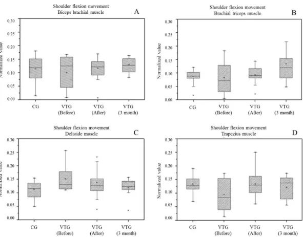
Figure 2 represents the values for the electromyographic activity of biceps (A), triceps
brachial (B), deltoid (middle ibers) (C), and upper trapezius (D) muscles during shoulder lexion movement
of both groups, VTG and CG.
When comparing the CG with VTG before treatment, it was possible to observe that the myoelectric activity
of deltoid muscle ibers had higher RMS signal
(p = 0.0013). However, the upper trapezius muscle had lower RMS signal (p = 0.0032) in the VTG. Comparing CG with VTG after treatment showed an increase in electrical activity of the biceps muscles and
middle deltoid, but no signiicant values. At 3 months
after the end of treatment, VTG triceps muscle had increased electrical activity when compared to the CG (p = 0.0019).
After treatment, the VTG had a significant increase in the RMS signal of the upper trapezius
muscle (p = 0.0164), no signiicant increase in
the triceps electrical activity, as well as decreased
electromyographic activity of the middle deltoid, when compared with the CG. However, it was not
possible to notice any signiicant changes in biceps.
Relative to VTG assessments before experimental protocol and 3 months later, the volunteers maintained some long-term results, with increased RMS signal of triceps (p = 0.0099) and decreased RMS signal of deltoid (p = 0.0006). The electromyographic
activity of the upper trapezius muscle ibers reduced
3 months after the second assessment, but remained higher than the signal before experimental protocol.
The result of triceps showed signiicant differences
(p = 0.0255) after treatment assessment compared to three months later.
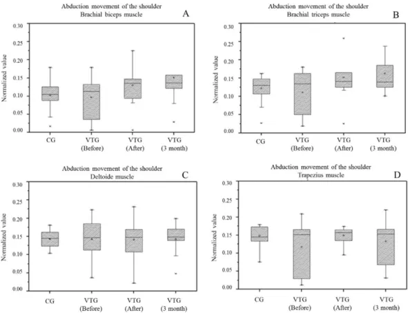
Figure 3 represents the values for the electromyographic activity of biceps muscle (A), triceps brachial (B), middle deltoid (C), and upper
trapezius (D) during the shoulder abduction motion
of VTG and CG.
When we compared the VTG before treatment with the GC during shoulder abduction movement, note that women after breast cancer had lower myoelectrical activity of muscles analyzed, but with
signiicant results only for the trapezoidal upper ibers
Table 1. Results obtained for range of motion measured by goniometry.
Movement
Media ± standard deviation p
Before After 3 months Before vs
after
Before vs 3 months
After vs 3 months
Flexion 135.7 ± 9.3 142 ± 9.6 143.5 ± 13.3 ns ns ns
Extension 33.2 ± 11.3 47.9 ± 11.8 44.2 ± 10.1 < 0.0001* 0.0008* ns
Abduction 124.6 ± 20 156.0 ± 18.8 134.2 ± 29.2 < 0.0001* ns 0.0002*
Horizontal adduction
24.2 ± 6.4 28.5 ± 6.6 17.8 ± 6.9 0.0317* < 0.0001* < 0.0001*
*Anova (p ≤ 0.05); ns: not signiicant.
(P = 0.0340). Soon after treatment, these women
showed a signiicant increase in the recruitment of muscle ibers of biceps (p = 0.0213) and triceps
(p = 0.0290) compared with healthy volunteers, and this remained three months later.
The VTG showed that after the experimental
protocol and 3 months later, there was a signiicant
increase in RMS signal of biceps (p = 0.0071) and triceps (p = 0.0017). The upper trapezius muscle
showed no signiicant increase in myoelectrical activity compared with the initial assessment. Deltoid medium iber muscle had no signal change.
It was not possible to observe signiicant RMS
values for VTG after treatment compared with three months later, but an increased myoelectric activity of the biceps along with the decreased signal of
trapezius upper ibers were noticeable. In addition,
triceps and deltoid muscles kept the average signal values obtained after experimental protocol.
Data regarding to shoulder dynamometry,
presented in Figure 4, were statistically signiicant, showing less muscle strength in after breast cancer patients than in the control group, in all situations:
before (p < 0.0001), after treatment (p = 0.0024), and 3 months after treatment (p = 0.0008).
The intra-group analysis of VTG showed increased muscle strength after treatment (p = 0.0005) compared to initial assessment, as well as before compared to three
Figure 3. Box plots of the mean and two standard errors of the RMS values of the RMS values from myoelectric signal during shoulder abduction movement of control group (CG) and vibration therapy group (VTG). (A) Biceps muscle; (B) Triceps muscle; (C) Deltoid muscle; (D) Trapezius muscle.
months after (p = 0.0006). There was no signiicant
difference after treatment compared to three months later, but the gained muscle strength remained.
Discussion
This study describes the alterations that can
inluence the biomechanics of the shoulder joint of
post-surgical breast cancer women. Usually these women have the clinical condition of pain, reduced range of motion and muscle function (Haddad et al., 2013). This study proposed the use of vibration as a therapeutic resource in order to minimize these symptoms.
Vibration is a non-pharmacological therapeutic technique used to alleviate pain by the activation of
supericial and deep mechanical receptors. The vibratory
stimuli are transmitted by the Meissner corpuscle, which is sensitive to about 40 Hz and by Vater-up Paccini corpuscle above 100 Hz (Rittweger et al., 2010).
In this study, we found that vibration therapy was effective to minimize the intensity of pain in post-surgical breast cancer women, after ten 15-minutes sessions
of experimental protocol. This inding corroborates
the study of Volz et al. (2013), which reported 70% reduction of chronic and acute pain after vibration therapy. They concluded that vibration stimulus increases neuronal conduction of signals, through
the large diameter myelinated ibers, and inhibits short ibers of spinal dorsal horn before the synapse.
This hypothesis may be associated with the reduction
of pain perception, in which the supericial receptors
interact with deep receptors in spinal cord synaptic transmission and in nociceptive processing, which decreases the pain threshold.
Nevertheless, Dahlin et al. (2006) applied vibration in healthy women for twenty minutes and said that
this time was suficient to promote pain relief. Other
studies have reported that the appropriate application time is still unknown, contradicting the time stated by the author above (Mikhael et al., 2010; Osawa et al., 2013).
On reevaluation after three months, increased pain intensity was observed, when compared to the end of treatment (10 sessions); however, this increase
was less intense than measured initially at the irst session. This inding corroborates with Bensmaia et al. (2005), which stated that the awareness of the nerve
ibers are reversible with time.
Another common complication relative to breast cancer is the reduced range of motion of the shoulder (Galiano-Castillo et al., 2013). Smoot et al. (2010) stated that the deicit in the range of motion for post-treatment of breast cancer results from the
formation of a scar tissue, radiation-induced ibrosis,
antalgic posture of shoulder, disuse, and pain. These limitations interfere directly in daily-life activities,
as dificulties in washing and combing their hair, for
example. Early interventions are necessary in order to minimize these kinds of complications.
Some women report sensation of heaviness in the upper limbs due to the development of lymphedema and limited range of motion, which can result in severe pain, bursitis, tendonitis, or chronic injury with rotator ischemia and cuff. According to Jeong et al. (2011), the muscles that make up the rotator cuff are responsible for stabilizing the shoulder joint complex; however, tendinitis of the rotator cuff can be a complication related to lymphoedema, which is
caused by internal derangement of the tendon ibers,
generating functional overload.
Adriaenssens et al. (2012) conducted a randomized study with 119 women post-breast cancer in order to evaluate the morbidity of ipsilateral surgery shoulder. They observed that in the period of 1-3 months after radiotherapy, the upper limb volume increased 4.1%, and the movement of abduction decreased 8.6%. The authors stated that the compensatory muscle activity is necessary to improve the stability of the shoulder joint, which is associated with pain and
muscle spasms. In this study, we observed a signiicant
increase in range of motion of extension, abduction, and horizontal shoulder adduction movements, after the experimental vibration protocol. The gain was maintained in Post-surgical breast cancer women (VTG) three months after completion of treatment.
According to Martínez et al. (2013), vibration
has beneicial effects on proprioception mechanisms and can inluence the muscle cycles of contraction
and relaxation, which can be detected by surface electromyography. This improvement is attributed
to the tonic vibration relex triggered by excitation
of the muscle spindles.
The control group in this study included healthy women who participated only for assessing the myoelectric activity and muscle strength, so that these values could be a reference to compare with post-breast cancer women.
The post-breast cancer women (VTG) had reduced RMS signal obtained by electromyography compared to healthy women (CG), probably due to differences in muscle activation mechanism, as well as decreased muscle strength. However, after the vibration therapy, we observed the increase in RMS signal for the biceps, triceps, and deltoids muscles, as well as increased muscle strength, near the normal parameters.
surgical treatment of breast cancer. The authors stated that the increase causes recruitment of muscle
ibers in the scapular stabilization of the trapezius muscle, and the increase occurs due to deicits of
other musculatures and the serratus anterior muscle. They found increased RMS value of the trapezius muscle due to a compensatory form for scapular stabilization; and increased electrical activity of the deltoid muscle as a strategy to keep the glenoid cavity of the humerus in a favorable position, as well as contribute to the complex stability of the shoulder during the execution of movements.
According to Foti et al. (2012), vibratory stimuli are responsible for activating spindle receivers that innervate the agonist and antagonist muscles when both work synchronously. Vibration stimuli increase
neurons iring and, subsequently, increase the excitability
of muscle spindles and muscle strength.
Based on the reported results, we found that vibration therapy could improve the musculoskeletal function,
because the vibratory stimuli generate increased iring
of neurons, and subsequently increase excitability of muscle spindles, which leads to an increase in
recruitment of muscle ibers, providing improved
muscular strength, increased range of motion and attenuation of the painful condition of post-surgical breast cancer women. Therefore, we suggest the inclusion of the vibration blanket proposed in this study as a therapeutic device, which may assist in reducing the morbidity after breast cancer.
In the course of this study, points were identiied
to be developed in future studies, which corroborate with the results described, among them are: the application of this therapeutic resource with a larger sample number and long-term treatment with vibration therapy for post-breast cancer complications.
Vibration therapy attenuated pain symptoms, improved joint movements of the shoulder, muscle activation mechanisms, and strength of post-surgical breast cancer women, as well as assisted in the long-term relief of symptoms. Therefore, vibration
blanket may be considered a beneicial therapeutic
resource to be inserted in rehabilitation programs of post-surgical breast cancer patients.
Acknowledgements
The authors would like to thank Coordenação de Aperfeiçoamento de Pessoal de Nível Superior
(CAPES) for the inancial support (PE 030/2008).
References
Adriaenssens N, Ridder M, Lievens P, Parijs HV, Vanhoeij M, Miedema G, Voordeckers M, Versmessen H, Storme G,
Lamote J, Pauwels S, Vinh-Hung V. Scapula alata in early breast cancer patients enrolled in a randomized clinical trial of postsurgery short-course image-guided radiotherapy. World Journal of Surgical Oncology. 2012; 10(86):1-12. http://dx.doi.org/10.1186/1477-7819-10-86. PMid:22214417.
Aman JE, Elangovan N, Yeh L, Konczak J. The effectiveness of proprioceptive training for improving motor function: a systematic review. Frontiers in Human Neuroscience. 2015; 8:1-18. http://dx.doi.org/10.3389/fnhum.2014.01075. PMid:25674059.
Atalay OT, Ozkir A, Calik BB, Baskan E, Taskin H. Effects of phase I complex decongestive physiotherapy on physical functions and depression levels in breast cancer related lymph edema. Journal of Physical Therapy Science. 2015; 27(3):865-70. http://dx.doi.org/10.1589/jpts.27.865. PMid:25931748.
Bensmaia SJ, Leung YY, Hsiao SS, Johnson KO. Vibratory adaptation of cutaneous mechanoreceptive afferents. Journal of Neurophysiology. 2005; 94(5):3023-36. http://dx.doi. org/10.1152/jn.00002.2005. PMid:16014802.
BioEstat. Institute Mamiraua: statistical institute of science and technology [internet]. Manaus, 2015 [cited 2015 Mar 28]. Available from: http://www.mamiraua.org.br/pt-br/ downloads/programas/
Brasil. Ministério da Saúde. Instituto Nacional do Cancer. Cancer palliative care: pain control [internet]. Rio de Janeiro: INCA; 2002 [cited 2015 Jan 04]. Available from: http:// bvsms.saude.gov.br/bvs/publicacoes/inca/manual_dor.pdf.
Brasil. Ministério da Saúde. Instituto Nacional do Cancer. Estimation: incidence of cancer in Brazil [internet]. Rio de Janeiro: INCA; 2014 [cited 2015 Mar 16]. Availabre from: http://www.inca.gov.br/estimativa/2014/
Dahlin L, Lund I, Lundeberg T, Molander C. Vibratory stimulation increases the electro-cutaneous sensory detection and pain thresholds in women but not in men. Bio Med Central Complementary and Alternative Medicine. 2006; 6(20):1-6. http://dx.doi.org/10.1186/1472-6882-6-20. Delsys. Wearable sensors for movement sciences [internet]. Massachusetts, 2015 [cited 2015 Mar 28]. Available from: http://www.delsys.com/products/emgworks-software/ download/.
Foti C, Laurini A, Tiberti S, Carli G, Tsarpela O, Adamidis K, Bonifazi M, Giombini A, Tihanyi J, Duvillard S, Vita M, Bosco C. Leg extension test, semg and vibratory stimuli to assess functional recovery following knee joint surgery. Muscles, Ligaments and Tendons Journal. 2012; 2(2):127-32. PMid:23738286.
Galiano-Castillo N, Ariza-García A, Cantarero-Villanueva I, Fernández-Lao C, Díaz-Rodríguez L, Legerén-Alvarez M, Sánchez-Salado C, Del-Moral-Avila R, Arroyo-Morales M. Telehealth system (e-CUIDATE) to improve quality of life in breast cancer survivors: rationale and study protocol for a randomized clinical trial. Trials. 2013; 14(187):1-10. http:// dx.doi.org/10.1186/1745-6215-14-187. PMid:23799886.
review. Bio Med Central Research International. 2015; 2015:1-18.
Haddad CAS, Saad M, Perez MCJ, Miranda F Jr. Assessment of posture and joint movements of the upper limbs of patients after mastectomy and lymphadenectomy. Einstein (Sao Paulo, Brazil). 2013; 11(4):426-34. http://dx.doi. org/10.1590/S1679-45082013000400004. PMid:24488379.
Hadi N, Soltanipour S, Talei A. Impact of modified radical mastectomy on health-related quality of life in women with early stage breast cancer. Archives of Iranian Medicine. 2012; 15(8):504-7. PMid:22827789.
International Society of Electrophysiolohy and Kinesiology – ISEK. Standards for reporting EMG data [internet]. Victoria: ISEK; 1999 [cited 2015 Mar 28]; Available from: http://earc.eng.niigata-u.ac.jp/links/ISEK_Standards.pdf.
Jeong HJ, Sim YJ, Hwang KH, Kim GC. Causes of shoulder pain in women with breast cancer-related lymphedema: a pilot study. Yonsei Medical Journal. 2011; 52(4):661-7. http://dx.doi.org/10.3349/ymj.2011.52.4.661.
Jung D, Moon DC. The effects of shoulder joint abduction angles on the muscle activity of the serratus anterior muscle and the upper trapezius muscle while vibrations are applied. Journal of Physical Therapy Science. 2015; 27(1):117-9. http://dx.doi.org/10.1589/jpts.27.117. PMid:25642052. Kim SS, Ju SB, Park GD. Changes in stress hormone levels with the application of vibrations before resistance exercises at different intensities. Journal of Physical Therapy Science. 2015; 27(9):2845-7. http://dx.doi.org/10.1589/jpts.27.2845. PMid:26504307.
Liao LR, Huang M, Lam FMH, Pang MYC. Effects of whole-body vibration therapy on whole-body functions and structures, activity, and participation poststroke: A systematic review. Physical Therapy. 2014; 94(9):1232-51. http://dx.doi. org/10.2522/ptj.20130366. PMid:24786940.
Londen GJ, Beckjord EB, Dew MA, Cooper KL, Davidson NE, Bovbjerg DH, Donovan HS, Thurston RC, Morse JQ, Nutt S, Rechis R. Associations between adjuvant endocrine therapy and onset of physical and emotional concerns among breast cancer survivors. Supportive Care in Cancer. 2014; 22(4):937-45. http://dx.doi.org/10.1007/s00520-013-2041-y. PMid:24271937.
Martín ML, Hernández MA, Avendaño C, Rodríguez F, Martínez H. Manual lymphatic drainage therapy in patients with breast cancer related lymphedema. BMC Cancer. 2011; 11(94):1-6. http://dx.doi.org/10.1186/1471-2407-11-94. PMid:21392372.
Martínez F, Rubio JA, Ramos DJ, Esteban P, Mendizábal S, Jiménez F. Effects of 6-week whole body vibration training on the reflex response of the ankle muscles: a randomized controlled trial. International Journal of Sports Physical Therapy. 2013; 8(1):15-24. PMid:23439725.
Mendes IS, Freitas STT, Lima FPS, Andrade AO, Lima MO. Vibrational Therapy after breast cancer: influence on the painful intensity and myoelectric activity. In: Proceedings of the 24th Brazilian Congress on Biomedical Engineering – CBEB; 2014 Out 13-17; Uberlândia, Brazil. Uberlândia: UFU; 2014. p. 945-8.
Mikhael M, Orr R, Amsen F, Greene D, Singh MAF. Effect of standing posture during whole body vibration training on muscle morphology and function in older adults: a randomised controlled trial. BMC Geriatrics. 2010; 10(74):1-13. http:// dx.doi.org/10.1186/1471-2318-10-74. PMid:20946685.
Mutrie N, Campbell A, Barry S, Hefferon K, McConnachie A, Ritchie D, Tovey S. Five-year follow-up of participants in a randomised controlled trial showing benefits from exercise for breast cancer survivors during adjuvant treatment: are there lasting effects? Journal of Cancer Survivorship: Research and Practice. 2012; 6(4):420-30. http://dx.doi. org/10.1007/s11764-012-0233-y. PMid:22836201. Naghii MR, Darvishi P, Ebrahimpour Y, Ghanizadeh G, Mofid M, Hedayati M, Asgari AR. Effect of combination therapy of fatty acids, calcium, vitamin D and boron with regular physical activity on Cardiovascular risk factors in rat. Journal of Oleo Science. 2012; 61(2):103-11. http:// dx.doi.org/10.5650/jos.61.103. PMid:22277894.
OriginLab Corporation. [internet]. Northampton; 2015 [cited 2015 Mar 16]. Availabre from: http://www.originlab.com/
Osawa Y, Oguma Y, Ishii N. The effects of whole-body vibration on muscle strength and power: a meta-analysis. Journal of Musculoskeletal & Neuronal Interactions. 2013; 13(3):380-90. PMid:23989260.
Pereira TB, Bergmann A, Ribeiro ACP, Silva JG, Dias R, Ribeiro MJP, Thuler LCS. Myoeletric activity pattern of scapular muscles after axillary lymphadenectomy in breast cancer. Journal of Obstetrics & Gynaecology. 2009; 31(5):224-9. PMid:19669029.
Rauch F, Sievanen H, Boonen S, Cardinale M, Degens H, Felsenberg D, Roth J, Schoenau E, Verschueren S, Rittweger J. Reporting whole-body vibration intervention studies: recommendations of the International Society of Musculoskeletal and Neuronal Interactions. Journal of Musculoskeletal & Neuronal Interactions. 2010; 10(3):193-8. PMid:20811143.
Rittweger J, Beller G, Felsenberg D. Acute physiological effects of exhaustive whole-body vibration exercise in man. Clinical Physiology (Oxford, England). 2010; 20(2):134-42. http://dx.doi.org/10.1046/j.1365-2281.2000.00238.x. PMid:10735981.
Rodrick JR, Poage E, Wanchai A, Stewart BR, Cormier JN, Armer JM. Complementary, alternative, and other noncomplete decongestive therapy treatment methods in the management of lymphedema: a systematic search and review. PM & R. 2013; 6(3):250-74. PMid:24056160.
Seniam. Surface ElectroMyoGraphy for the non-invasive assessment of muscles. Biomedical Health and Research Program (BIOMED II) of the European Union [internet]. 2015 [cited 2015 Jan 30]. Available from: http://www. seniam.org/.
Silva JM, Lima MO, Paula AR Jr. Acute effect of vibratory stimulation in spastic hemiparetic after a stroke. Revista Brasileira de Engenharia Biomédica. 2011; 27(4):224-30. http://dx.doi.org/10.4322/rbeb.2011.018.
without lymphedema following breast cancer treatment. Journal of Cancer Survivorship: Research and Practice. 2010; 4(2):167-78. http://dx.doi.org/10.1007/s11764-010-0118-x. PMid:20373044.
Souza GAS, Mendes IS, Oliveira F, Borges ACL, Lima MO, Lima FPS. Effect of vibration therapy on Lymphoedema surgical after care of breast cancer. In: Proceedings of the 18th Latin American Meeting of Scientific Initiation, 14th Latin American Postgraduate and 4th Meeting of Initiation to Teaching University of Vale do Paraiba; 2014 Out 23-24; São José dos Campos, Brazil. 2014. p. 1-4.
Springer BA, Levy E, McGarvey C, Pfalzer LA, Stout NL, Gerber LH, Soballe PW, Danoff J. Pre-operative assessment enables early diagnosis and recovery of shoulder function in patients with breast cancer. Breast Cancer Research and Treatment. 2010; 120(1):135-47. http://dx.doi.org/10.1007/ s10549-009-0710-9. PMid:20054643.
Stillman BC. Vibratory motor stimulation. The Australian Journal of Physiotherapy. 1970; 16(3):118-23. http://dx.doi. org/10.1016/S0004-9514(14)61096-5. PMid:25028186.
Stout NL, Binkley JM, Schmitz KH, Andrews K, Hayes SC, Campbell KL, McNeely ML, Soballe PW, Berger AM, Cheville AL, Fabian C, Gerber LH, Harris SR, Johansson K, Pusic AL, Prosnitz RG, Smith RS. A prospective surveillance model for rehabilitation for women with breast cancer. Cancer. 2012; 118(8 Suppl):2191-200. http://dx.doi. org/10.1002/cncr.27476. PMid:22488693.
The MathWorks Inc. [internet]. Novi: MathWorks; 2015 [cited 2015 Mar 28]; Available from: http://www.mathworks. com/index.html?s_tid=gn_logo.
Tovar JR, Zandonade E, Amorim MHC. Factors associated with the incidence of local recurrences of breast cancer in women who underwent conservative surgery. International Journal of Breast Cancer. 2014; 2014:1-9. http://dx.doi. org/10.1155/2014/639534.
Veqar Z, Imtiyaz S. Vibration therapy in management of delayed onset muscle soreness (DOMS). Journal of Clinical and Diagnostic Research. 2014; 8(6):1-4. http://dx.doi. org/10.7860/JCDR/2014/7323.4434.
Volz MS, Suarez-Contreras V, Mendonca ME, Pinheiro FS, Merabet LB, Fregni F. Effects of sensory behavioral tasks on pain threshold and cortical excitability. PLoS One. 2013; 8(1):e52968. http://dx.doi.org/10.1371/journal. pone.0052968. PMid:23301010.
Walczyńska-Dragon K, Baron S. The biomechanical and functional relationship between temporomandibular dysfunction and cervical spine pain. Acta of Bioengineering and Biomechanics. 2011; 13(4):93-8. PMid:22339095.
Xu X, McGorry RW, Lin JH. A regression model predicting isometric shoulder muscle activities from arm postures and shoulder joint moments. Journal of Electromyography and Kinesiology. 2014; 2014(24):419-29. http://dx.doi. org/10.1016/j.jelekin.2014.02.004. PMid:24618104. Yang J, Seo D. The effects of whole body vibration on static balance, spinal curvature, pain, and disability of patients with low back pain. Journal of Physical Therapy Science. 2015; 27(3):805-8. http://dx.doi.org/10.1589/jpts.27.805. PMid:25931735.
Authors
Izabela dos Santos Mendes1*, Fernanda Pupio Silva Lima1, Sergio Takeshi Tatsukawa de Freitas1, Tamires de Souza
Moreira Prianti1, Adriano Oliveira Andrade2, Mario Oliveira Lima1
1 Laboratório de Engenharia de Reabilitação Sensório Motora, Universidade do Vale do Paraíba – UNIVAP, Avenida Shishima Hifumi, 2911, Urbanova, CEP 12244-000, São José dos Campos, SP, Brazil.