A
RTIGO
C
IENTÍFICO
Revista Brasileira de FisioterapiaEffects of neuromuscular electrical stimulation
on tibialis anterior muscle of spastic
hemiparetic children
Efeitos de estimulação elétrica neuromuscular no músculo tibial anterior em
crianças hemiparéticas espásticas
Nunes LCBG1, Quevedo AAF1, Magdalon EC2
Abstract
Objective: This study evaluated the effects of neuromuscular electrical stimulation (NMES) on muscle strength, range of motion (ROM)
and gross motor function, among spastic hemiparetic children while standing, walking, running and jumping. Methods: Ten children
were divided into two groups of five. The children who were normally receiving physical therapy sessions twice a week had two 30-minute NMES sessions per week (group 1), while those who were having one physical therapy session per week had one 30-30-minute NMES session per week (group 2), for seven weeks in both groups. The children were evaluated three times: before beginning the NMES protocol (initial), right after the end of the protocol (final) and eight weeks after the final evaluation (follow-up). The evaluations included manual goniometry on ankle dorsiflexion, manual muscle strength of the tibialis anterior and gross motor function (measurements while standing, walking, running and jumping). The statistical analysis was performed using the Wilcoxon and Mann-Whitney tests, considering a p level of 0.05. Results: There were significant increases in muscle strength, gross motor function and passive ROM of ankle dorsiflexion, in both groups, and in active dorsiflexion in the first group. No significant differences were found between the groups.
Conclusions: The improvements in ROM, muscle strength and gross motor function demonstrated that the use of NMES was effective in both groups, since no significant differences were found between the groups. This study suggests that NMES may be a useful therapeutic tool, even when applied once a week. Further studies are needed to confirm these findings.
Key words: electrical stimulation; hemiparetic children; spasticity; cerebral palsy; tibialis anterior.
Resumo
Objetivo: Este estudo avaliou os efeitos da estimulação elétrica neuromuscular (EENM) na força, amplitude de movimento (ADM) e
função motora grossa (FMG) em pé, andando, correndo e pulando de crianças hemiparéticas espásticas. Métodos: Dez crianças
foram divididas em dois grupos de cinco. As que realizavam sessões de fisioterapia duas vezes por semana tiveram duas sessões semanais de EENM de 30 minutos cada (grupo 1), enquanto as que compareciam à uma sessão tiveram uma sessão semanal (grupo 2), ambas por sete semanas. As crianças foram avaliadas três vezes: antes do início do protocolo de EENM (inicial), ao final do protocolo (final) e oito semanas após a avaliação final (tardia). As avaliações englobaram goniometria manual da dorsiflexão de tornozelo, força muscular manual do tibial anterior e função motora grossa, (Gross Motor Function Measure em pé, andando correndo
e pulando). A análise estatística foi feita pelos testes de Wilcoxon e Mann-Whitney, com p adotado de 0,05. Resultados: Houve
aumentos significativos na força muscular, na FMG e na ADM passiva da dorsiflexão de tornozelo em ambos os grupos, assim como na dorsiflexão ativa no primeiro grupo. Nenhuma diferença significativa foi encontrada entre os grupos. Conclusões: As melhoras obtidas na ADM, força muscular e FMG demonstram que o uso da EENM foi eficaz nos dois grupos, não tendo sido encontradas diferenças significativas entre os mesmos. Este estudo sugere que a EENM pode ser útil no auxílio à terapia, mesmo em baixas freqüências, como uma vez por semana. Estudos adicionais são necessários para confirmar estes achados.
Palavras-chave: estimulação elétrica; crianças hemiparéticas; espasticidade; paralisia cerebral; tibial anterior.
Recebido: 01/08/07 – Revisado: 08/11/07 – Aceito: 08/04/08
1Pediatrics Investigation Center, School of Medical Sciences; Department of Biomedical Engineering, School of Electrical and Computer Engineering; Biomedical Engineering Center, Universidade
Estadual de Campinas (Unicamp) – Campinas (São Paulo), Brazil
2Department of Biomedical Engineering, School of Electrical and Computer Engineering; Biomedical Engineering Center, Unicamp – Campinas (SP), Brazil
Correspondence to: Ligia Christina Borsato Guimarães Nunes, Centro de Investigação em Pediatria, Faculdade de Ciências Médicas; Departamento de Engenharia Biomédica, Faculdade de Engenharia Elétrica e de Computação; Centro de Engenharia Biomédica, Universidade Estadual de Campinas, Rua Padre Vieira, 565, apto. 41, CEP 13015-301, Campinas (SP), Brasil, e-mail: [email protected]
Introduction
Cerebral palsy (CP) may be deined as static encephalopa-thy, and some of its possible consequences are non-progressive
movement and posture disturbances1. Stroke in children is a
consequence of encephalic arterial blockade or rupture that starts abruptly and, within minutes or hours, develops into a neurological syndrome that varies in intensity and
conse-quences2. One of the most common impairments due to CP or
stroke is spastic hemiparesis, which is characterized by imbal-ance between agonist and antagonist muscles that may lead to walking disabilities, muscle contractures and deformities like
equinus foot1-3. Even among children with mild hemiparesis,
equinus foot is a common impairment that afects gait, due to weakness of the tibialis anterior and triceps surae muscles. An inefective tibialis anterior may decrease foot clearance, which
may cause stumbles and falls4.
Neurodevelopmental treatment is the most commonly used treatment technique for cerebral palsy. Although this approach acknowledges functional independence as an important treat-ment goal, the means of obtaining function is based on inhi-bition of abnormal posture and movement and on improving the child’s quality and eiciency of movement by encouraging
typical patterns of movement5.
Scientiic advances have allowed new technologies to be used to help rehabilitation of patients who sufer from neuro-logical problems. Interactions between health sciences and engineering have contributed towards improving quality of life, through promoting functional independence for otherwise de-pendent patients. Neuromuscular electrical stimulation (NMES) has been shown to be useful in the rehabilitation of neurological
patients6-8. However, in neuropediatric Physical herapy (PT)
this kind of procedure has not yet been widely explored, since therapists fear increasing spasticity through electrical stimula-tion. For this reason, NMES is not a common practice for CP
patients9,10, although it has been used for research on CP
sub-jects, usually with high weekly frequencies of treatment, or up
to twice a day by some authors7,11-13. hese protocols are used
mainly for research, and they do not relect what can be done in large-scale therapy. Although NMES is applied to speciic muscles, increases in overall functioning after its use have been documented, because increases in strength and range of
move-ment (ROM) can lead the child to use the limb more efectively12.
Speciic use of NMES on the tibialis anterior muscle can increase ankle ROM and dorsilexion strength, which may produce more eicient dorsilexion and clearance during gait.
In most countries, there are huge demands from patients on the healthcare system. he demands are greater than the human and material resources available, thus making it impos-sible to treat patients daily or even three times a week. Children
usually have physical therapy only once or twice a week and it is very diicult to implement a protocol based on what can be found in the scientiic literature. Furthermore, in addition to the resource limitations of the system, most families are unable to take their children to and from the physical therapy center more than once or twice a week.
he main aim of the present study was to investigate and compare the efects of NMES on the strength of the tibialis anterior muscle and on active and passive range of motion (ROM) of ankle joint dorsilexion, and in relation to the more sophisticated aspects of gross motor function (GMF), between children undergoing NMES once and twice a week.
Materials and methods
he research protocol was approved by the institutional eth-ics committee of the School of Medical Sciences, Universidade Estadual de Campinas (Unicamp), Brazil, under the procedure number 468/2002. A written informed consent form giving agree-ment to participation and publication of results was signed by the children’s parents. All children at the neuropediatric physical ther-apy outpatient clinic of Unicamp’s teaching hospital who met the selection criteria and whose parents agreed to their participation were recruited as a convenience sample. he selection criteria were that the patients should be users of conventional physical therapy (mainly based on Bobath’s neurodevelopmental approach), have spastic hemiparesis (due to CP or stroke), be aged between seven and 15 years, be able to walk independently, have no cognitive im-pairments, be collaborative, have surface sensitivity preserved in their legs, have no ankle deformities, have had no botulinum toxin application for at least six months before the study and have had no previous triceps surae tendon surgery.
Ten children aged seven to ifteen years were chosen: eight with CP and two with stroke. hey were divided into two groups of ive (with one children with stroke in each group), accord-ing to their usual previous therapy frequencies. hus, children who were having physical therapy twice a week were placed in group 1 and children who were having physical therapy once a week were placed in group 2. Descriptive data from these chil-dren were: age range 7 to 14.8 years (mean 11.34 years); seven males and three females; ive afected on the left side and ive afected on the right side.
he patients underwent NMES sessions for seven weeks, twice a week in group 1 (total of 14 sessions) and once a week in group 2 (total of seven sessions). Each session had a length of 30 minutes. All the children completed the protocol.
electrical stimulators, respectively. It is important to emphasize
that all the children continued to attend their conventional physical therapy sessions at the neuropediatric physical therapy outpatient clinic of Unicamp’s teaching hospital.
he study was single-blinded and there were three NMES evaluations: irstly, one week before beginning the protocol (initial); secondly, right after the end of the protocol ( inal); and thirdly, eight weeks after the inal evaluation ( follow-up). he examiner was blind to the group in which each child was placed. he evaluation tests consisted of passive and active manual ankle dorsilexion goniometry, measured in degrees using a handheld goniometer, and the Research Medical Coun-cil manual muscle strength test for the tibialis anterior and plantar lexors14. Goniometry was performed with the subject
in supine position with extended knees, and the measurement was made at the neutral position between dorsal lexion and plantar lexion,id esttt, 0º of dorsal lexion. hese tests were
chosen because of their easy applicability, low cost and wide application in clinical practice. Another test was gross motor function (GMF) measurement, which evaluates capabilities in some functional activities performed by children. In the present study, only the GMF dimensions of standing, walking and climbing were used, which were the dimensions that were expected to change after applying the NMES protocol15.
he NMES sessions (30 minutes each) took place at Unicamp’s Center of Investigations in Pedriatrics (CIPED). During these ses-sions, the children were seated on a comfortable chair, with knees positioned at 90º of lexion. hey were barefoot, with their heels placed in contact with loor (Figure 1). For patients whose heels did not reach the ground, a small support box was placed under their feet. NMES electrodes were placed on the skin surface of the paretic side, over the tibialis anterior muscle and near its motor point. he parameters used were: pulse frequency of 50Hz, pulse width of 250µs and on/of ratio of ive seconds stimulation and ten seconds rest. hese parameters were chosen based on electri-cal stimulation studies in the literature16-20 and on pilot tests
per-formed on seven normal adults (because of the lack of children to act as volunteers). hese parameters were shown to evoke a muscle response with minimal discomfort to the patients. NMES was expected to induce a visible muscle contraction, without pain. he current intensity applied was chosen during the ses-sions, according to the patient’s sensitivity. he maximum inten-sity reached during therapy ranged from 28 to 44mA.
he Wilcoxon nonparametric test was used to compare diferent dependent variables within a group, and the Mann-Whitney test was applied for comparisons between groups. AD of 0.05 was chosen as the level of statistical signiicance. Nonparametric tests are more suitable than parametric tests for analyzing small sample sizes and in evaluations that at-tribute scores instead of absolute values21.
Figure 1. Subject positioning during neuromuscular electrical stimulation (NMES) protocol.
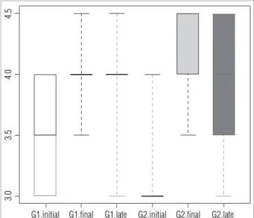
Figure 2. Median, minimum and maximum strength scores and 25 and 75 percentiles (edges of the boxes) from initial, final and follow-up evaluations of group one (G1) and group two (G2).
4.
5
4.
0
3.
5
3.
0
G1.initial G1.final G1.late G2.initial G2.final G2.late
Results
Strength
For anterior tibialis muscle strength (paretic side), the me-dian scores and the 25 and 75 percentiles for the initial, inal and follow-up evaluations are shown in Figure 2. here was a signiicant diference between the initial and inal evaluations of the strength test for paretic side ankle dorsilexion (p=0.05) in group 1. Diferences were also found in strength tests for paretic side ankle dorsilexion between the initial and inal
scores (p<0.05) and between the initial and follow-up evalua-tions for group 2. No signiicant diferences were found between the initial and follow-up evaluations for group 1. However, the group 1 follow-up evaluation values were found to be interme-diate between the initial and inal values. Comparison using the Mann-Whitney test did not show any signiicant diference between groups 1 and 2.
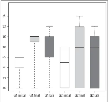
Figure 3. Active range of motion boxplot with median, minimum and maximum scores and 25 and 75 percentiles (edges of the boxes) from initial, final and follow-up evaluations of group one (G1) and group two (G2).
14
12
10
8
6
4
2
0
G1.initial G1.final G1.late G2.initial G2.final G2.late
Figure 4. Passive range of motion boxplot with median, minimum and maximum scores and 25 and 75 percentiles (edges of the boxes) from initial, final and follow-up evaluations of group one (G1) and group two (G2).
20
15
10
5
G1.initial G1.final G1.late G2.initial G2.final G2.late
Figure 5. Gross motor function (GMF) measurement boxplot with median, minimum and maximum scores and 25 and 75 percentiles (edges of the boxes) from initial, final and follow-up evaluations of group one (G1) and group two (G2).
104
102
100
98
96
G1.initial G1.final G1.late G2.initial G2.final G2.late
ROM measures
he goniometric results shown here are from the afected side. Figures 3 and 4 show the medians and percentiles obtained from active and passive ankle dorsal lexion ROM. Statistical analysis found signiicant changes (p=0.05) between initial and inal and between initial and follow-up for both active and pas-sive dorsal lexion in group 1, and between initial and inal and between initial and follow-up for passive dorsal lexion (p<0.05) in group 2. Comparison using the Mann-Whitney test did not show any signiicant diference between groups 1 and 2.
Gross motor function (GMF) measurement
his measurement scale attempts to quantify function and functional changes over time and is divided into ive dimensions: lying and rolling; sitting; kneeling and crawling; standing; and
walking, running and jumping15. If the dimensions of standing
and walking, running and jumping are combined, the maximum score that can be achieved is 105. he medians obtained by groups 1 and 2 are shown in Figure 5. Group 1 reached 94.28% of the total score possible in the initial evaluation and 97.14% in the inal and follow-up assessments. Group 2 reached a mean of 95.23% of the total score possible in the initial evaluation, 98.09% in the inal assessment and 97.14% in the follow-up assessment.
follow-up results were higher than the initial scores. The Mann-Whitney test did not show any differences between groups 1 and 2.
Discussion
In the scientific literature, some studies on NMES for hemiparetic spastic children have shown that it is possible to increase muscle strength and range of motion, and to promote functional improvements in motor tasks among
CP children10,12. Although NMES is a therapeutic resource
directed specifically to one muscle or muscle group, its re-sults can be reflected in overall improvements in function, because children can reach better results in their overall functioning through improved strength and range of move-ment. However, the studies that have shown this result used high-frequency therapies.
In our study, NMES was applied only once or twice a week. Increases in tibialis anterior muscle strength were observed in both groups, with no diference between groups. In addition, the values reached in the follow-up evaluation were higher than the initial values. his suggests that, with the aid of NMES, even with weekly sessions, satisfactory progress can be obtained in relation to hemiparetic spastic muscle strength. In a review of the literature conducted by Kerr, McDowell
and McDonough17, the quality of the electrical stimulation
protocols was analyzed, as well as the results obtained, and it was noticed that there was a signiicant increase in anterior tibialis muscle strength. herefore, our indings conirm what is shown in the literature.
Furthermore, neuronal plasticity mediated by NMES is still a subject of research. Some authors have suggested theories about plasticity mechanisms, and have observed that children
have better recovery capacity than adults22-26. Neuronal
plastic-ity was not directly evaluated here, but the changes in GMF indicate that it may have occurred, because function was im-proved after NMES.
Hazlewood et al.27 found increased tibialis anterior muscle
strength after 35 days of treatment with NMES, for one hour per day. Other muscle groups have also been found to strengthen as a result of NMES protocols, for example the intrinsic hand
muscles28 and the gluteus maximus29. According to Damiano,
Dodd and Taylor9, intense electrical stimulation is one way to
increase muscle strength in CP cases.
Regarding GMF, there were increases in the scores tween the initial and inal evaluations in both groups, and be-tween the initial and follow-up evaluations in group 1. hese increases seem to indicate that the children’s functional performance was better after NMES than before it. In group
1 (two sessions a week), this increase was also present eight weeks after the end of the sessions. However, the compari-sons between groups 1 and 2 did not show any statistically signiicant diferences in any of the evaluations. Some studies have also shown functional increases in GMF due to
electri-cal stimulation12,17,30. Bertoti et al30 found that GMF improved
after electrical stimulation protocol during gait in some mus-cles, including the tibialis anterior. hey used percutaneous intramuscular electrical stimulation in diplegic children. Kerr
et al31 found no statistically signiicant diferences in GMF
be-tween evaluations before and after a 16-week NMES protocol, in which NMES was applied to a group of 18 CP children for one hour per day, ive days per week.
Finally, with regard to range of movement (ROM) measure-ments, it was observed that there was an increase in active dorsilexion ROM in group 1, but group 2 did not show this result in active ROM. his can be explained by the way that the measurements were made: from the neutral position be-tween dorsilexion and plantar lexion. his method may have masked possible ROM increases in patients who did not reach the neutral position actively. Another possible reason was the high percentiles observed in group 2, thus showing large variability in ROM in this group. In passive dorsilexion ROM, both groups had improvement between the initial and inal evaluations and between the initial and follow-up evaluations. here were no diferences between groups 1 and 2 in any of the evaluations. Some studies seem to conirm our indings with regard to improvements in passive ROM after electrical
stimulation therapies17, 30.
his study presented limitations due to the small sample size, and therefore it is not conclusive. Nevertheless, it was the irst study to consider the inluence of therapy frequency and it focused on low weekly frequencies, which are closer to reality.
Conclusions
Neuromuscular electrical stimulation on the anterior tibialis muscle was an efective coadjuvant for the rehabili-tation of hemiparetic spastic children in this study, thereby increasing their strength, range of motion and standing, walking, running and jumping function. In our study, weekly therapy frequencies still enabled positive results. he children exposed to one weekly NMES session had almost the same results as those who had NMES twice a week, and the groups were not statistically diferent regarding the data obtained in all evaluations. he results thus justify the use of NMES protocols once a week, which is feasible within the realities of physical therapy practice, given all the public healthcare problems described earlier. It is important to emphasize that
1. Nelson WE. Tratado de Pediatria. 14ª. ed. (Translated by Marcio Moacir de Vasconcelos). Rio de Janeiro: Guanabara Koogan; 1997.
2. Rowland LP. Merrit: Tratado de Neurologia. 7ª ed. Rio de Janeiro: Guanabara Koogan; 1986.
3. Perry J. Determinants of muscle function in the spastic lower extremity. Clin Orthop Relat Res. 1993;288:10-25.
4. Orlin MN, Pierce SR, Stackhouse CL, Smith BT, Johnston T, Shewokis PA et al. Immediate effect of percutaneous intramuscular stimulation during gait in children with cerebral palsy: a feasibility study. Dev Med Child Neurol. 2005;47(10):684-90.
5. Law M, Darrah J, Pollock N, Rosenbaum P, Russell D, Walter SD et al. Focus on function - a randomized controlled trial comparing two rehabilitation interventions for young children with cerebral palsy. BMC Pediatr. 2007;7:31.
6. Bower E, Michell D, Burnett M, Campbell MJ, McLellan DL. Randomized controlled trial of physiotherapy in 56 children with cerebral palsy followed for 18 months. Dev Med Child Neurol. 2001;43(1):4-15.
7. Chae J, Fang ZP, Walker M, Pourmehdi S, Knutson J. Intramuscular electromyographically controlled neuromuscular electrical stimulation for ankle dorsiflexion recovery in chronic hemiplegia. Am J Phys Med Rehabil. 2001;80(11):842-7.
8. Giuffrida JP, Crago PE. Reciprocal EMG control of elbow extension by FES. IEEE Trans Neural Syst Rehabil Eng. 2001;9(4):338-45.
9. Damiano DL, Dodd K, Taylor NF. Should we be testing and training muscle strength in cerebral palsy? Dev Med Child Neurol. 2002;44(1):68-72.
10. Wiley ME, Damiano DL. Lower-extremity strength profiles in spastic cerebral palsy. Dev Med Child Neurol. 1998;40(2):100-7.
11. Scheker LR, Chesher SP, Ramirez S. Neuromuscular electrical stimulation and dynamic bracing as a treatment for upper-extremity spasticity in children with cerebral palsy. J Hand Surg [Br]. 1999;24(2):226-32.
12. Wright PA, Granat MH. Therapeutic effects of functional electrical stimulation of the upper limb of eight children with cerebral palsy. Dev Med Child Neurol. 2002;42(11):724-7.
13. Sommerfelt K, Markestad T, Berg K, Saetesdal I. Therapeutic electrical stimulation in cerebral palsy: a randomized, controlled, crossover trial. Dev Med Child Neurol. 2001;43(9):609-13.
14. Kendall FP, McCreary EK, Provance PG. Músculos: Provas e Funções. 4ª ed. São Paulo: Manole; 1995. p. 4-5.
in order to beneit from NMES therapy, children must comply with the instructions given (to stay seated or in a certain posi-tion) and help in controlling the current intensity. herefore, their skin sensitivity must be preserved, and they should also not present any signiicant cognitive impairment. Further studies, with larger sample sizes, need to be conducted to conirm the current results and to support the use of NMES at low frequencies of therapy.
Acknowledgements
he authors would like to thank Coordination for the Improve-ment of High Education Personnel (CAPES), for inancial support, Quark Medical Devices for providing the Functional Electrical Stimulation (FES) equipment; Center of Investigations in Pedriat-rics (CIPED) for providing a place to perform NMES and the evalu-ations, and especially the volunteers and their parents.
15. Russell DJ, Rosenbaum PL, Cadman DT, Gowland C, Hardy S, Jarvis S. The gross motor function measure: a means to evaluate the effects of physical therapy. Dev Med Child Neurol. 1989;31(3):341-52.
16. Scott O. Efeitos estimulantes. In: Kitchen S, Bazin, S, editores. Eletroterapia de Clayton. 10ª ed. São Paulo: Manole; 1998. p. 111-9.
17. Kerr C, McDowell B, McDonough S. Electrical stimulation in cerebral palsy: a review of effects on strength and motor function. Dev Med Child Neurol. 2004;46(3):205-13.
18. Scott OM, Vrbová G, Hyde SA, Dubowitz V. Effects of chronic low frequency electrical stimulation on normal human tibialis anterior muscle. J Neurol Neurosurg Psychiatry. 1985;48(8):774-81.
19. Guirro ECO, Guirro RRJ. Fisioterapia em Estética: fundamentos, recursos e patologias. 2ª ed. São Paulo: Manole; 1996. p.91-117.
20. Mylon Tech Health Technologies Inc (homepage on internet). Ottawa: What is N.M.E.S.? (cited 2008 Apr 15). Available from: http://www.mylontech. com/faq2.asp
21. IATROS - Estatística em ciências da saúde (homepage on internet). Recife: Escolha de testes estatísticos (cited 2004 Apr 20). Available from: http:// www.vademecum.com.br/iatros/Testes.htm
22. Daly JJ, Marsolais EB, Mendell LM, Rymer WZ, Stefanovska A, Wolpaw JR et al. Therapeutic neural effects of electrical stimulation. IEEE Trans Rehabil Eng. 1996;4(4):218-30.
23. Kroon JR, Ijzerman MJ, Lankhorst GJ, Zivold G. Electrical stimulation of the upper extremity in stroke: comparison of two stimulation strategies [monograph on internet]. Roessing Research and Development [Accessed on 2004 Mar 10]. Available from: http://www.rrd.nl/www/publications/ Esetueis.pdf.
24. Piovesana AMSG. Paralisia cerebral hemiparética: aspectos evolutivos, plasticidade cerebral, e especialização hemisférica (tese de Doutorado). Campinas (SP): Unicamp; 1999.
25. Villar FAS. Alterações Centrais e Periféricas Após Lesão do Sistema Nervoso Central. Considerações e Implicações para a Fisioterapia. Braz J Phys Ther. 1997;2(1):19-34.
26. Rushton DN. Functional electrical stimulation and rehabilitation - an hypothesis. Med Eng Phys. 2003;25(1):75-8.
27. Hazlewood ME, Brown JK, Rowe PJ, Salter PM. The use of therapeutic electrical stimulation in the treatment of hemiplegic cerebral palsy. Dev Med Child Neurol. 1994;36(8):661-73.
28. Carmick J. Use of neuromuscular electrical stimulation and dorsal wrist splint to improve the hand function of a child with spastic hemiparesis. Phys Ther. 1997;77(6):661-71.
29. van der Linden ML, Hazlewood ME, Aitchison AM, Hillman SJ, Robb JE. Electrical stimulation of gluteus maximus in children with cerebral palsy: effects on gait characteristics and muscle strength. Dev Med Child Neurol. 2003;45(6):385-90.
30. Bertoti DB, Stanger M, Akers JM, Betz RR. Investigation of the efficacy of percutaneous intramuscular electrical stimulation for children with cerebral palsy, spastic diplegia. Gait & Posture. 1996;4:191-2.
31. Kerr C, McDowell B, Cosgrove A, Walsh D, Bradbury I, McDonough S. Electrical stimulation in cerebral palsy: a randomized controlled trial. Dev Med Child Neurol. 2006;48(11):870-6.