ISSN 0100-879X
BIOMEDICAL SCIENCES
AND
CLINICAL INVESTIGATION
www.bjournal.com.br
www.bjournal.com.br
Volume 43 (11) 1010-1134 November 2010
Institutional Sponsors
The Brazilian Journal of Medical and Biological Research is partially financed by
Hotsite of proteomics metabolomics developped by:
Braz J Med Biol Res, November 2010, Volume 43(11) 1027-1033
Lipopolysaccharide-induced dental pulp cell apoptosis and the
expression of Bax and Bcl-2
in vitro
Lipopolysaccharide-induced dental pulp
cell apoptosis and the expression of
Bax and Bcl-2
in vit
ro
H. Yang
1, Y.T. Zhu
1,2, R. Cheng
1, M.Y. Shao
1, Z.S. Fu
1,3, L. Cheng
1,
F.M. Wang
1and T. Hu
11State Key Laboratory of Oral Diseases, West China College of Stomatology, Sichuan University, Chengdu, China 2Stomatology Hospital of Guangzhou Medical University, Guangzhou,
Guangdong Province, China 3The Affiliated Dental Hospital, Chongqing Medical University,
Chongqing, China
Abstract
Lipopolysaccharide exerts many effects on many cell lines, including cytokine secretion, and cell apoptosis and necrosis. We investigated the in vitro effects of lipopolysaccharide on apoptosis of cultured human dental pulp cells and the expression of Bcl-2 and Bax. Dental pulp cells showed morphologies typical of apoptosis after exposure to lipopolysaccharide. Flow cytom-etry showed that the rate of apoptosis of human dental pulp cells increased with increasing lipopolysaccharide concentration. Compared with controls, lipopolysaccharide promoted pulp cell apoptosis (P < 0.05) from 0.1 to 100 µg/mL but not at 0.01 µg/ mL. Cell apoptosis was statistically higher after exposure to lipopolysaccharide for 3 days compared with 1 day, but no differ-ence was observed between 3 and 5 days. Immunohistochemistry showed that expression of Bax and Bcl-2 was enhanced by lipopolysaccharide at high concentrations, but no evident expression was observed at low concentrations (0.01 and 0.1 µg/mL) or in the control groups. In conclusion, lipopolysaccharide induced dental pulp cell apoptosis in a dose-dependent manner, but apoptosis did not increase with treatment duration. The expression of the apoptosis regulatory proteins Bax and Bcl-2 was also up-regulated in pulp cells after exposure to a high concentration of lipopolysaccharide.
Key words: Dental pulp cells; Lipopolysaccharide; Apoptosis; Bax; Bcl-2
Introduction
Correspondence: T. Hu, State Key Laboratory of Oral Diseases, West China College of Stomatology, Sichuan University, No. 14, Third Section, Renmin Nan Road, 610041 Sichuan, Chengdu, China. Fax: +86-028-8558-2167. E-mail: [email protected]
Received February 7, 2010. Accepted September 22, 2010. Available online October 8, 2010. Published November 12, 2010.
Apoptosis is a form of programmed cell death that eliminates specific cells without disturbing tissue structure or function and is pivotal for the development and main-tenance of multicellular organisms (1). It is necessary for morphogenesis during tooth development (2), and in the dentin formation process, odontoblasts undergo apoptosis to maintain an appropriate dentin deposition rate (3). Apop-tosis also occurs in mature dental pulp (4) when the pulp is exposed to extrinsic stimuli such as bacterial infection, ischemia, mechanical stimuli, or dental material (5,6).
Numerous studies have demonstrated that members of the Bcl-2 protein family are involved in cell apoptosis (7-9). The Bcl-2 protein family, which includes pro- and antiapoptotic proteins such as Bax and Bcl-2, respectively, is encoded by the bcl-2 gene family (10). The balance
be-tween pro- and antiapoptotic members of the Bcl-2 protein family determines whether a cell will die or survive. Thus, Bcl-2 family members function as checkpoints through which survival and death signals must pass before the fate of cells is determined (11).
1028 H. Yang et al.
occur (18). Previous studies have demonstrated that LPS affects the odontoblastic properties of odontoblast lineage cell lines (19), vascular endothelial growth factor expression in odontoblasts (20), and gene (21) and adhesion molecule expression (22) in human dental pulp cells. However, there are no reports about the direct effect of LPS on human dental pulp cell apoptosis.
To understand whether LPS can induce apoptosis of human dental pulp cells and the potential changes of protein expression in this process, we investigated the temporal and dose-related effects of LPS on dental pulp cell apoptosis, Bcl-2 and Bax expression, and the probable interaction between Bcl-2 and Bax expression and dental pulp cell apoptosis in vitro.
Material and Methods
Cell culture
Impacted human third molars with no clinical caries or severe abrasions were collected from adults aged 18-25 years with the consent of the patients. Dental pulp cells were extracted and cultured as previously described (23). The cells were cultured in Dulbecco’s modified Eagle’s medium (DMEM; Hyclone, USA) supplemented with 10% fetal calf serum (Hyclone), 100 U/mL penicillin (Invitrogen, USA) and 100 U/mL streptomycin (Invitrogen), and then incubated in a humidified atmosphere of 5% CO2 at 37°C.
When the cells reached 80%-90% confluence, they were trypsinized with 0.25% trypsin-EDTA (Sigma-Aldrich, USA) and then replated into culture flasks. The cells were con -firmed as dental pulp cells using keratin and vimentin as cell markers by immunohistochemistry. Escherichia coli LPS (Sigma L6529, USA) was used to stimulate cells between passages five and eight at concentrations of 100, 10, 1, 0.1, and 0.01 µg/mL for 1, 3, or 5 days.
Cell apoptosis
Cells (1 x 10⁵) were seeded into six-well plates for 24
h and starved for a further 24 h in serum-free DMEM. After exposure to different concentrations of LPS for 24, 72, and 120 h, respectively, the cells were washed three times with PBS and immediately fixed in 4% paraformaldehyde for 15 min. The cells were then treated with PBS containing 1 mL/L Triton X-100 for 5 min at room temperature and incubated with FITC-phallotoxin (Sigma) for 30 min in the dark at room temperature. Cell nuclei were stained using 10 µg/mL Hoechst 33258 (Sigma) for 5-10 min. Morphological changes were observed using a confocal laser scanning microscope (MRC1024ES; Bio-Rad, USA). Experiments were conducted in triplicate.
Flow cytometry analysis
The annexin V-FITC/propidium iodide (PI) apoptosis assay was conducted using a KeyGEN apoptosis detection kit (China) following manufacturer instructions. Dental pulp
cells were harvested, washed, and resuspended in 500 µL of binding buffer. They were then stained with 5 µL annexin V-FITC and 5 µL PI for 15 min at room temperature. Samples were analyzed by flow cytometry using a FACScan instru -ment (Epics Elite ESP; Beckman Coulter, USA). Frag-ments of dead cells and debris were gated out by forward- and side-scatter analysis. Cells with negative PI staining and positive annexin V staining were considered to be actively undergoing apoptosis, and the number of these cells and the total number of cells analyzed were determined. Experi-ments were repeated three times.
Immunohistochemistry
Cells (1 x 10⁵) were seeded onto glass slides and treated
with various concentrations of LPS for 3 days. The slides were washed three times in PBS for 5 min and subsequently exposed to a fixative containing 4% formaldehyde. After treatment with 0.2% Triton X-100 for 10 min at room tem-perature, the slides were immersed in 5% bovine serum albumin for 30 min at 37°C. The slides were then incubated with anti-Bax antibody (bs-0127R; Beijing Biosynthesis and Biotechnology, China) or anti-Bcl-2 antibody (bs-0032R; Beijing Biosynthesis and Biotechnology) for 24 h at 4°C. They were then incubated with a biotin-labeled anti-rabbit secondary antibody for 3 h at room temperature followed by incubation with an avidin-biotin-peroxidase complex for 1.5 h at room temperature. Finally, they were exposed to a 3,3’-diaminobenzidine tetrahydrochloride solution and the immunoreactivities of Bax and Bcl-2 were visualized as brown staining. Evaluation of staining intensity and digital documentation were performed using an inverted phase contrast microscope and a photomechanical system (IX50; Olympus, Japan). Histological staining was classified as negative, moderately positive (light brown), or strongly positive (dark brown).
Statistical analysis
Data are reported as means ± SD of 3 replicates. Sta-tistical analyses were performed using one-way analysis of variance (ANOVA), with the level of significance set at P < 0.05. Differences between variables within the ANOVA were compared by the Student t-test.
Results
Cell morphology
1030 H. Yang et al.
rows) and membrane shrinkage (Figure 1E, red arrows) were frequently observed after exposure to LPS. The nuclear fragmentation enclosed by an intact cell membrane (Figure 1E, red arrows) might indicate the early formation of an apoptotic body.
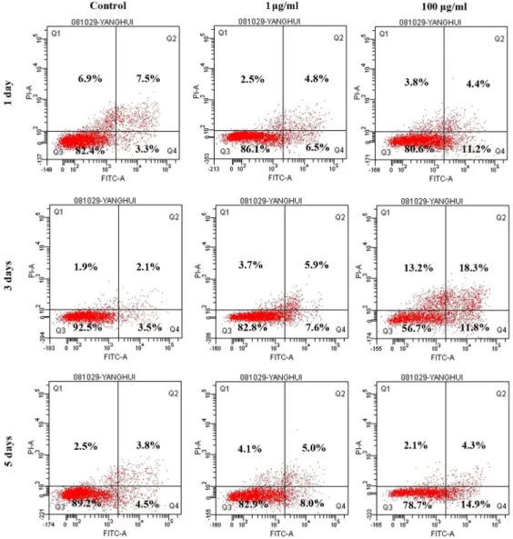
Flow cytometry
Flow cytometry dot plots (Figure 2) showed the percent-age of differently staining cells. Cells with positive staining
for annexin V-FITC, but negative staining for PI, were considered to be undergoing active apoptosis, and the lower right quadrant of each plot shows the percentage of apoptotic cells. Annexin V-FITC and PI-positive cells in the upper right quadrants revealed the late apoptotic cells or necrotic cells, and to avoid the interference of confounding factors (e.g., necrosis) these cells were excluded from the apoptosis counts. The percentages of viable cells with nega-tive staining for both annexin V-FITC and PI are presented
in the lower left quadrants. Apoptosis was enhanced with increasing concentrations of LPS at different times (Figure 2). The increasing trend of apoptosis in the 1- and 100-µg/mL groups was notable compared with control groups. The effects of stimu-lation concentration and stimustimu-lation time were also analyzed. Paired comparisons between the LPS-treated groups at each time resulted in significant differences (P < 0.05). Although most cells in the 0.01-µg/ mL groups and control groups were viable, the number of apoptotic cells increased in a dose-dependent manner with increas-ing LPS concentration. Apoptosis was greater than that of the control group in all LPS groups except for the 0.01-µg/ mL groups (Figure 3A). Apoptosis on the third and fifth days differed from that on the first day after exposure to LPS, but no significant difference was observed between that on the third and fifth days (Figure 3B). Apoptosis did not increase with stimulation time. The effect of LPS on dental pulp cell apoptosis was not time dependent.
Immunohistochemistry
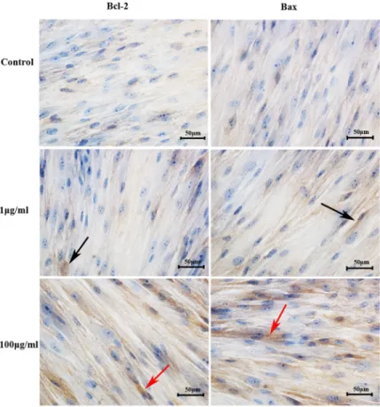
Bcl-2 and Bax immunostaining was mainly present in the cytosol and was occa-sionally observed on the nuclear membrane. According to the results of flow cytometry, pulp cells were exposed to LPS for 3 days. In the control groups (Figure 4), immunos-taining for Bcl-2 and Bax was weak, and the staining in the 0.01- and 0.1-µg/mL groups was also very weak (data not shown). In the 1-µg/mL groups (Figure 4), some light brown staining appeared, but was not very strong. However, strongly positive dark brown im-munostaining appeared in the 100-µg/mL groups (Figure 4), indicating an enhanced expression of Bcl-2 and Bax. We expected that Bcl-2 and Bax expression might differ between control groups and low LPS con-centration groups (0.01 and 0.1 µg/mL), and the expression level might be different between Bcl-2 and Bax in apoptotic cells, but our data did not demonstrate this. It may be that the detection method was not
sufficiently sensitive to differentiate small
changes in protein expression. Further stud-ies should be conducted to obtain accurate information related to the expression of the target proteins.
Figure 4. Bcl-2 and Bax immunoreactivity in dental pulp cells. Serum-starved cells were stimulated with different concentrations of lipopolysaccharide (LPS) for 3 days. The upper two panels show the basal expression level of Bcl-2 and Bax in control groups. The immunoreactivity of Bcl-2 and Bax in the 1-µg/mL groups (black arrows) was weakly positive. Increased dark brown staining indicated that the expression of both Bcl-2 and Bax was enhanced (red arrows) in cells receiving 100 µg/mL. Scale bar = 50 µm.
1032 H. Yang et al.
Discussion
Dental pulp can be invaded by bacterial LPS through dentinal tubules following caries infection (18). Apoptosis of dental pulp cells can be triggered by a wide variety of internal and external stimuli (5,6). In the present study, LPS-stimulated dental pulp cells underwent morphological and ultrastructural changes typical of apoptosis. Apoptotic bodies, a special characteristic of apoptosis, were formed after exposure to LPS, which would facilitate the elimination of unnecessary components to stabilize the internal pulp environment (24). Irritation of pulp releases growth factors that regulate pulp cell apoptosis, migration, and differentiation (25). Apoptosis of dental pulp cells plays a pivotal role when the pulp is insulted by harmful stimuli. Apoptotic cells may transmit a death signal to facilitate prompt clearance of cell debris to prevent inflammation. Dental pulp cell apoptosis induces subsequent changes in the pulp and the secretion of growth factors after the death signal, which may activate or inactivate signaling pathways that promote mesenchymal cell migration and differentiation to initiate the pulp reparative process (25). Therefore, pulp cell apoptosis could be con-sidered to be cytoprotective against bacterial infection and clearance of infected cells could be regarded as a means of restricting the spread of infection (26). Cell apoptosis in dental pulp may actively reduce pulp injury to a minimum. This may explain why pulp does not undergo irreversible changes in the presence of bacteria and their metabolites in some cases of slow, progressive caries. However, further studies are needed to confirm this conclusion.
Moreover, flow cytometry demonstrated that LPS-induced apoptosis occurs in a dose-dependent manner. This is con-sistent with the results of Hamada et al. (27) who showed that LPS induced a dose-dependent increase in the number of TUNEL-positive hepatocytes. Another study demonstrated that LPS indirectly induced apoptosis in osteoblasts and periodontal ligament cells in a dose-dependent manner (28). In our study, the rate of apoptosis differed in each treatment group after exposure to LPS. When compared with the con-trol group, a notable difference also existed except for the 0.01-µg/mL group. However, a previous study initiated by Kitamura et al. (17) found that LPS induced the proliferation of pulp cells in a dose-dependent manner. In our study, pulp cells preexposed to a low concentration (0.01 µg/mL) of LPS showed the baseline apoptosis rate, but higher concentra-tions (0.1, 1, 10, and 100 µg/mL) induced obvious apoptosis. Further studies are needed to verify the cytoprotective or cytotoxic role of LPS at concentrations lower than 0.01 µg/ mL in human dental pulp cells. Kitamura et al. (17) conducted an experiment using clonal dental pulp cells (RPC-C2A). Thus, a different biological response and sensitivity of cells exposed to LPS might have occurred because of species specificity. Our study also found that the rate of apoptosis differed at different times. The 3rd day and the 5th day rates were much higher than that on the first day, but no significant
difference was observed between the 3rd and 5th day. The results indicated that LPS-induced dental pulp cell apoptosis did not show a time-dependent effect for different exposure times, and that apoptosis did not increase constantly with stimulation time. Although apoptosis was enhanced on the 3rd day when compared with the 1st day, the evidence was not strong enough to illustrate a temporal effect of LPS in our study.
To obtain further information regarding apoptotic modu-lation, we examined the immunoreactivity of Bax and Bcl-2 in dental pulp cells after exposure to LPS. The two Bcl-2 family proteins acted as pro- and antiapoptotic modulators in cell apoptosis, respectively. Our findings indicate that both proteins were up-regulated after stimulation with high LPS concentrations. This was consistent with a previous report that showed increased expression of Bcl-2 and Bax accom-panying increased apoptosis (29). Several studies (29-31) have demonstrated that pro- and antiapoptotic members of the Bcl-2 family might interact with each other to form homodimers and heterodimers in regulating apoptosis. Our results showed that high concentrations of LPS corresponded to an increased expression of Bcl-2 and Bax. Although the exact dose-response relationship could not be inferred from the results, the tendency for LPS-induced apoptosis to be increased with LPS concentration was obvious. However, the evidence of direct involvement of Bcl-2 and Bax in dental pulp cell apoptosis in our study is insufficient, but may indicate a role for the two proteins in mediating LPS-induced dental pulp cell apoptosis and provide some clues for understanding the mechanisms of dental pulp cell apoptosis after exposure to LPS. Moreover, Bcl-2 and Bax expression was not differ-ent between control groups and low concdiffer-entration groups; it may be that immunohistochemistry could not detect the small changes in protein expression and immunoreactivity could only be observed after strong expression in groups treated with high LPS concentrations.
Interestingly, in our study, the apoptosis rate did not differ between the 0.01-µg/mL LPS group and the control group. Therefore, the lowest concentration of LPS was insufficient to induce notable dental pulp cell apoptosis. This may indicate that pathogenic material and bacteria of restricted virulence may not reach the intensity required to induce meaningful pulp repair responses such as apoptosis. Considering the limited repair capacity of pulp, acquiring information regarding the threshold level of irritation to induce biological pulp responses appears to be crucial in understanding the modulation of the pulp repair process.
Acknowledgments
We thank X.Y. Li, Y.R. Liu, and M. Zhou for assistance with confocal microscopy and flow cytometry. Research sup
-ported by the National Natural Science Foundation of China (grant #30973322), the Research Fund for Higher Education Doctoral Program in China (grant #20090181110089 and #20070610056).
References
1. Cohen JJ, Duke RC, Fadok VA, Sellins KS. Apoptosis and programmed cell death in immunity. Annu Rev Immunol
1992; 10: 267-293.
2. Kim JY, Cha YG, Cho SW, Kim EJ, Lee MJ, Lee JM, et al. Inhibition of apoptosis in early tooth development alters tooth shape and size. J Dent Res 2006; 85: 530-535.
3. Mitsiadis TA, De Bari C, About I. Apoptosis in developmental and repair-related human tooth remodeling: a view from the inside. Exp Cell Res 2008; 314: 869-877.
4. Vermelin L, Lecolle S, Septier D, Lasfargues JJ, Goldberg M. Apoptosis in human and rat dental pulp. Eur J Oral Sci
1996; 104: 547-553.
5. Mitsiadis TA, Rahiotis C. Parallels between tooth develop-ment and repair: conserved molecular mechanisms following carious and dental injury. J Dent Res 2004; 83: 896-902. 6. Tziafas D, Smith AJ, Lesot H. Designing new treatment
strategies in vital pulp therapy. J Dent 2000; 28: 77-92. 7. Haendeler J, Zeiher AM, Dimmeler S. Vitamin C and E
prevent lipopolysaccharide-induced apoptosis in human endothelial cells by modulation of Bcl-2 and Bax. Eur J Pharmacol 1996; 317: 407-411.
8. Kim KY, Shin HK, Choi JM, Hong KW. Inhibition of lipopoly-saccharide-induced apoptosis by cilostazol in human umbili-cal vein endothelial cells. J Pharmacol Exp Ther 2002; 300: 709-715.
9. Messmer UK, Briner VA, Pfeilschifter J. Tumor necrosis factor-alpha and lipopolysaccharide induce apoptotic cell death in bovine glomerular endothelial cells. Kidney Int
1999; 55: 2322-2337.
10. Cory S, Huang DC, Adams JM. The Bcl-2 family: roles in cell survival and oncogenesis. Oncogene 2003; 22: 8590-8607.
11. van Delft MF, Huang DC. How the Bcl-2 family of proteins interact to regulate apoptosis. Cell Res 2006; 16: 203-213. 12. Alikhani M, Alikhani Z, He H, Liu R, Popek BI, Graves DT. Li-popolysaccharides indirectly stimulate apoptosis and global induction of apoptotic genes in fibroblasts. J Biol Chem
2003; 278: 52901-52908.
13. Xaus J, Comalada M, Valledor AF, Lloberas J, Lopez-Soriano F, Argiles JM, et al. LPS induces apoptosis in macrophages mostly through the autocrine production of TNF-alpha. Blood
2000; 95: 3823-3831.
14. Hull C, McLean G, Wong F, Duriez PJ, Karsan A. Lipopoly-saccharide signals an endothelial apoptosis pathway through TNF receptor-associated factor 6-mediated activa-tion of c-Jun NH2-terminal kinase. J Immunol 2002; 169: 2611-2618.
15. Hachiya O, Takeda Y, Miyata H, Watanabe H, Yamashita T, Sendo F. Inhibition by bacterial lipopolysaccharide of sponta-neous and TNF-alpha-induced human neutrophil apoptosis
in vitro. Microbiol Immunol 1995; 39: 715-723.
16. Akgul C, Moulding DA, Edwards SW. Molecular control of neutrophil apoptosis. FEBS Lett 2001; 487: 318-322. 17. Kitamura C, Nishihara T, Ueno Y, Chen KK, Morotomi T,
Yano J, et al. Effects of sequential exposure to lipopolysac -charide and heat stress on dental pulp cells. J Cell Biochem
2006; 99: 797-806.
18. Matsushita K, Motani R, Sakuta T, Nagaoka S, Matsuyama T, Abeyama K, et al. Lipopolysaccharide enhances the pro-duction of vascular endothelial growth factor by human pulp cells in culture. Infect Immun 1999; 67: 1633-1639. 19. Nomiyama K, Kitamura C, Tsujisawa T, Nagayoshi M,
Mo-rotomi T, Terashita M, et al. Effects of lipopolysaccharide on newly established rat dental pulp-derived cell line with odontoblastic properties. J Endod 2007; 33: 1187-1191. 20. Botero TM, Shelburne CE, Holland GR, Hanks CT, Nor JE.
TLR4 mediates LPS-induced VEGF expression in odonto-blasts. J Endod 2006; 32: 951-955.
21. Tokuda M, Nagaoka S, Torii M. Interleukin-10 receptor ex-pression in human dental pulp cells in response to lipopoly-saccharide from Prevotella intermedia. J Endod 2003; 29: 48-50.
22. Lee JC, Yu MK, Lee R, Lee YH, Jeon JG, Lee MH, et al. Terrein reduces pulpal inflammation in human dental pulp cells. J Endod 2008; 34: 433-437.
23. Wang FM, Hu T, Tan H, Zhou XD. p38 Mitogen-activated protein kinase affects transforming growth factor-beta/Smad signaling in human dental pulp cells. Mol Cell Biochem 2006; 291: 49-54.
24. Steller H. Mechanisms and genes of cellular suicide. Sci-ence 1995; 267: 1445-1449.
25. Goldberg M, Smith AJ. Cells and extracellular matrices of dentin and pulp: a biological basis for repair and tissue engineering. Crit Rev Oral Biol Med 2004; 15: 13-27. 26. Benedict CA, Norris PS, Ware CF. To kill or be killed: viral
evasion of apoptosis. Nat Immunol 2002; 3: 1013-1018. 27. Hamada E, Nishida T, Uchiyama Y, Nakamura J, Isahara
K, Kazuo H, et al. Activation of Kupffer cells and caspase-3 involved in rat hepatocyte apoptosis induced by endotoxin.
J Hepatol 1999; 30: 807-818.
28. Thammasitboon K, Goldring SR, Boch JA. Role of mac-rophages in LPS-induced osteoblast and PDL cell apoptosis.
Bone 2006; 38: 845-852.
29. Filippovich IV, Sorokina NI, Lisbona A, Cherel M, Chatal JF. Radiation-induced apoptosis in human myeloma cell line increases BCL-2/BAX dimer formation and does not result in BAX/BAX homodimerization. Int J Cancer 2001; 92: 651-660. 30. Adams JM, Cory S. Life-or-death decisions by the Bcl-2
protein family. Trends Biochem Sci 2001; 26: 61-66. 31. Kelekar A, Thompson CB. Bcl-2-family proteins: the role of