ISSN 0100-879X
BIOMEDICAL SCIENCES
AND
CLINICAL INVESTIGATION
www.bjournal.com.br
www.bjournal.com.br
Volume 42 (10) 870-992 October 2009
Institutional Sponsors
The Brazilian Journal of Medical and Biological Research is partially financed by
Braz J Med Biol Res, October 2009, Volume 42(10) 958-962
Does hepatocellular carcinoma in non-alcoholic steatohepatitis
exist in cirrhotic and non-cirrhotic patients?
Does hepatocellular carcinoma in
non-alcoholic steatohepatitis exist
in
cirrhotic and non-cirrhotic patients?
A.L. Chagas
1, L.O.O. Kikuchi
1, C.P.M.S. Oliveira
1, D.C.P. Vezozzo
1,
E.S. Mello
2, A.C. Oliveira
1, L.C. Cella
1, P. Herman
1, T. Bachella
1,
S.H. Caldwell
3, V.A.F. Alves
2and F.J. Carrilho
11Departamento de Gastroenterologia (LIM 07/37), 2Departamento de Patologia (LIM 14),
Faculdade de Medicina, Universidade de São Paulo, São Paulo, SP, Brasil
3Department of Gastroenterology and Hepatology, University of Virginia, Charlottesville, VA, USA
Abstract
Non-alcoholic steatohepatitis (NASH) has been associated with hepatocellular carcinoma (HCC) often arising in histologically advanced disease when steatohepatitis is not active (cryptogenic cirrhosis). Our objective was to characterize patients with HCC and active, histologically defined steatohepatitis. Among 394 patients with HCC detected by ultrasound imaging over 8 years and staged by the Barcelona Clinic Liver Cancer (BCLC) criteria, we identified 7 cases (1.7%) with HCC occurring in the setting of active biopsy-proven NASH. All were negative for other liver diseases such as hepatitis C, hepatitis B, autoimmune hepatitis, Wilson disease, and hemochromatosis. The patients (4 males and 3 females, age 63 ± 13 years) were either overweight (4) or obese (3); 57% were diabetic and 28.5% had dyslipidemia. Cirrhosis was present in 6 of 7 patients, but 1 patient had well-differentiated HCC in the setting of NASH without cirrhosis (fibrosis stage 1) based on repeated liver biopsies, the absence of portal hypertension by clinical and radiographic evaluations and by direct surgical inspection. Among the cirrhotic patients, 71.4% were clinically staged as Child A and 14.2% as Child B. Tumor size ranged from 1.0 to 5.2 cm and 5 of 7 patients were classified as early stage; 46% of all nodules were hyper-echoic and 57% were <3 cm. HCC was well differentiated in 1/6 and moderately differentiated in 5/6. Alpha-fetoprotein was <100 ng/mL in all patients. HCC in patients with active steatohepatitis is often multifocal, may precede clinically advanced disease and occurs without diagnostic levels of alpha-fetoprotein. Importantly, HCC may occur in NASH in the absence of cirrhosis. More aggressive screening of NASH patients may be warranted.
Key words: Hepatocellular carcinoma; Liver cancer; Fatty liver; Steatohepatitis; Cirrhosis
Introduction
Correspondence: C.P.M.S. Oliveira, Departamento de Gastroenterologia, Faculdade de Medicina, Universidade de São Paulo, Av. Dr. Arnaldo, 455, 3º andar, Sala 3117, 01246903 São Paulo, SP, Brasil. Fax: +55-11-3069-7830. E-mail: [email protected]
Received February 20, 2009. Accepted June 29, 2009. Available online September 4, 2009.
Non-alcoholic fatty liver disease (NAFLD) is a clinical and pathological condition whose spectrum can range from steatosis to steatohepatitis and cirrhosis in patients without a history of alcohol abuse (1). Non-alcoholic steatohepatitis (NASH), the severe form of NAFLD, has emerged as a clinically important type of chronic liver disease character-ized pathologically by hepatocellular ballooning, Mallory’s
hyaline, scattered inflammation, and perisinusoidal fibrosis
(2). NASH associated with cirrhosis progresses to hepa-tocellular cancer and reoccurs post-transplantation (3,4). This disorder is now considered to be the major cause of cryptogenic cirrhosis in many regions of the world (5).
More recently, NASH has been increasingly associated with hepatocellular carcinoma (HCC), often arising in the late
stage of the disease with non-specific histology ascribed to
cryptogenic cirrhosis (6). Thus, NASH-related HCC is often thought to occur primarily in patients with advanced disease,
when hepatocellular ballooning, inflammation and steatosis
(e.g., NASH) have subsided. For example, Marrero et al. (7) studied the etiology of liver disease in 150 patients with HCC wherein NAFLD-related cryptogenic cirrhosis accounted for
at least 13% of the cases. However, fibrosis progression in
NAFLD appears to be typically slow (8,9)and the clinical features of HCC arising in earlier stages of NASH are not
well-defined, although obesity is a recognized risk factor
Hepatocellular carcinoma in nonalcoholic steatohepatitis 959
and histologically defined NASH with or without cirrhosis.
Patients and Methods
Patients
Between April 1998 and August 2006, 394 consecutive patients with HCC were evaluated at a single center (Depart-ment of Gastroenterology of Clinic Hospital of University of São Paulo School of Medicine). According to etiology,
hepatitis C virus was the most frequent in 58%, followed by hepatitis B virus in 16%, alcohol in 10% and cryptogenic
disease in 9%. We identified 7 patients (1.7%) with histo
-logically proven NASH. Six of 7 associated hepatocellular
cancers were confirmed histologically. The protocol was
approved by the Internal Review Board of the University of São Paulo (São Paulo, SP, Brazil).
Diagnostic criteria
The diagnosis of NASH was based on the following criteria:
1) a liver biopsy specimen showing steatosis (>10% of hepa
-tocytes), hepatocellular ballooning and pericellular fibrosis as
assessed by a liver pathologist with expertise in NAFLD; 2) the exclusion of liver diseases such as hepatitis B, hepatitis
C, autoimmune hepatitis, alpha1-antitrypsin deficiency, Wilson
disease, and hemochromatosis; 3) the exclusion of patients who had a >100 g/week alcohol intake determined by a detailed personal history, questioning of family members, and an inves-tigation of previous medical records, and 4) the exclusion of patients with steatohepatitis accompanying other liver diseases, or systemic diseases other than obesity, hyperlipidemia, and diabetes or with an intake of hepatotoxic drugs or lipid-lowering
agents. HCC diagnostic criteria included coincident findings by
at least two imaging techniques showing characteristic features in a focal hepatic lesion >2 cm, with arterial hypervasculariza-tion (Barcelona Clinic Liver Cancer (BCLC) criteria) (12)and
histologically confirmed in 6 of 7 patients.
Laboratory assays
Laboratory examinations included tests for hepatitis B surface antigen, hepatitis B surface antibody (anti-HBs), hepatitis B core antibody (anti-HBc) by enzyme immunoas-say (Dinabot, Japan), HCV antibody by third-generation en-zyme immunoassay (Ortho Diagnostics, Japan), HCVRNA by the polymerase chain reaction (Amplicor HCV, Roche Diagnostic Systems, Japan), anti-nuclear antibody and
anti-smooth muscle antibody by immunofluorescence on
Hep 2 cells. Tests for alpha-fetoprotein (AFP) were done by EIA. Aminotransferase studies and other routine tests were performed by standard methods using automated techniques (Modular P800, Roche/Hitachi, Switzerland).
Histology
The liver tissue was fixed in 4% formaldehyde and
processed for hematoxylin-eosin, Masson trichrome and picrosyrius red stains, Gomori’s silver impregnation for
extracellular matrix and Perls staining for pigments. All specimens were scored blindly for individual histological parameters by a liver pathologist with expertise in NAFLD: macro- and microvacuolar fatty change, zonal
distribu-tion, foci of necrosis, portal and perivenular fibrosis, and inflammatory and fibrotic infiltrate with zonal distribution
and the composite NASH activity score were calculated (13).Fibrosis was assessed using a 4-grade scale. In this
scheme, F1 denotes foci of perivenular/pericellular fibrosis in zone 3, F2 represents the changes defining F1 plus focal or extensive periportal fibrosis, F3 denotes bridging
fibrosis (focal or widespread), and F4 indicates the pres
-ence of cirrhosis. The grade of tumor differentiation was assessed as best differentiated (grade I), well differentiated (grade II), moderately differentiated (grade III), or poorly differentiated (grade IV), according to the Edmonson &
Steiner classification.
Results
There were 4 males and 3 females with a mean age of
63 ± 13.9 years. Four patients (5%) were overweight (BMI
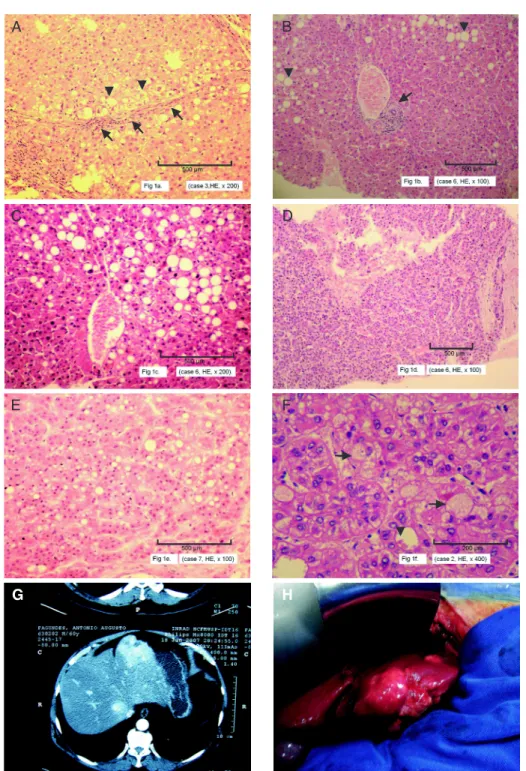
25-29.9 kg/m2) and 3 were obese (BMI ≥30) (43%); 57% had type II diabetes and 28.5% had dyslipidemia. Cirrhosis was
present in 6 of the 7 patients (Figure 1A) and 1 had HCC and NASH without cirrhosis, with a liver biopsy showing
only perivenular and pericellular fibrosis (stage 1; Figure 1B,C). Clinical and radiologic parameters confirmed the
absence of evidence of portal hypertension in this patient
(see details below). Among the cirrhotic patients, 71.4% were clinically staged as Child A and 14.2% as Child B.
Lesions were visualized by ultrasound in all patients. These data are summarized in Table 1.
Four patients had only one HCC nodule, 1 patient had 2 tumors, 1 patient had 3 tumors, and 1 had 4 tumors (Table 1). Tumor size ranged from 1.0 to 5.2 cm and 8 of
14 nodules (57%) were <3 cm. Most lesions (46%) were
hyper-echoic by ultrasound. The HCC was well differentiated
in 2/6 (33.3%; Figure 1D,E), moderately differentiated in 4/6 (66.6%; Figure 1F) and poorly differentiated in none. An interesting finding was the presence of histological features
of NASH in 4 of 7 cases. In several neoplastic cells, we observed focal to moderate steatosis (Figure 1E,F), balloon-ing and Mallory bodies (Figure 1F). In 2 cases, we found also peliosis. These intracellular lesions were apparently
associated with milder histological grades. AFP was <100
ng/mL in all patients (Table 1).
HCC in NAFLD in the absence of cirrhosis
Hepatocellular carcinoma in nonalcoholic steatohepatitis 961
liver disease. Using ultrasound, computed tomography, and magnetic resonance imaging, we detected two hypoechoic tumors measuring 52 mm in the greatest dimension in segment II and 10 mm in segment V, and steatosis in the background, without signs of cirrhosis or portal hypertension (Figure 1G,H). An ultrasound-guided biopsy from the largest nodule showed a grade-I trabecular HCC (Figure 1D). The non-cancerous areas showed NASH stage 1, characterized
by minimal perivenular fibrosis, moderate steatosis with
mild hepatocellular ballooning and mild necroinflamma
-tion (Figure 1C). Portal tracts were normal-sized, without
fibrous septa (Figure 1B). Therefore, surgical resection (left
hepatectomy) for the segment 2 lesion was indicated and at the time of surgery radioablation of the tumor in segment V was planned. However, since intra-operatory ultrasound showed 12 nodules, both in the right and in left hepatic lobes, it was decided not to do any hepatic resection or radioablation. The macroscopic liver aspect on surgery revealed only steatosis, without signs of cirrhosis or portal hypertension. The patient was therefore intra-operatively submitted again to a liver and tumor biopsy.
The histological findings confirmed a diagnosis of grade I
HCC and underlying NASH stage 1, without cirrhosis. The pa-tient was treated with transcatheter arterial chemoembolization. He is alive without symptoms, 24 months after diagnosis.
Discussion
Hepatocellular carcinoma is a well-recognized end-point of the progression of NASH although the exact risk remains
to be defined. In recent years, NASH has been proposed
as an important cause of HCC especially in industrialized
countries where obesity rates and associated fatty liver disease have become a common problem. However, most cases have been found in the setting of cryptogenic cirrhosis thought to be due to progression of NASH. In the present
paper, we describe 7 cases of HCC in histologically defined
NASH patients including one case without cirrhosis. The clinical features of NASH patients with HCC studied herein differ in some respects from those of HCC patients with cirrhosis associated with HCV or HBV infection. In the series of 93 cirrhotic patients who developed HCC during the screening program in our group (Campos-Cella LT, Paranagua-Vezozzo DC, Farias AQ, Ono-Nita SK, Matielo CH, Alves VAF, et al., unpublished data), we found that 57 years was the average age of patients with HCC. In the present study of NASH patients, the mean age was considerably higher with an average of 63 years, similar to a previous study reporting later HCC development in patients with NASH (14).
The most important factors for the development of HCC in most literature reports were hepatitis B and C infections. In the present series, viruses were ruled out in patients
with a background of overweight in all cases, 57% of the
patients had non-insulin-dependent diabetes mellitus and
28.5% had dyslipidemia. Obesity itself is a recognized risk
factor for both NASH and HCC (10) and isolated cases of HCC arising from non-cirrhotic NAFLD have been reported (11). In our series, there was 1 patient of 7 with non-cirrhotic NASH who was obese.
The histological features of NASH complicated by HCC were somewhat different from HCC associated with other cirrhotic causes. The livers were cirrhotic in 6 patients and non-cirrhotic in 1 patient and the histological characteristics
Table 1. Demographic, clinical, biochemical, and histological parameters of the 7 patients studied.
1 2 3 4 5 6 7
Gender F M M M F M F
Age (years) 59 71 61 77 35 65 73
Diabetes mellitus Y Y N N Y N Y
Overweight Y Y Y Y Y Y Y
Dyslipidemia N Y N N N Y N
Cirrhosis Y Y Y Y Y N Y
Child-Pugh A6 A5 A5 A5 B9 - A6
AFP (ng/mL) 6 10 21 10 3 7 17
HCC Rx TACE Resection PEI + TX Resection TACE TACE PEI
HCC differentiation NA GII GII GIII NA GI GI
Number of nodules 1 1 3 1 4 2 1
Echo pattern High High High NA Low Mixed High
HCC size (mm) 33 34 30 43 28 52 33
Stage (BCLC) Early Early Early Early Intermediate Intermediate Early
of HCC showed a predominance of well to moderately differ-entiated HCC, similar to previously reported data regarding NASH-related HCC (15). No case of undifferentiated HCC was detected. Intriguingly, morphologic features of NASH such as fatty change, ballooning and Mallory bodies were observed in neoplastic hepatocytes, a fact that must be further studied in prospective series. All patients studied herein had been selected for presenting histological fea-tures of non-alcoholic steatohepatitis in liver parenchyma distant from the tumor.
The pathogenesis of HCC in NASH remains uncer-tain, although cirrhosis itself may be seen as a promoter
of neoplastic transformation. However, the finding of one case of HCC in the absence of relevant fibrosis or cirrhosis
shows that cirrhosis is not mandatory for HCC appearance in NASH patients. The mechanisms of pathogenesis of
HCC in the non-fibrotic liver have not been elucidated.
NASH-associated insulin resistance causes inhibition of hepatic mitochondrial fatty acid oxidation and increased intracellular fatty acids may lead to oxidative DNA damage by stimulating microsomal peroxidases.
Another interesting aspect differed from the results
reported by Iannaccone et al. (16), i.e., most patients were
classified as early stage according to the BCLC staging
system and most of them were uninodular and smaller than 5 cm. This discrepancy may be due to the fact that, in our service, patients are submitted to screening programs with serum AFP and ultrasound every 6-12 months. The non-cirrhotic patient discovered a multinodular HCC in a routine ultrasound. The fact that, although multinodular, the tumor was so well-differentiated also requires further study to search for a possible carcinogenic pathway for this intriguing case.
Cancer in our NASH patients preceded clinically ad-vanced disease, occurred with non-diagnostic levels of AFP and can be diagnosed in the early stage in a screen-ing program. Moreover, the association of NASH with HCC is not limited to patients with NASH-related cirrhosis. We observed HCC in 1 of 7 patients without cirrhosis by multiple parameters including repeated biopsy, radiographic imag-ing and direct surgical inspection. Further investigation of these relationships is warranted to determine relative risk and the possible need for more aggressive screening in NASH patients.
References
1. Clark JM. The epidemiology of nonalcoholic fatty liver disease in adults. J Clin Gastroenterol 2006; 40 (Suppl 1): S5-S10.
2. Brunt EM. Nonalcoholic steatohepatitis. Semin Liver Dis 2004; 24: 3-20.
3. Bugianesi E, Leone N, Vanni E, Marchesini G, Brunello F, Carucci P, et al. Expanding the natural history of nonalco-holic steatohepatitis: from cryptogenic cirrhosis to hepato-cellular carcinoma. Gastroenterology 2002; 123: 134-140. 4. Ong J, Younossi ZM, Reddy V, Price LL, Gramlich T, Mayes
J, et al. Cryptogenic cirrhosis and posttransplantation nonal-coholic fatty liver disease. Liver Transpl 2001; 7: 797-801. 5. Caldwell SH, Oelsner DH, Iezzoni JC, Hespenheide EE,
Battle EH, Driscoll CJ. Cryptogenic cirrhosis: clinical charac-terization and risk factors for underlying disease. Hepatology 1999; 29: 664-669.
6. Bugianesi E, Vanni E, Marchesini G. NASH and the risk of cirrhosis and hepatocellular carcinoma in type 2 diabetes. Curr Diab Rep 2007; 7: 175-180.
7. Marrero JA, Fontana RJ, Su GL, Conjeevaram HS, Emick DM, Lok AS. NAFLD may be a common underlying liver dis-NAFLD may be a common underlying liver dis-ease in patients with hepatocellular carcinoma in the United States. Hepatology 2002; 36: 1349-1354.
8. Dam-Larsen S, Franzmann M, Andersen IB, Christoffersen P, Jensen LB, Sorensen TI, et al. Long term prognosis of fatty liver: risk of chronic liver disease and death. Gut 2004; 53: 750-755.
9. Adams LA, Sanderson S, Lindor KD, Angulo P. The histologi-cal course of nonalcoholic fatty liver disease: a longitudinal study of 103 patients with sequential liver biopsies. J Hepatol
2005; 42: 132-138.
10. Calle EE, Rodriguez C, Walker-Thurmond K, Thun MJ. Over-weight, obesity, and mortality from cancer in a prospectively studied cohort of U.S. adults. N Engl J Med 2003; 348: 1625-1638.
11. Cuadrado A, Orive A, Garcia-Suarez C, Dominguez A, Fernandez-Escalante JC, Crespo J, et al. Non-alcoholic steatohepatitis (NASH) and hepatocellular carcinoma. Obes Surg 2005; 15: 442-446.
12. Bruix J, Sherman M, Llovet JM, Beaugrand M, Lencioni R, Burroughs AK, et al. Clinical management of hepatocellular carcinoma. Conclusions of the Barcelona-2000 EASL con-ference. European Association for the Study of the Liver. J Hepatol 2001; 35: 421-430.
13. Kleiner DE, Brunt EM, Van Natta M, Behling C, Contos MJ, Cummings OW, et al. Design and validation of a histological scoring system for nonalcoholic fatty liver disease. Hepatol-ogy 2005; 41: 1313-1321.
14. Hai S, Kubo S, Shuto T, Tanaka H, Takemura S, Yamamoto T, et al. Hepatocellular carcinoma arising from nonalcoholic steatohepatitis: report of two cases. Surg Today 2006; 36: 390-394.
15. Shimada M, Hashimoto E, Taniai M, Hasegawa K, Okuda H, Hayashi N, et al. Hepatocellular carcinoma in patients with non-alcoholic steatohepatitis. J Hepatol 2002; 37: 154-160. 16. Iannaccone R, Piacentini F, Murakami T, Paradis V, Belghiti