Thermodynamic evaluation and
modeling of proton and water exchange
associated with benzamidine and
berenil binding to ß-trypsin
1Centro de Desenvolvimento da Tecnologia Nuclear,
2Departamento de Bioquímica e Imunologia, Instituto de Ciências Biológicas, 3NEQUIM, Departamento de Química, Instituto de Ciências Exatas,
Universidade Federal de Minas Gerais, Belo Horizonte, MG, Brasil M.T. Pereira1,2,
J.M. Silva-Alves2,
A. Martins-José3,
J.C.D. Lopes3
and M.M. Santoro2
Abstract
Serine-proteases are involved in vital processes in virtually all species. They are important targets for researchers studying the relationships between protein structure and activity, for the rational design of new pharmaceuticals. Trypsin was used as a model to assess a possible differential contribution of hydration water to the binding of two synthetic inhibitors. Thermodynamic parameters for the association of bovine ß-trypsin (homogeneous material, observed 23,294.4 ± 0.2 Da, theoretical 23,292.5 Da) with the inhibitors benzamidine and berenil at pH 8.0, 25ºC and with 25 mM CaCl2, were determined using
isothermal titration calorimetry and the osmotic stress method. The association constant for berenil was about 12 times higher compared to the one for benzamidine (binding constants are K = 596,599 ± 25,057 and 49,513 ± 2,732 M-1, respectively; the number of binding
sites is the same for both ligands, N = 0.99 ± 0.05). Apparently the driving force responsible for this large difference of affinity is not due to hydrophobic interactions because the variation in heat capacity (∆Cp), a characteristic signature of these interactions, was similar in both systems tested (-464.7 ± 23.9 and -477.1 ± 86.8 J K-1 mol-1 for
berenil and benzamidine, respectively). The results also indicated that the enzyme has a net gain of about 21 water molecules regardless of the inhibitor tested. It was shown that the difference in affinity could be due to a larger number of interactions between berenil and the enzyme based on computational modeling. The data support the view that pharmaceuticals derived from benzamidine that enable hydrogen bond formation outside the catalytic binding pocket of ß-trypsin may result in more effective inhibitors.
Correspondence
M.M. Santoro
Departamento de Bioquímica e Imunologia, UFMG Av. Antonio Carlos, 6627 31270-901 Belo Horizonte, MG Brasil
E-mail: [email protected]
Received September 22, 2004 Accepted July 28, 2005
Key words
•Benzamidine •Berenil •Calorimetry •Protein modeling •Trypsin
•Osmotic stress
Introduction
The objectives of the study of the interac-tions of proteins with ligands are to increase our understanding of their function, their importance in physiological processes, the
infor-mation about the relationship between pro-tein structure and activity has been obtained in X-ray crystallography studies on many proteins and with modern bioinformatic tech-niques (1). Complex molecular organiza-tions can now be studied at the atomic level. The notion that proteins craft well-defined binding sites that are custom-tailored to their ligand remains central to analyses of bio-logical recognition and drug discovery (2). Nevertheless,the contact between a protein and its coupling with any other co-solute cannot be explained based on random steric interactions. Different forms of electrostatic forces necessarily act in intermolecular or interatomic interactions (3), reflecting on the catalytic efficacy of an enzyme or the specificity of any protein.
ß-Trypsin was chosen for the present study due to its importance as a protease model for the serine-protease class. Proteins of this class play an important role in physi-ological and pathphysi-ological processes (4). Fur-thermore, trypsin is reasonably stable and relatively easy to obtain and purify. It was considered to be an exclusively digestive enzyme expressed in the pancreas (5), where it is produced as the zymogen trypsinogen. Four trypsinogen isoforms are known so far: trypsinogen 1 or cationic trypsinogen, tryp-sinogen 2 or anionic tryptryp-sinogen, gen 3 or mesotrypsinogen (6), and trypsino-gen 4, identified in brain cells (7). Several diseases are associated with deregulation of the cationic form, especially pancreatitis (8). Recent studies have shown thattrypsin is widely expressed at low levels in various non-pancreaticepithelial tissues, vascular endothelial cells, leukocytes, andneurons, and is overexpressed in some kinds of can-cer cellsand tissues (9,10).
Cationic pancreatic trypsinogen origi-nates the ß-trypsin isoform after a hydrolysis that liberates the first six residues of the peptide chain. Additional cleavages produce the isoforms ψ, α, γ, δ, and others with decreasing amidase activity (11). This
au-tolytic process can regulate the activity of the enzyme which is also exerted through natural inhibitors such as bovine pancreatic trypsin inhibitor and several others described in the literature. After zymogen cleavage, the active ß-trypsin form acquires higher plasticity, maintaining the spatial distribu-tion of the several segments practically in-tact (12). Differential scanning calorimetry characterization at acid pH plus the pH titra-tion of native and unfolded forms allowed the estimation of the stability of this enzyme at acid and neutral pH (13).
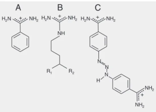
amino acids that represent the primary speci-ficity of this enzyme (23). The results ob-tained for berenil are compared to those for benzamidine. The involvement of water molecules and protons associated with the binding of these inhibitors is also described.
Material and Methods
Purification of ß-trypsin
A commercial bovine trypsin (Sigma®,
St. Louis, MO, USA) was submitted to chro-matography (730 ml on a 50-cm high col-umn, 4 cm in inner diameter) with SE-Sepha-dex-C50 and eluted with 50 mM sodium formiate. The pH was adjusted to 3.50 with formic acid. The enzyme was eluted with a linear increasing NaCl gradient (0.1 to 0.3 M), containing 25 mM CaCl2 and 1 mM
benzamidine. The active fractions contain-ing ß-trypsin were identified (24) and com-bined. The pool was lyophilized, resuspended in 1 mM HCl, dialyzed for 4 h against the same solution, and for another 2 h in milliQ water at 4ºC in both cases. All low-molecu-lar weight components were thus eliminated from the pooled effluent. The pooled efflu-ent was lyophilized once more and 2.5- to 3.0-mg aliquots of the enzyme were stored at -10ºC until use. When necessary, the frac-tion was dissolved in the desired buffer in an ice bath immediately before each experi-ment, in volumes reaching final concentra-tions of about 1.0 to 3.0 mg/ml. Concentra-tions were determined by absorbance meas-urements at 280 nm with the molar extinc-tion coefficient of 37,000 M-1 cm-1 (25).
Ti-tration of active sites were performed with p-nitrophenyl-p-guanidinobenzoate-HCl (26). One sample was then submitted to mass spectrometry to confirm ß-trypsin homoge-neity.
Microcalorimetry
Titration of enzyme-inhibitor interactions
was carried out with an isothermal titration calorimeter (VP-ITC, MicroCal LLC, North-ampton, MA, USA) (usable cell volume of 1.42 ml), using Pipes, HEPES, tricine and Tris buffers (50 mM, with 25 mM CaCl2, pH
8.0). Benzamidine or berenil concentrations were determined during the experiment by the injection and data acquisition procedure according to the equipment manual. Briefly: [L] >30[Enz]; an initial injection of 1 µl was discarded, and 20 to 30 injections of 3-5 µl resulted in the complete saturation of the enzyme binding site. The data were ana-lyzed by fitting a single-site binding iso-therm (27). After a single experiment at controlled temperature, the following pa-rameters were determined: the reaction equi-librium constant and consequently the varia-tion of Gibbs energy (∆Gbind), the
stoichio-metric relationship between the reagent spe-cies, or the number of binding sites concern-ing the association between proteins and ligands, and the enthalpy variation of the association (∆Hbind). The entropy variation
of the association (∆Sbind) was then
calcu-lated.
Osmotic stress
Osmotic stress experiments were carried out with ß-trypsin and the inhibitors benza-midine and berenil (50 mM Tris, with 25 mM CaCl2, pH 8.0, at 25ºC). Two sets of
isothermal titration calorimetry experiments were performed for each inhibitor with con-centrations of glucose or glycine ranging
from 0.25 to 1.00 osm, here used as compat-ible osmolites.
Docking and minimization
The complexes between ß-trypsin and the ligands berenil, p-aminobenzamidine and benzamidine were formed by replacing the benzamidine contained in the crystallo-graphic structure deposited in the Protein Data Bank (<http://www.rscb.org> PDB ID 1CE5), using the superimposal procedure of the INSIGHTII program (MSI). The ligand charges were calculated by the MNDO method from the MOPAC package (28). The complexes thus constructed were studied with the AutoDock program, version 3.0 (29), using a method based on the Genetic Algorithm to search for the best position for the ligands at the active site of ß-trypsin. One hundred genetic algorithm runs were used, with a population of 50 individuals, a gene mutation rate of 0.02 and crossover rates of 0.8. The affinity grid was centered at Ser195 with dimensions of 48 x 48 x 48 Å and
a grid spacing of 0.5 Å, including the cata-lytic site and its surroundings (16). The most stable conformation according to the dock-ing energy was transferred to the SYBYL program (30) in which optimization of the geometry of the complexes and of the iso-lated ligands and trypsin was performed. The 4.0 value was used for the distance-dependent dielectric constant, with a cutoff of 14.0 Å.
Results and Discussion
The participation of water molecules not as a solvent but as an adjuvant in protein action has deserved special attention in the last few years (31). The degree of hydration seems to define states of higher or lower structural flexibility for the protein (17). It was proposed that about 300 water mol-ecules are associated with trypsin (32). A recent study from our laboratory (33)
dem-onstrated, on the basis of the variation of the Stokes radius, the occurrence of an interme-diate state between native and denatured forms, a molten globule. It was estimated that 770 associated water molecules occur if the variation in the volume of the protein is due only to included/excluded water mol-ecules.
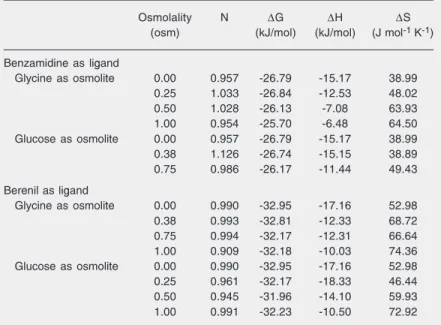
Titrations of the enzyme with the ligands benzamidine and berenil were performed on purified ß-trypsin. A typical isotherm of these titrations is shown in Figure 2. ∆G, ∆H, ∆S and the stoichiometric relationships for the experiments in Tris buffer are shown in Table 1. These results refer to 0.0 osm (without any addition of co-solute or osmolite). The re-sults for ∆G in the titration with benzami-dine confirm literature data regarding this parameter (23,25,34). It is noteworthy that the value of the association constant for berenil was 12 times higher than the con-stant for benzamidine.
Some energetic contribution (absorbed or released heat) specific for the ionization/ association process may contribute to the effects of the enzyme-ligand association it-self. Such a contribution, or dependence of the observed result, can be a consequence of association/dissociation processes of ioniz-able groups in the enzyme, exchanged with the buffer. The variation in the protonation state of the residues involved in the associa-tion process can be obtained from the num-ber of protons exchanged between the en-zyme and the buffer by the linked Wyman equation (19,35): ∆Hobs = ∆Hbind + nH+ x ∆Hdis, where ∆Hobs is the value measured in
the experiment, ∆Hbind is the enthalpy value
of the enzyme-inhibitor association itself, nH+ corresponds to the number of protons
exchanged, and ∆Hdis corresponds to the
values of the variation in the enthalpy of the ionization of the buffers. ∆Hbind and nH+ can
be calculated from the intercept and the slope of the curve in the graphic representation of
∆Hobs versus ∆Hdis. The titrations were
wide range of enthalpy of the proton disso-ciation (∆Hdis) interval: Pipes (∆Hdis = 11.45
kJ/mol); HEPES (∆Hdis = 21.01 kJ/mol);
tricine (∆Hdis = 31.96 kJ/mol), and Tris (∆Hdis
= 47.53 kJ/mol) (36). For the experiments (graph not shown) with benzamidine, we found that ∆Hbind = -16.78 ± 0.80 and nH+ =
0.037 ± 0.026 kJ/mol, and for the experi-ments with berenil ∆Hbind = -18.20 ± 3.78
and nH+ = 0.007 ± 0.12 kJ/mol. These results
indicate that there is no association or disso-ciation of acidic groups during binding of the two ligands.
The results shown in Table 1 (lines corre-sponding to 0.00 osm) indicate that there is a favorable enthalpic and entropic contribu-tion to the formacontribu-tion of the ß-trypsin-inhib-itor complex. The enthalpic contribution can be analyzed on the basis of the equation δ∆H = ∆Cp x δT which, after integration, be-comes: ∆H = ∆Cp x T + one constant, for
∆Cp constant in the temperature interval studied. The calculated values for the change in the heat capacity for the ß-trypsin-inhibi-tor associations in Tris buffer are (graph not shown): ∆Cp = -477.1 ± 86.8 J K-1 mol-1 for
benzamidine, and ∆Cp = -464.7 ± 23.9 J K-1
mol-1 for berenil. It should be observed that
the value of ∆S obtained in the absence of the osmolite (Table 1) is about 10 to 20 times lower than these ∆Cp values.
As a consequence of the relationships of
∆H, ∆S and T with ∆G, and of ∆H with T,
δ∆H/δT = ∆Cp and δ(TAS)/δT = ∆Cp + ∆S.
When ∆S becomes negligible with respect to
∆Cp the dependence of ∆H and T∆S be-comes a constant factor equal to ∆Cp, and
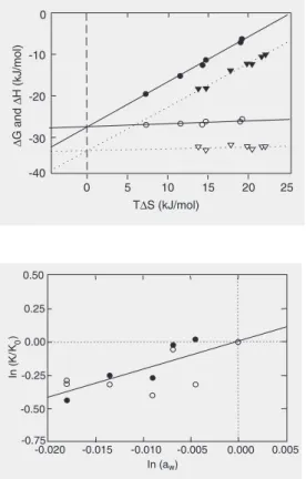
∆G becomes relatively independent of T. This is shown both by the benzamidine and berenil associations with ß-trypsin, as can be observed in Figure 3. The temperatures at which the ∆H and ∆S parameters become zero, TH and TS respectively, were
calcu-lated from the graphs: TH = -3.6ºC and TS =
62.3ºC for benzamidine, and TH = -11.9ºC
and TS = 58.4ºC for berenil.
The well-known effect of water
mol-Figure 2. Isothermal titration calorimetry of ß-trypsin with berenil. The data in this figure were obtained in 50 mM Tris buf-fer, with 25 mM CaCl2, atpH 8.0, 25oC. A, The lower trace shows the raw data for the titra-tion and the upper trace shows the heat of dilution of the berenil.
B, Net isotherm of the titration obtained by subtracting the gross value of heat at each titra-tion point by the linear regres-sion of the dilution heat data of berenil.
Table 1. Energetic parameters of binding of the inhibitors benzamidine and berenil to ß-trypsin.
Osmolality N ∆G ∆H ∆S
(osm) (kJ/mol) (kJ/mol) (J mol-1 K-1)
Benzamidine as ligand
Glycine as osmolite 0.00 0.957 -26.79 -15.17 38.99
0.25 1.033 -26.84 -12.53 48.02
0.50 1.028 -26.13 -7.08 63.93
1.00 0.954 -25.70 -6.48 64.50
Glucose as osmolite 0.00 0.957 -26.79 -15.17 38.99
0.38 1.126 -26.74 -15.15 38.89
0.75 0.986 -26.17 -11.44 49.43
Berenil as ligand
Glycine as osmolite 0.00 0.990 -32.95 -17.16 52.98
0.38 0.993 -32.81 -12.33 68.72
0.75 0.994 -32.17 -12.31 66.64
1.00 0.909 -32.18 -10.03 74.36
Glucose as osmolite 0.00 0.990 -32.95 -17.16 52.98
0.25 0.961 -32.17 -18.33 46.44
0.50 0.945 -31.96 -14.10 59.93
1.00 0.991 -32.23 -10.50 72.92
ecules on the structure (15,16) and function of proteins, influencing the macromolecular dynamics, allows additional insight into the energetic aspects involved in the enzyme-inhibitor interaction. This was done using the osmotic stress method (19-21). With the Gibbs-Duhem equation it can be demon-strated that, for a protein at infinite dilution (21) d∆G = -∆Newdµw. In this equation ∆New
is the variation in the number of water mol-ecules in the final macromolecular state mi-nus the number in the initial state, ew is the deficit or excess of water molecules, and µw
is the chemical potential of the water or another equivalent parameter. The equation can be written as dlnK/dlnaw = ∆New = NEIw
-(NE w + N
I
w). In this equation N EI
w is the number
of water molecules associated with the com-plex formed and NE
w and NwI are the number
of water molecules associated with the en-zyme and with the inhibitor in the bath, respectively. The results obtained from the osmotic stress analyses are shown in Table
1. The utilization of glucose and glycine as osmolites follows the strategy of Colombo and Bonilla-Rodriguez (37), in order to ex-clude a possible interference due to a change in the dielectric constant of the solution, since the two osmolites have opposite ef-fects on this property. The data for each inhibitor as a function of the chemical poten-tial of water were plotted and resulted in the same slopes within the experimental error. Consequently they resulted in an equal num-ber of water molecules, regardless of the osmolite and the inhibitor tested. Because of this similarity and for a better statistical align-ment of the results the values for the equilib-rium constants were normalized and plotted; only one regression was calculated, as shown in Figure 4. From the slope of this curve the number of water molecules taken up from the bulk water during binding was estimated at 21.1 ± 3.4. Surfaces topologically distant from the catalytic site (16) are considered to be active participants in this inclusion. Ac-cording to Parsegian et al. (20), “this result is characteristic of reactions with changes in the number of waters that are sequestered in sterically inaccessible pockets or cavities”. Enzymes are known to be more stable when substrates and competitive inhibitors are pres-ent, and a tightening of the enzyme is linked to the formation of the corresponding ad-ducts. In fact, the result observed, inclusion, involves the participation of distant surfaces in the process of inhibitor binding to the catalytic pocket per se since the exclusion of at least five water molecules would be ex-pected to occur. According to computational modeling (38), the volume occupied by the side chain of arginine corresponds to the volume of six water molecules, while the corresponding volume for the lysine side chain is equivalent to five water molecules; in the latter case one water molecule stays in the catalytic pocket to compensate for a smaller side chain (16).
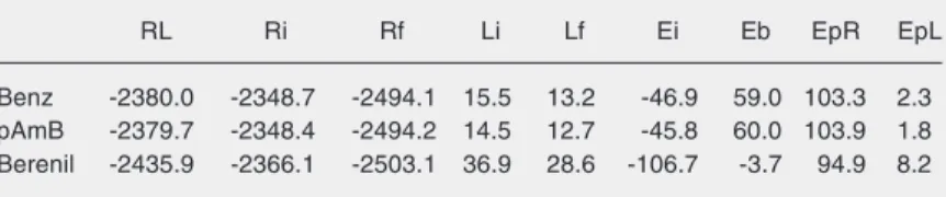
Theoretical studies of the binding of these two inhibitors to the enzyme were carried
out hoping to use modeling data to explain the difference in the affinities of benzami-dine and berenil for ß-trypsin. Furthermore, p-aminobenzamidine was included in the analysis because some experimental data are available in the literature (25,34), and be-cause its structure is related to those of the two ligands under study. Using the AutoDock and the SYBYL programs, the complexes formed between ß-trypsin (R) and the men-tioned inhibitors (L) were optimized (RL); the energies of the enzyme and the ligands isolated in the pharmacophoric conforma-tion were evaluated (Ri and Li, respectively) as also were the energies after optimization of the geometries (Rf and Lf, respectively). These results are shown in Table 2, which also shows the values for energies of pertur-bation (Ep), interaction (Ei), and binding (Eb), calculated according to the following equations: Ep = EpR + EpL = (Rf - Ri) + (Lf - Li); Ei = RL - Ri - Li and Eb = Ei + Ep.
The calculated energies (Ep, Ei and Eb) for benzamidine and p-aminobenzamidine are reasonably coincident, which is the re-sult of the structural similarity of the two compounds. Among the calculated binding energies, only berenil had a negative value. This is due to the kind of perturbations caused by the other two ligands at the binding site of trypsin (EpR), according to the force field applied. The calculated enegies reflect only the intermolecular interaction between the ligand and enzyme, after optimization with the SYBYL package. It is also worth noting that berenil exhibits the highest interaction energy. The fact that the benzamidine inter-action energy was slightly more favorable than that of p-aminobenzamidine can be ex-plained on the dispersion of the positive charge of the amidine group of the latter. This occurs because the amino group, an excellent electron donor, decreases the posi-tive charge of the amidine group, thus reduc-ing the interaction of this group with the carboxylate of Asp177 present at the bottom
of the pocket of the S1 site.
However, calorimetric determinations contradict this result since the measured af-finity of p-aminobenzamidine is about 4 times higher than that of benzamidine (25,34). This discrepancy may be due to the method-ology used in the calculations that does not take into account the effect of differential solvation of the complexes or of the isolated ligands and receptor. The variation in the heat capacity, ∆Cp, for both inhibitors sug-gests that the contribution of the hydropho-bic effect should be negative for both com-pounds (39). ∆Cp for benzamidine binding is -469.4 ± 51.4 J mol-1 K-1 (this work) and
-519 J mol-1 K-1 for p-aminobenzamidine
(25). The solvation and desolvation effects of the ligands, receptor and the complexes, i.e., the changes in free energies to transfer the ligands from the bulky solution to the binding site, are exergonic. The fact that the positive charge of the amidine is more dis-persed in the p-aminobenzamidine group should decrease the interaction force be-tween this inhibitor and the water molecules. Consequently, the energetic cost related to the release of the water molecules around the ligand molecule, necessary to the bind-ing, will be smaller for p-aminobenzamidine compared to benzamidine. Thus, p-ami-nobenzamidine is expected to present a more favorable binding constant than benzami-dine, although the specific interaction inside the binding site is weakened by charge dis-persion.
Table 2. Theoretical energetic components involved in the binding of ß-trypsin to inhibitors (kJ/mol).
RL Ri Rf Li Lf Ei Eb EpR EpL
Benz -2380.0 -2348.7 -2494.1 15.5 13.2 -46.9 59.0 103.3 2.3
pAmB -2379.7 -2348.4 -2494.2 14.5 12.7 -45.8 60.0 103.9 1.8
Berenil -2435.9 -2366.1 -2503.1 36.9 28.6 -106.7 -3.7 94.9 8.2
The effects of charge dispersion on free energy of binding are two-fold and opposite: at the same time they weaken the interaction between the amidine group and the aspartic residue at the bottom of the pocket, that hinders the binding interaction, but they also weaken the interaction between the amidine group and solvent molecules, which favors the binding interaction. The free energy of binding from isothermal titration calorim-etry studies indicates that the second effect should be more pronounced. However, these effects need to be investigated by computing solvent accessible surface areas and calcu-lating free solvation energy in order to gain
some insight into the solvation and desolva-tion effects.
The participation of hydrogen bonds is another important factor in the stabilization of inhibitor-enzyme complexes. The present study evaluates the possibilities of this type of interaction among the molecules analyzed. In the simulations we observed that p-ami-nobenzamidine can form an additional hy-drogen bond with the OH group of the Ser177
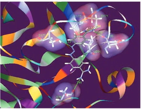
residue. Figure 5 shows the arrangement of berenil in the pocket of the catalytic S1 site of ß-trypsin after optimization and minimi-zation of energies with the SYBYL pro-gram. Berenil shows several interactions with trypsin, some in the interior of the pocket with residues Asp171 and Ser195, and also
with the Ser177, His40 and Ser78 residues
lo-cated outside the catalytic site. Berenil shows a much more negative interaction energy than the two benzamidines and, despite psenting several contact points with the re-ceptor, it shows a less positive perturbation energy. These results explain quantitatively the calorimetric data obtained and, most im-portantly, they show that benzamidine and berenil are promising molecules as starting points for the synthesis of chemical deriva-tives that can act as improved ligands or inhibitors for trypsin and other related serine proteases.Aditionally, many physiologically important serine proteases are in fact “tryp-sin-like” enzymes and the information de-veloped here can have direct applications.
Acknowledgments
We thank one anonymous referee for useful suggestions and comments.
Figure 5. Interactions of berenil with residues located at or near the S1 catalytic site of ß-trypsin. Only hydrogen bonds are shown. Berenil forms hydrogen bonds with Asp171, Ser195, Gly204, Ser177, and Ser78 residues. The residues Asp171, Ser195 and Gly204 are located at the bottom of S1 pocket. The Ser177 residue is located at the entrance and the Ser78 residue is located outside the catalytic site.
References
1. Czapinska H & Otlewski J (1999). Structural and energetic determi-nants of the S1-site specificity in serine proteases. European Jour-nal of Biochemistry, 260: 571-595.
2. Mu T, Lester HA & Dougherty DA (2003). Different binding orienta-tions for the same agonist at homologous receptors: a lock and key
or a simple wedge? Journal of the AmericanChemical Association, 125: 6850-6851.
36: 44-53.
4. Netzel-Arnett S, Hooper JD, Szabo R et al. (2003). Membrane anchored serine-proteases: a rapidly expanding group of cell sur-face proteolytic enzymes with potential roles in cancer. Cancer and Metastasis Reviews, 22: 237-258.
5. Rothman S, Liebow C & Isenman L (2002). Conservation of diges-tive enzymes. Physiological Reviews, 82: 1-18.
6. Rinderknecht H, Renner IG, Abramson SB et al. (1984). Mesotrypsin: a new inhibitor-resistant protease from a zymogen in human pancre-atic tissue and fluid. Gastroenterology, 86: 681-692.
7. Wiegand U, Corbach S, Minn A et al. (1993). Cloning of the cDNA encoding human brain trypsinogen and characterization of its prod-uct. Gene, 136: 167-175.
8. Kukor Z, Tóth M & Tóth-Sahin M (2003). Human anionic trypsino-gen: Properties of autocatalytic activation and degradation and im-plications in pancreatic diseases. European Journal of Biochemis-try, 270: 2047-2058.
9. Koshikawa N, Hasegawa S, Nagashima Y et al. (1998). Expression of trypsin by epithelial cells of various tissues, leukocytes, and neurons in human and mouse. American Journal of Pathology, 153: 937-944.
10. Stenman UH (2002). Tumor-associated trypsin inhibitor. Clinical Chemistry, 48: 1206-1209.
11. Foucault G, Seydoux F & Yon J (1974). Comparative kinetic proper-ties of α, ß and ψ forms of trypsin. European Journal of Biochemis-try, 295: 295-302.
12. Kossiakoff AA, Chambers JL, Kay LM et al. (1977). Structure of bovine trypsinogen at 1.9 Å resolution. Biochemistry, 16: 654-664. 13. Bittar ER, Caldeira FR, Santos AM et al. (2003). Characterization of
ß-trypsin at acid pH by differential scanning calorimetry. Brazilian Journal of Medical and Biological Research, 36: 1621-1627. 14. Santoro MM, Liu Y, Khan SMA et al. (1992). Increased thermal
stability of proteins in the presence of natural occurring osmolytes.
Biochemistry, 31: 278-283.
15. Fischer S & Verma CS (1999). Binding of buried structural water increases the flexibility of proteins. Proceedings of the National Academy of Sciences, USA, 96: 9613-9615.
16. Helland R, Otlewski J, Sundheim O et al. (1999). The crystal struc-ture of the complexes between bovine ß-trypsin and ten P1 variants of BPTI. Journal of Molecular Biology, 287: 923-942.
17. Camacho CJ, Kimura KR, DeLisi C et al. (2000). Kinetics of desolva-tion-mediated protein-protein binding. Biophysical Journal, 78: 1094-1105.
18. Salvay AG, Grigera JR & Colombo MF (2003). The role of hydration on the mechanism of allosteric regulation: in situ measurements of the oxygen-linked kinetics of water binding to hemoglobin. Biophysi-cal Journal, 84: 564-570.
19. Colombo MF, Rau DC & Parsegian VA (1992). Protein solvation in allosteric regulation: a water effect on hemoglobin. Science, 256: 655-659.
20. Parsegian VA, Rand RP & Rau DC (2000). Osmotic stress, crowd-ing, preferential hydration, and binding: A comparison of perspec-tives. Proceedings of the National Academy of Sciences, USA, 97: 3987-3992.
21. LiCata VJ & Allewell NM (1998). Measuring hydration changes of
proteins in solution: applications of osmotic stress and structure-based calculations. Methods in Enzymology, 295: 42-62.
22. Rand RP (2004). Probing the role of water in protein conformation and function. Philosophical Transactions of the Royal Society of London, Series B. Biological Sciences, 359: 1277-1284.
23. Mares-Guia M & Shaw E (1965). Studies on the active center of trypsin. The binding of amidines and guanidines as models of the substrate side chain. Journal of Biological Chemistry, 240: 1579-1585.
24. Erlanger BF, Kokowsky N & Cohen W (1961). The preparation and properties of two new chromogenic substrates of trypsin. Archives of Biochemistry and Biophysics, 95: 271-278.
25. Talhout R & Engberts JBFN (2001). Thermodynamic analysis of binding of p-substituted benzamidines to trypsin. European Journal of Biochemistry, 268: 1554-1560.
26. Chase Jr T & Shaw E (1967). p-Nitrophenyl-p-guanidinobenzoate-HCl: a new active site titrant for trypsin. Biochemical and Biophysi-cal Research Communications, 29: 508-514.
27. Wiseman T, Williston S, Brandt JF et al. (1989). Rapid measurement of binding constants and heats of binding using a new calorimeter.
Analytical Biochemistry, 179: 131-135.
28. Stewart JJ (1990). MOPAC: A semi empirical molecular orbital program. Journal of Computer-Aided Molecular Design, 4: 1-105. 29. Goodsell DS, Morris GM & Olson AJ (1996). Automated docking of
flexible ligands: applications of AutoDock.Journal of Molecular Rec-ognition, 9: 1-5.
30. Sybyl 6.5 (1999). Tripos Associates, St. Louis, MO, USA.
31. Papoian GA, Ulander J, Eastwood MP et al. (2004). From the cover: water in protein structure prediction. Proceedings of the National Academy of Sciences, USA, 101: 3352-3357.
32. Suurkuusk J (1974). Specific heat measurements on lysozyme, chymotrypsinogen, and ovalbumin in aqueous solution and in solid state. Acta Chemica ScandinavicaB, 28: 409-417.
33. Martins NF, Ferreira E, Torres KC et al. (2003). The denaturation of alpha, beta and psi bovine trypsin at pH 3.0: evidence of intermedi-ates. Protein and Peptide Letters, 10: 73-81.
34. Oliveira MGA, Rogana E, Rosa JC et al. (1993). Tyrosine 151 is part of the substrate activation binding site of bovine trypsin. Journal of Biological Chemistry, 268: 26893-26903.
35. Wyman Jr J (1964). Linked functions and reciprocal effects in hemo-globin: a second look. Advances in Protein Chemistry, 19: 223-286. 36. Fukada H & Takahashi K (1998). Enthalpy and heat capacity changes for the proton dissociation of various buffer components in 0.1 M potassium chloride. Proteins, 33: 159-166.
37. Colombo MF & Bonilla-Rodriguez GO (1996). The water effect on allosteric regulation of hemoglobin probed in water/glucose and water/glycine solutions. Journal of Biological Chemistry, 271: 4895-4899.
38. Gerstein M, Tsai J & Levitt M (1995). The volume of atoms on the protein surface: Calculated from simulation, using Voronoi polyhe-dra. Journal of Molecular Biology, 249: 955-966.