ISSN 0100-879X
BIOMEDICAL SCIENCES
AND
CLINICAL INVESTIGATION
www.bjournal.com.br
www.bjournal.com.br
Volume 43 (4) 268-380 April 2011
Braz J Med Biol Res, April 2011, Volume 44(4) 319-326
doi: 10.1590/S0100-879X2011007500027
Evidence for eosinophil recruitment, leukotriene B production and
4
mast cell hyperplasia following
Toxocara canis
infection in rats
D. Carlos, E.R. Machado, L. De Paula, A. Sá-Nunes, C.A. Sorgi, M.C. Jamur, C. Oliver, W.T. Lima and
L.H. Faccioli
Faculdade de Medicina de Ribeirão Preto Campus
Ribeirão Preto
Institutional Sponsors
The Brazilian Journal of Medical and Biological Research is partially financed by
analiticaweb.com.br S C I E N T I F I C
Evidence for eosinophil recruitment, leukotriene
B
4
production and mast cell hyperplasia
following
Toxocara canis
infection in rats
D. Carlos
1, E.R. Machado
1, L. De Paula
1,
A. Sá-Nunes
4,
C.A. Sorgi
1,
M.C. Jamur
2, C. Oliver
2, W.T. Lima
3and L.H. Faccioli
11Departamento de Análises Clínicas, Toxicológicas e Bromatológicas,
Faculdade de Ciências Farmacêuticas de Ribeirão Preto, Universidade de São Paulo, Ribeirão Preto, SP, Brasil
2Departamento de Biologia Molecular e Celular, Faculdade de Medicina de Ribeirão Preto,
Universidade de São Paulo, Ribeirão Preto, SP, Brasil
3Departamento de Farmacologia, 4Departamento de Imunologia, Instituto de Ciências Biomédicas,
Universidade de São Paulo, São Paulo, SP, Brasil
Abstract
It is well known that eosinophilia is a key pathogenetic component of toxocariasis. The objective of the present study was to
de-termine if there is an association between peritoneal and blood eosinophil influx, mast cell hyperplasia and leukotriene B4 (LTB4)
production after Toxocara canis infection. Oral inoculation of 56-day-old Wistar rats (N = 5-7 per group) with 1000 embryonated eggs containing third-stage (L3) T. canis larvae led to a robust accumulation of total leukocytes in blood beginning on day 3 and peaking on day 18, mainly characterized by eosinophils and accompanied by higher serum LTB4 levels. At that time, we also
noted increased eosinophil numbers in the peritoneal cavity. In addition, we observed increased peritoneal mast cell number in the peritoneal cavity, which correlated with the time course of eosinophilia during toxocariasis. We also demonstrated that mast cell hyperplasia in the intestines and lungs began soon after the T. canis larvae migrated to these compartments, reach-ing maximal levels on day 24, which correlated with the complete elimination of the parasite. Therefore, mast cells appear to
be involved in peritoneal and blood eosinophil infiltration through an LTB4-dependent mechanism following T. canis infection
in rats. Our data also demonstrate a tight association between larval migratory stages and intestinal and pulmonary mast cell hyperplasia in the toxocariasis model.
Key words: Mast cells; Leukotriene B4; Eosinophils; Eosinophil peroxidase; Toxocara canis; Rat
Introduction
Correspondence: L.H. Faccioli, Departamento de Análises Clínicas, Toxicológicas e Bromatológicas, FCFRP, USP, Av. do Café, s/n, 14040-903 Ribeirão Preto, SP, Brasil. Fax: +55-16-3602-4725. E-mail: [email protected]
Received October 10, 2010. Accepted February 16, 2011. Available online March 4, 2011. Published April 11, 2011.
Intestinal nematodes cause some of the most prevalent parasitic infections in humans. Toxocara canis is an intestinal parasite of dogs, and is the etiologic agent of visceral larva migrans syndrome (VLMS). In non-compatible hosts such as rodents and humans, VLMS results from the ingestion of T. canis-embryonated eggs that eclode in the small in-testine. After ingestion, the larvae migrate to other tissues, inducing inflammation in reaction to the excretory/secretory products produced by the larvae (1). In general, helminthic parasites infect vertebrate hosts and typically promote a Th2-type inflammatory response that is marked by eosino-philia, high levels of immunoglobulin E (IgE) and mast cell hyperplasia (2). Studies from our laboratory have shown an accumulation of eosinophils and increased total serum
IgE during the course of T. canis infection in guinea pigs (3) and Wistar rats (4). Despite the presence of elevated levels of circulating IgE during eosinophilic inflammation (5,6) and the capacity of mast cell secretory products to promote the traffic of leukocytes to the challenge sites (7,8), the relative importance of the IgE-mast cell system for the maintenance of the chronic inflammatory response to helminthic infections is controversial.
320 D. Carlos et al.
a variety of preformed mediators, including β-hexosaminidase and tumor necrosis factor-alpha (TNF-α); in addition, de novo synthesis of proinflammatory mediators such as pros-taglandin, leukotrienes (LTs) and cytokines occurs (9,10). LTs are generated by the metabolism of arachidonic acid through the 5-lipoxygenase pathway (11). 5-LO expression is generally restricted to myeloid cells, in particular neutro-phils, eosinoneutro-phils, monocytes, macrophages, and mast cells (12,13). Leukotriene B4 (LTB4) was originally discovered as an arachidonate metabolitethat stimulates neutrophils (14), and has been described to be a chemoattractant for eosinophils (15,16).It has been shown that IgE-driven in-flammation may lead to eosinophil accumulation through a mechanism dependent on eotaxin, platelet-activating factor and LTs (17). Moreover, it has been shown that eosinophil migration to the rat peritoneal cavity is mediated by mast cells, which release chemotactic factors such as LTB4 after stimulation (18).
A previous study showed clearly that the eosinophil response induced by T. canis infection was significantly lower in mast cell-deficient W/Wv mice when compared to normal littermates, suggesting that mast cells play a pivotal role in blood eosinophilia during infection with T. canis (19). The present study was undertaken to evaluate the kinet-ics of peripheral blood and tissue eosinophil recruitment, eosinophil peroxidase (EPO) activity, and the levels of LTB4 in the serum of T. canis-infected rats. We also deter-mined if changes in peritoneal mast cell number correlated with eosinophilia and increased LTB4 release. In addition, parasitological parameters, such as the number of T. canis larvae in the lungs and intestine as well as the inflammatory consequences of larval migration in intestinal and lung mast cell hyperplasia, were also assessed following infection of Wistar rats with T. canis.
Material and Methods
Animals
Female Wistar rats (200-250 g) were obtained from the animal facility of the School of Pharmaceutical Sciences of Ribeirão Preto, University of São Paulo, Brazil. They were maintained under standard laboratory conditions, and all experiments were approved and conducted in accordance with the guidelines of the Animal Care Committee of the University of São Paulo.
Infection of animals
T. canis eggs were obtained according to the method of Olson and Schulz (20), modified by Faccioli et al. (3). Briefly, female worms were recovered from young dogs and the eggs were removed from the uteri, washed and allowed to develop to the infective stage (L3) in shallow dishes containing 0.5% formalin at 37°C. Before use, the eggs were thoroughly washed with saline. Infective doses of 1000 embryonated eggs were prepared in 1 mL saline. Rats were infected with
the embryonated eggs by gastric intubation using a metal cannula. Controls received 1 mL saline without eggs.
Collection of peritoneal cavity fluid, blood and serum
At 3, 6, 18, or 24 days after infection, the animals were euthanized with a lethal dose of 2.5% tribromoethanol ip (Acros Organics, USA), and blood samples were obtained by cardiac puncture. Peritoneal cells were obtained by inject-ing rats with 5 mL PBS containinject-ing 0.5% sodium citrate. The peritoneal wash was gently collected with a Pasteur pipette after laparotomy and placed in plastic tubes. Total blood or peritoneal cavity fluid cell counts were performed immediately using a Neubauer chamber. Differential counts were obtained using Rosenfeld-stained cytospin preparations. After blood coagulation, sera were collected and stored at -20°C.
Detection of LTB4 in the sera
Quantification of serum LTB4 was performed by enzyme immunoassay (Cayman Chemical, USA) according to the method of Pradelles et al. (21). Supernatant dilutions were incubated with conjugated eicosanoid-acetylcholinesterase and with antiserum in 96-well plates precoated with anti-rabbit immunoglobulin G antibodies. After overnight incubation at 4°C, plates were washed and enzyme substrate (Ellman’s reagent) was added for 60 to 120 min at 25°C. Sample ab-sorbance was determined at 412 nm in a microplate reader, and concentrations of eicosanoids were calculated based on a standard curve.
Histological examination
For histological examination of the intestine (ileum) and lungs, fragments of tissue were fixed in buffered formalin, dehydrated, and embedded in paraffin. Tissues were sec-tioned and stained with 0.1% Toluidine blue, pH 2.8, for 20 min. For staining of mucosal mast cells, the ileum was fixed in Carnoy’s fixative and stained with 1% Alcian blue at pH 2.5 for 30 min. All sections were analyzed, and mast cell numbers were determined using an Olympus BX50 micro-scope equipped with a Nikon DXM1200 digital camera and the Image-Pro Plus software (Media Cybernetics, USA). A counting field was superimposed on the image obtained with a 40X objective. Each counting point corresponds to a field area of 5000 µm2 on the section.
Eosinophil peroxidase activity
microplates were then centrifuged at 200 g at 4°C for 10 min. The supernatants were carefully removed, 100 µL substrate solution (2.4 mM o-phenylenediamine dihydrochloride in 50 mM Tris-HCl, pH 8.0, with 6.6 mM H2O2)was added to each well, and the plates were incubated at room temperature for 15 min. This substrate is specific for EPO and does not recognize myeloperoxidase (23). The reaction was stopped by addition of 50 µL 4 M H2SO4, and the absorbance of the samples was determined at 490 nm.
Recovery of larvae
Ratswere orally infected with 1000 embryonated T. canis eggs. On days 1, 3, 6, 18, and 24 after inoculation, 3 rats were euthanized, and larvae were recovered from the lungs and small intestine by digesting the tissue in 0.5% HCl for 24 h at 37°C. The sedimental liquid was poured into a tube and centrifuged for 2 min at 400 g. After centrifugation, 2 mL of the sediment was collected and mixed thoroughly, and 100 µL of the samples was observed with a light microscope to count the number of larvae (24).
Statistical analyses
Each experiment was performed twice. Data are reported as means ± SEM. Statistical differences were analyzed by the Student t-test and the level of significance was set at P < 0.05.
Results
Mast cell and eosinophil accumulation after T. canis
infection
During the course of T. canis infection, an intense inflam -matory reaction characterized by an increase in the total number of leukocytes in the blood and peritoneal cavity was observed (Figure 1A and B). Also, we found increased numbers of blood eosinophils in T. canis-infected rats from day 3 to day 18, with decreasing numbers thereafter (Figure 1C). A massive infiltration of eosinophils into the peritoneal cavity was first noted at 6 days and peaked within 18 days of infection. This correlated with higher EPO activity in the peritoneal cavity (Figure 2A and B). In infected rats, the peritoneal mast cell number was significantly higher than in control rats from day 3 to day 24 after infection (Figure 2C). In fact, eosinophil recruitment into the peritoneal cavity occurred in parallel to increased peritoneal mast cell accumulation in this compartment (Figure 2A and C). These data show that eosinophil extravasation from the blood into the peritoneal cavity is related to alterations in mast cell numbers during toxocariasis.
Increased LTB4 levels in serum after T. canis
infection
The production of LTB4 was assessed in the serum of T.
canis-infected rats and -uninfected rats that received only saline (control). Levels of LTB4 in the serum from T. canis
-infected rats were significantly higher on the 3rd, 18th, and 24th days after infection compared to control rats. Although not expected, LTB4 values for controls almost tripled at day 24 (Figure 3). These results indicate that release of the LTB4 chemotactic factor correlates with mast cell accumulation and appears to contribute, at least in part, to eosinophil recruit-ment following T. canis infection.
Figure 1. Kinetics of inflammatory cell increase following
Toxo-cara canis infection. The numbers of blood leukocytes (A),
peri-toneal cavity fluid leukocytes (B) and blood eosinophils (C) were
322 D. Carlos et al.
Mast cell hyperplasia in the small intestine and lungs following T. canis infection
The major site of mast cell accumulation during infec-tion was the small intestine, where mast cell counts per field increased 3-fold compared to uninfected rats on the
1st day after infection. Quantification of mast cell numbers established that maximal accumulation occurred on the 24th day after infection, when a 915-fold increase over uninfected controls was observed (Table 1 and Figure 4B-F). A slight but significant increase of the mast cell population was observed on the 1st and 3rd days after infection in the lung. On the 6th and 18th days, there was a marked increase in the number of mast cells in the lungs of infected rats. A maximum was reached on the 24th day, when mast cell counts per field increased 5-fold compared to uninfected controls (Table 2 and Figure 4H-L).
Larval counts in the lungs and small intestines of rats infected with T. canis
In order to determine whether migration of the larvae
Figure 2. Kinetics of inflammatory cell increase following
Toxo-cara canis infection. The number of peritoneal cavity fluid eosino
-phils (A), peritoneal cavity fluid eosinophil peroxidase (B) and peritoneal cavity fluid mast cells (C) were assessed. Samples
were collected 3, 6, 18, and 24 days after po inoculation of 1000 infective T. canis eggs (filled bars). Control uninfected rats re -ceived only 1 mL saline po (open bars). Data are reported as means ± SEM of two independent experiments (N = 8). *P < 0.05 vs control group (Student t-test).
Figure 3. Kinetics of leukotriene B4 (LTB4) release into the blood
induced by Toxocara canis infection. Enzyme immunoassay of serum leukotriene concentration was performed 3, 6, 18, and 24 days after po inoculation of 1000 T. canis eggs (filled bars). Con -trol uninfected rats received only 1 mL saline po (open bars). Data are reported as means ± SEM of at least 4 animals. *P < 0.05 vs control group (Student t-test).
Table 1. Differences in mast cell numbers in the intestinal
mu-cosa from uninfected and Toxocara canis-infected rats.
Days after infection Mast cell numbers
Uninfected Infected
1 34.7 ± 4.2 93.3 ± 0.6*
3 47.3 ± 9.3 175.3 ± 12.5*
6 70.7 ± 2.3 333.7 ± 20.7*
18 76.3 ± 5.5 532.7 ± 18.6*
24 76.3 ± 5.5 775.0 ± 33.0*
was associated with mast cell hyperplasia, we quantified the number of larvae recovered from the small intestine and lungs 1, 3, 6, 18, and 24 days after inoculation. The great-est number of larvae was seen 1 day after infection in the
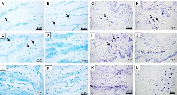
Figure 4. Light microscopy analysis of mast cell hyperplasia in the intestine or lung 1 (B, H), 3 (C, I), 6 (D, J), 18 (E, K), and 24 (F, L)
days after infection, respectively. The analysis was conducted on Alcian blue- or Toluidine blue-stained tissue sections from the ileum and lung of Toxocara canis-infected rats or uninfected rats (A and G). Mast cells were readily detected in the intestinal mucosa and pulmonary parenchyma after 1 day and progressively increased until day 24. Mast cells are indicated by arrows.
small intestine. After the first day, the number of larvae in this compartment was drastically decreased. In the lungs, the number of larvae recovered increased only slightly on days 3, 6, and 18 after infection (Figure 5).
Figure 5. Kinetics of Toxocara canis-migrating larvae
recov-ered from lung and intestine. Larvae were recovrecov-ered from the lungs (triangles) and small intestines (inverted triangles) of rats inoculated with 1000 infective T. canis eggs. Larval number was counted in a pool of 4 animals killed at 1, 3, 6, 18, and 24 days after infection. Data are reported as means ± SEM.
Table 2. Differences in mast cell numbers in the pulmonary
pa-renchyma from uninfected and Toxocara canis-infected rats.
Days after infection Mast cell numbers
Uninfected Infected
1 46.7 ± 8.1 61.3 ± 2.3*
3 36.0 ± 4.0 65.3 ± 5.0*
6 31.3 ± 2.3 114.7 ± 7.6*
18 32.0 ± 6.9 130.0 ± 19.1*
24 39.3 ± 6.2 217.3 ± 18.0*
324 D. Carlos et al.
Discussion
To gain insight into the mechanisms underlying in-creased eosinophilia during infection with T. canis, we compared some features of the inflammatory response in infected Wistar rats.Three major effects were observed: 1) increased peripheral blood and tissue eosinophil influx and EPO activity, 2) significant peritoneal mast cell ac-cumulation and LTB4 production in the serum following T.
canis infection, and 3) mast cell hyperplasia in the lungs and intestinal tissues probably induced by the passage of larvae into these compartments.
Increased eosinophilia was observed in the blood and peritoneal cavities of rats infected with T. canis. The peri-toneal eosinophil accumulation reached a peak on day 18 of infection and occurred in parallel to a marked increase in the number of mast cells in the peritoneal cavity. Consis-tent with these data, previous findings from our laboratory demonstrated elevated serum IgE levels on the 18th day after infection (4). We also evaluated EPO activity, a specific marker for eosinophils, and detected higher amounts of EPO in the peritoneal exudates of infected rats compared to uninfected rats. Our results also showed that mast cell accumulation in the peritoneal cavity correlates with blood eosinophil mobilization and extravasation to the peritoneal cavity during toxocariasis. In this context, a study by Nawa et al. (19) reported that production of eosinophils and their re-lease from bone marrow into peripheral blood was impaired in mast cell-deficient W/Wv mice compared to their normal littermates after T. canis infection. For instance, the results of the present study agree with previous investigations that have shown that IgE-dependent mast cell-mediated mechanisms play a crucial role in eosinophilia and in the immunological control induced in rats by other nematodes, such as Angiostrongylus cantonensis (25), Strongyloides venezuelensis (26), Nippostrongylus brasiliensis (27), and Trichinella spiralis (28).
It is not clear why eosinophil infiltration persists in T. canis-infected mice after the decrease in the number of inciting larvae, but the involvement of various eosinophil chemotactic factors has been suggested (29). A recent report established a tight correlation between tissue lesions caused by larval migration and plasma cytokine production. Moreover, the authors mentioned eotaxin and RANTES as potential factors responsible for the marked eosinophilic response that is a hallmark of T. canis infection (30). In fact, our group described an increased concentration of eotaxin in lung homogenates of T. canis-infected mice, which also correlated with eosinophil recruitment to this organ (31). Although the in vivo factors responsible for eosinophil ac-cumulation at inflammatory sites of parasitic infections are known to be produced by mast cells, only a single study has reported that cultured mast cells obtained from mice infected by T. canis released eosinophil chemotactic factors after stimulation with calcium ionophore A23187 or IgE-antigen.
These investigators also suggested that these factors might be arachidonic acid metabolites (32). In agreement with the results of these studies, treatment of T. canis-infected rats with a specific LTB4 receptor antagonist significantly reduced eosinophil chemotactic activity in bronchoalveolar fluid, suggesting that LTB4 also contributes to the accumulation of eosinophils in the lungs (33). In another study, treatment of mice infected with the nematode S. venezuelensis with the leukotriene inhibitor MK886 significantly inhibited the recruitment of eosinophils in the bronchoalveolar space, peritoneal cavity and blood (34). To determine the in vivo relevance of mast cell-derived LTB4 in eosinophilia, we examined the release of this mediator in rats following T. canis infection. We observed increased levels of LTB4 in the serum of infected rats compared to control, with the maximal response occurring 24 days after infection. These data suggest that LTB4 released by mast cells is a potent inflammatory mediator probably involved in eosinophil ac-cumulation in blood and maybe in the peritoneal cavity in this experimental model. Another study also demonstrated a role for endogenous stem cell factor production in mediating eosinophil recruitment in an allergic pleurisy model in mice (35). Moreover, the authors also showed that the effects of stem cell factor were dependent on the release of LTB4, which was most likely produced by mast cells.
The biological cycle of T. canis has common features with many nematodes; the larvae penetrate the small in-testine, enter the circulation and are free to migrate to the liver, lungs and brain (36). Excretory/secretory products released by the larvae interact with molecules expressed on the immune system cells, control their activity and induce inflammation in several tissues (37). We observed that most of the T. canis larvae penetrated the intestinal mucosa within 24 h and migrated to the lungs after 72 h of the L3 larvae inoculation. In fact, these larvae are metabolically active and inflammatory mediators are secreted due to activation of the host’s immune cells by larval products, resulting in an increased influx of leukocytes (38). In our study, mast cell hyperplasia was already greater in the small intestine after 1 day of infection, which coincided with the presence of the T. canis larvae in this organ. In addition, we also showed that in T. canis-infected rats, L3 larvae were most prevalent in the lungs on the 3rd day, although a second cycle of larval invasion could be detected on the 18th day of infection. Prominent mast cell recruitment is a common feature of infections by nematodes in most host species. The timing and degree of mast cell hyperplasia can vary between species and with the intensity of infection. Infection with the nematode Trichinella spiralis is associated with early and intense recruitment of intestinal mast cells (39). However, with other parasites, such as Schistosoma mansoni, chronic infection causes only a slight increase in mast cell numbers in the lungs and small intestine of rats (40).
References
1. Glickman LT, Schantz PM. Epidemiology and pathogenesis of zoonotic toxocariasis. Epidemiol Rev 1981; 3: 230-250. 2. Maizels RM, Yazdanbakhsh M. Immune regulation by
helminth parasites: cellular and molecular mechanisms. Nat Rev Immunol 2003; 3: 733-744.
3. Faccioli LH, Mokwa VF, Silva CL, Rocha GM, Araujo JI, Nahori MA, et al. IL-5 drives eosinophils from bone marrow to blood and tissues in a guinea-pig model of visceral larva migrans syndrome. Mediators Inflamm 1996; 5: 24-31. 4. Carlos D, Sá-Nunes A, de Paula L, Matias-Peres C, Jamur
MC, Oliver C, et al. Histamine modulates mast cell de-granulation through an indirect mechanism in a model IgE-mediated reaction. Eur J Immunol 2006; 36: 1494-1503. 5. Elsheikha HM, El-Beshbishi SN, El-Shazly AM, Hafez AO,
Morsy TA. Kinetics of eosinophilia and IgE production in ex-perimental murine toxocariasis. J Egypt Soc Parasitol 2008; 38: 53-64.
6. Machado ER, Carlos D, Lourenço EV, Souza GE, Sorgi CA, Silva EV, et al. Cyclooxygenase-derived mediators regulate the immunological control of Strongyloides venezuelensis infection. FEMS Immunol Med Microbiol 2010; 59: 18-32. 7. Stone KD, Prussin C, Metcalfe DD. IgE, mast cells,
baso-phils, and eosinophils. J Allergy Clin Immunol 2010; 125: S73-S80.
8. Temkin V, Pickholtz D, Levi-Schaffer F. Tumor necrosis fac-tors in a murine model of allergic peritonitis: effects on
eo-sinophil accumulation and inflammatory mediators’ release.
Cytokine 2003; 24: 74-80.
9. Gordon JR, Burd PR, Galli SJ. Mast cells as a source of mul-tifunctional cytokines. Immunol Today 1990; 11: 458-464. 10. Metcalfe DD, Baram D, Mekori YA. Mast cells. Physiol Rev
1997; 77: 1033-1079.
11. Funk CD. Prostaglandins and leukotrienes: advances in eicosanoid biology. Science 2001; 294: 1871-1875. 12. Werz O. 5-Lipoxygenase: cellular biology and molecular
pharmacology. Curr Drug Targets Inflamm Allergy 2002; 1: 23-44.
13. Peters-Golden M, Brock TG. 5-Lipoxygenase and FLAP. Prostaglandins Leukot Essent Fatty Acids 2003; 69: 99-109.
14. Serezani CH, Aronoff DM, Jancar S, Peters-Golden M. Leu-kotriene B4 mediates p47phox phosphorylation and mem-brane translocation in polyunsaturated fatty acid-stimulated
neutrophils. J Leukoc Biol 2005; 78: 976-984.
15. Cheraim AB, Xavier-Elsas P, de Oliveira SH, Batistella T, Russo M, Gaspar-Elsas MI, et al. Leukotriene B4 is essen-tial for selective eosinophil recruitment following allergen challenge of CD4+ cells in a model of chronic eosinophilic
inflammation. Life Sci 2008; 83: 214-222.
16. Miyahara N, Ohnishi H, Miyahara S, Takeda K, Matsubara S, Matsuda H, et al. Leukotriene B4 release from mast cells
in IgE-mediated airway hyperresponsiveness and inflamma -tion. Am J Respir Cell Mol Biol 2009; 40: 672-682. 17. Calheiros AS, Aguiar Pires AL, Pereira da Silva J, Cordeiro
RS, Martins MA, Lima MC. Role of the IgE-mediated sys-tem in eosinophil recruitment triggered by two consecutive cycles of sensitisation and challenge in rats. Int Arch Allergy Immunol 2001; 126: 325-334.
18. Oliveira SH, Costa CH, Ferreira SH, Cunha FQ. Sephadex induces eosinophil migration to the rat and mouse peritoneal cavity: involvement of mast cells, LTB4, TNF-alpha, IL-8 and PAF. Inflamm Res 2002; 51: 144-153.
19. Nawa Y, Owhashi M, Imai J, Abe T. Eosinophil response in
mast cell-deficient W/WV mice. Int Arch Allergy Appl Immu-nol 1987; 83: 6-11.
20. Olson LJ, Schulz CW. Nematode induced hypersensitivity reactions in guinea pigs: onset of eosinophilia and positive Schultz-Dale reactions following graded infections with Toxocara canis. Ann N Y Acad Sci 1963; 113: 440-455. 21. Pradelles P, Antoine C, Lellouche JP, Maclouf J. Enzyme
immunoassays for leukotrienes C4 and E4 using acetylcho-linesterase. Methods Enzymol 1990; 187: 82-89.
22. Strath M, Warren DJ, Sanderson CJ. Detection of eosino-phils using an eosinophil peroxidase assay. Its use as an as-say for eosinophil differentiation factors. J Immunol Methods 1985; 83: 209-215.
23. Denzler KL, Borchers MT, Crosby JR, Cieslewicz G, Hines EM, Justice JP, et al. Extensive eosinophil degranulation and peroxidase-mediated oxidation of airway proteins do not occur in a mouse ovalbumin-challenge model of pulmonary
inflammation. J Immunol 2001; 167: 1672-1682.
24. Luo ZJ, Cheng SW, Liao L. A method for isolation and
puri-fication of nematode larvae. J Parasitol 1999; 85: 573-574. 25. Serra MF, Barreto EO, Silva JP, Azevedo V, Mota EM,
Pelajo-Machado M, et al. Kinetics of eosinophil and IgE-mast cell changes following infection with Angiostrongylus
infiltration and in the release of LTB4 during infection with
T. canis in rats. In parallel, mast cell numbers were histo-logically assessed in the lung and intestine after staining with Toluidine and Alcian blue, respectively. An important correlation was established between mast cell hyperpla-sia and the time course of larval migration into the small intestine and lungs. These data showed that infection with T. canis in rats can be considered an alternative model to delineate the crucial features of the immune response evoked by T. canis. In addition, this model may contribute to our understanding of the interplay between the activa-tion and accumulaactiva-tion of mast cells, LTB4 production and
eosinophil recruitment.
Acknowledgments
326 D. Carlos et al.
costaricensis in Wistar rats. Parasite Immunol 2003; 25: 169-177.
26. Kimura K, Song CH, Rastogi A, Dranoff G, Galli SJ, Lantz CS. Interleukin-3 and c-Kit/stem cell factor are required for normal eosinophil responses in mice infected with Strongy-loides venezuelensis. Lab Invest 2006; 86: 987-996. 27. Watanabe N, Miura K, Fukuda Y. Chymase inhibitor
ame-liorates eosinophilia in mice infected with Nippostrongylus brasiliensis. Int Arch Allergy Immunol 2002; 128: 235-239. 28. Shin K, Watts GF, Oettgen HC, Friend DS, Pemberton AD,
Gurish MF, et al. Mouse mast cell tryptase mMCP-6 is a criti-cal link between adaptive and innate immunity in the chronic phase of Trichinella spiralis infection. J Immunol 2008; 180: 4885-4891.
29. Owhashi M, Arita H, Niwa A. Production of eosinophil chemotactic factor by CD8+ T-cells in Toxocara canis -infected mice. Parasitol Res 1998; 84: 136-138.
30. Pecinali NR, Gomes RN, Amendoeira FC, Bastos AC,
Mar-tins MJ, Pegado CS, et al. Influence of murine Toxocara canis infection on plasma and bronchoalveolar lavage fluid eosinophil numbers and its correlation with cytokine levels. Vet Parasitol 2005; 134: 121-130.
31. Sá-Nunes A, Rogerio AP, Medeiros AI, Fabris VE, Andreu GP, Rivera DG, et al. Modulation of eosinophil generation and migration by Mangifera indica L. extract (Vimang). Int Immunopharmacol 2006; 6: 1515-1523.
32. Abe T, Nawa Y. Eosinophil chemotactic factor released from murine bone marrow derived cultured mast cells. Arerugi 1990; 39: 69-74.
33. Okada K, Fujimoto K, Kubo K, Sekiguchi M, Sugane K.
Eo-sinophil chemotactic activity in bronchoalveolar lavage fluid
obtained from Toxocara canis-infected rats. Clin Immunol Immunopathol 1996; 78: 256-262.
34. Machado ER, Ueta MT, Lourenco EV, Anibal FF, Sorgi CA, Soares EG, et al. Leukotrienes play a role in the control of parasite burden in murine strongyloidiasis. J Immunol 2005; 175: 3892-3899.
35. Klein A, Talvani A, Silva PM, Martins MA, Wells TN, Proud-foot A, et al. Stem cell factor-induced leukotriene B4 produc-tion cooperates with eotaxin to mediate the recruitment of eosinophils during allergic pleurisy in mice. J Immunol 2001; 167: 524-531.
36. Taira K, Permin A, Kapel CM. Establishment and migration pattern of Toxocara canis larvae in chickens. Parasitol Res 2003; 90: 521-523.
37. Loukas A, Maizels RM. Helminth C-type lectins and host-parasite interactions. Parasitol Today 2000; 16: 333-339.
38. Lescano SZ, Queiroz ML, Chieffi PP. Larval recovery of
Toxocara canis in organs and tissues of experimentally infected Rattus norvegicus. Mem Inst Oswaldo Cruz 2004; 99: 627-628.
39. Tuohy M, Lammas DA, Wakelin D, Huntley JF, Newlands GF, Miller HR. Functional correlations between mucosal mast cell activity and immunity to Trichinella spiralis in high and low responder mice. Parasite Immunol 1990; 12: 675-685. 40. Miller HR, Newlands GF, McKellar A, Inglis L, Coulson PS,