O R I G I N A L P A P E R
Miche`le Milili
Æ
Henedina Antunes
Carla Blanco-Betancourt
Æ
Ana Nogueiras
Euge´nia Santos
Æ
Ju´lia Vasconcelos
Æ
Joa˜o Castro e Melo
Claudine Schiff
A new case of autosomal recessive agammaglobulinaemia
with impaired pre-B cell differentiation due to a large deletion
of the IGH locus
Received: 30 January 2002 / Accepted: 22 May 2002 / Published online: 12 July 2002
Springer-Verlag 2002
Abstract
Males withX-linked agammaglobulinaemia
(XLA) due to mutations in the Bruton tyrosine kinase
gene constitute the major group of congenital
hypo-gammaglobulinaemia withabsence of periph
eral B
cells. In these cases, blockages between the pro-B and
pre-B cell stage in the bone marrow are found. The
remaining male and female cases clinically similar to
XLA represent a genotypically heterogeneous group of
diseases. In these patients, various autosomal recessive
disorders have been identified such as mutations
af-fecting
IGHM,
CD79A,
IGLL1
genes involved in the
composition of the pre-B cell receptor (pre-BCR) or
the
BLNK
gene implicated in pre-BCR signal
trans-duction. In this paper, we report on a young female
patient characterised by a severe non-XLA
agamma-globulinaemia that represents a new case of Ig
l
defect.
We show that the B cell blockage at the pro-B to
pre-B cell transition is due to a large homologous deletion
in the
IGH
locus encompassing the
IGHM
gene
leading to the inability to form a functional pre-BCR.
The deletion extends from the beginning of the
di-versity (D) region to the
IGHG2
gene, withall
JH
segments and
IGHM,
IGHD,
IGHG3
and
IGHG1
genes missing.
Conclusion:
alteration in Ig
l
expression
seems to be relatively frequent and could account for
most of the reported cases of autosomal recessive
agammaglobulinaemia.
Keywords
Agammaglobulinaemia
Æ
B cell blockage
Æ
IGH
locus
Æ
Immunoglobulin gene deletion
Æ
Pre-B cell
Abbreviations
BET: ethidium bromide
Æ
BTK: Bruton
tyrosine kinase gene
Æ
IGH: immunoglobulin heavy
chain locus
Æ
pre-BCR: pre-B cell receptor
Æ
XLA: X-linked agammaglobulinaemia
Introduction
Recurrent infections, mostly respiratory, withpyogenic
bacteria are the predominant manifestations of children
suffering antibody deficiencies. In some of these patients,
early B cell development in the bone marrow is arrested
and hypogammaglobulinaemia results from the absence
of peripheral B cells. In these cases, chronic diarrhoea
could also be observed and constitutes a serious clinical
problem. This occurs in X-linked agammaglobulinaemia
(XLA), also known as Bruton disease, characterised by
defects in the Bruton tyrosine kinase gene (BTK) [13,14]
that encodes the cytoplasmic tyrosine kinase btk,
in-volved in signal transduction. In these cases, B cell
de-velopment is blocked in the bone marrow at the pre-B
cell stage, resulting in the accumulation of CD34+
CD19+ pro-B cells and in the presence of variable
numbers of CD34-CD19+ pre-B cells. Females witha
phenotype indistinguishable from XLA have also
been described and Conley et al. [2] have estimated
that these immunodeficiencies represent 10% of patients
with congenital hypogammaglobulinaemia. The defects
behind such autosomal recessive disorders have been
recently identified and shown to affect predominantly
the pre-B cell receptor (pre-BCR), which is an absolute
prerequisite for pro-B to pre-B cell transition and to
allow further B cell differentiation [3,12]. This receptor,
expressed on pre-B cells is composed of the Ig
l
, th e
Y
L
chain (made of
k
-like and VpreB) and the Ig
a
/Ig
b
transducing complex [5]. For example, mutations
affecting
IGHM
[12,15],
CD79A
[10],
IGLL1
[9] or
DOI 10.1007/s00431-002-0994-9
M. MililiÆC. Blanco-BetancourtÆC. Schiff (&)
Centre d’Immunologie de Marseille-Luminy, case 906, 13288 Marseille cedex 09, France
E-mail: [email protected] Tel.: +33-4-91269448
Fax: +33-4-91269430 H. AntunesÆA. Nogueiras
Gastroenterology and Nutrition Unit, Paediatric Department, Sa˜o Marcos Hospital, Braga, Portugal
E. SantosÆJ. VasconcelosÆJ. Castro e Melo
BLNK
that encodes a B cell linker protein essential for
Ig
l
signal transduction [11], have been reported.
In this paper we describe a new case of autosomal
recessive agammaglobulinaemia due to a large deletion
of the immunoglobulin heavy chain locus (IGH),
including the
IGHM
gene, that results in the absence of
a functional pre-BCR.
Case report
The patient, a 35-month-old girl, was the second child of healthy parents born in the same small village, near Braga, Portugal, who were thought to be non-consanguineous (see below). She had a healthy 6-year older brother. Apart from symmetrical intrauterine growthretardation due to uteroplacental insufficiency, no other significant abnormalities related to family history, pregnancy and delivery were detected. An echocardiogram confirmed a congenital cardiopathy, secundum ASD (atrium secundum, intra-auricular communication) which resolved spontaneously without surgery. At 9 months of age she was referred to the Paediatric Gastroenterol-ogy Department because of failure to thrive and chronic diarrhoea during the previous month. Her weight was 5 640 g (P<3), her height 60 cm (P<3) and her head circumference 40.8 cm (P<5). Physical examination showed that she was not dehydrated, she had no detectable tonsils and no palpable adenomegaly and revealed that she had elongated fingers (data not shown). According to the Kanawati-McLaren index (0.31), she had no clinical signs of mal-nutrition.
A routine blood examination showed leucocyte and platelet counts of 27500/mm3and 345000/mm3, respectively, and haemo-globin at 13.2 g/dl. Total protein was 5.2 g/dl and albumin 3.7 g/dl. Blood, urine and stool cultures were sterile. TORCH serology and sweat tests were negative. Searchin stools for virus, parasites, re-ducing sugar and fat was negative. Alpha-1 anti-trypsin in stools was <1.7 mg/g dry weight (normal <5.0 mg/g). Histology of a duodenal biopsy showed no histological changes but immunocy-tochemistry revealed a complete absence of B cells and normal numbers of T cells. The patient responds to BCG and oral polio-myelitis vaccines without any complication.
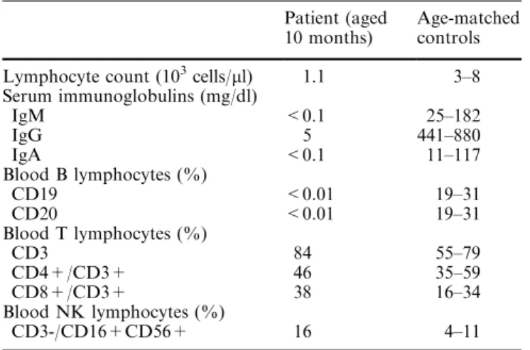
Her peripheral blood immunological data, determined at 10 months of age, are presented in Table 1. A clear agammaglobuli-naemia was evident witha serum IgG level of 5 mg/dl and unde-tectable IgM and IgA. Lymphocyte phenotyping showed the absence of circulating B cells (CD19 or CD20 positive cells), nor-mal numbers of CD4 T lymphocytes and increased numbers and proportions of NK and CD8 T cells, this latter population showing increased expression of HLA-DR, CD45RO and CD95 activation markers (data not shown).
Since the age of 10 months, she has been treated with intrave-nous replacement immunoglobulin therapy (400 mg/kg every 3 weeks, withpre-administration levels of 300–500 mg/dl). During the 25 months follow-up, two episodes of gastroenteritis, a viral rhinitis and an ear infection were diagnosed, which, however, did not require anti-microbial therapy.
The parents gave informed consent for this investigation.
Materials and methods
Immunoglobulin measurement and cell phenotyping
Serum immunoglobulin IgG, IgA, and IgM were determined by rate nephelometry. Whole peripheral blood and bone marrow mononuclear cells isolated by Ficoll-Hypaque gradient were used for phenotyping. Cell surface antigens were stained with conjugated anti-human monoclonal antibodies and were analysed by flow cytometry. Bone marrow mononuclear cells were stained with FITC-conjugated monoclonal antibodies against CD10, CD20, CD34, CD3, CD4 or phycoerythrin-conjugated monoclonal anti-bodies against CD19, CD8, CD16 and CD56, from Immunotech (France). Phycoerythrin-conjugated anti-Igd and APC-conjugated anti-CD19 were from Pharmingen (France). Biotin-conjugated anti-human Igl chain from Southern Biotechnology (USA) was
revealed withPerCP-conjugated Streptavidin (Becton and Dickin-son, USA). Peripheral blood B cells were stained with FITC-con-jugated monoclonal antibodies against CD19, CD20, CD4, CD8, phycoerythrin-conjugated monoclonal antibodies against CD8, CD16, CD56, PE/Cy5-conjugated monoclonal antibodies against CD3, from Immunotech(France) and from Dako, SA (Denmark), and were haemolysed and fixed (T–prep, Beckman-Coulter) before flow cytometric analysis.
RNA and reverse transcriptase polymerase chain reaction Total RNA was extracted from total bone marrow cells using TRIzol Reagent (Gibco BRL) as described by Chomczynski and Sacchi [1]. RNA (2lg) was reverse transcribed using the reverse
transcriptase Superscript II (Gibco BRL), 1lg of random hexamer
(dN6), 1 mM dNTPs and the supplied buffer. For reverse tran-scriptase-PCR, 1ll of cDNA was amplified for 30 cycles of 30 s at
94C, 1 min at the appropriate temperature and 2 min at 72C with
a final 10 min extension at 72C, using Taq DNA polymerase (BRL) and the primers already described [12].
DNA and DNA-polymerase chain reaction
Genomic DNA was extracted from peripheral blood mononuclear cells with proteinase K, sodium dodecyl sulphate and phenol-chloroform extractions. PCR was performed, as above, using 100 ng of DNA. For semi quantitative analysis, PCRs were carried out during the exponential phase of the DNA amplification. In that case, the exponential phase of each PCR was determined by titra-tion of cycle numbers and by quantities of total DNA. PCR products were analysed on 2% agarose gels and revealed by ethi-dium bromide (BET) staining. Quantification was performed using the MacBas software.
Results
Phenotypic analysis of bone marrow cells
Peripheral blood phenotype of the patient revealed a
pure B cell defect since a profound
agammaglobulina-emia witha total absence of periph
eral B cells was
observed and since the levels and phenotype of T and
Table 1. Immunological data
Patient (aged 10 months)
Age-matched controls Lymphocyte count (103cells/ll) 1.1 3–8
Serum immunoglobulins (mg/dl)
IgM <0.1 25–182
IgG 5 441–880
IgA <0.1 11–117
Blood B lymphocytes (%)
CD19 <0.01 19–31
CD20 <0.01 19–31
Blood T lymphocytes (%)
CD3 84 55–79
CD4+/CD3+ 46 35–59
CD8+/CD3+ 38 16–34
Blood NK lymphocytes (%)
NK cells did not indicate a T-B-combined
immunode-ficiency. Thus, we analysed bone marrow B cell
sub-populations. A bone marrow sample was obtained when
the patient (from now referred as the Ig
l
Dpatient, see
below) was 24 months old. Total CD19+ patient’s B
cells were diminished more than three-fold (13% versus
43% in age matched control) and the population of large
B cells, mainly corresponding to proliferating pre-B
cells, was nearly absent (Fig. 1A). Gated CD19+ cells
were completely negative for surface Ig
l
expression
(Fig. 1B) and for Ig
d
and CD40 expression (data not
shown), indicating a total absence of immature and
mature B cells in the patient’s bone marrow. In contrast,
CD34+ precursors and CD34+CD19+ pro-B cells
were within normal limits as compared to an
age-mat-ched control (Fig. 1C). Altogether these data indicate
that the patient suffered from an early bone marrow B
cell blockage, between pro-B and pre-B cell stages.
Molecular analysis
RT-PCR analysis was performed on total mononuclear
bone marrow cells for transcripts expressed during the
pro-B to pre-B cell transition, including
k
-like, VpreB,
Ig
a
, Ig
b
, and Blnk. The resulting PCR products were
directly sequenced and were normal (data not shown). In
contrast, no transcripts were detected for IgC
l
specific
amplification. This result was confirmed at the DNA
level since no amplification could be detected using the
l
CH1 and
l
CH2 oligonucleotides (Table 2, Fig. 2) for
the patient’s DNA. These data indicate that a deletion
within the
IGH
locus, carried by bothalleles, was
re-sponsible for the patient’s phenotype.
To analyse the extent of the DNA deletion, we
per-formed a detailed analysis of the
IGH
locus by PCR. By
using JH5-JH6 and
d
CH2-
d
CH3 primers, we showed
that the
JH
cluster and the
IGHD
gene respectively were
completely missing. Moreover, we mapped the 5’ end of
the deletion between the
IGHD2–2
and the
IGHD3–10
genes within the
IGHD
locus, since we could amplify the
IGHD2–2
and not the
IGHD3–10
gene (Fig. 2). Using
c
CH2 and
c
CH3 oligonucleotides that are common to
different
IGHG
gene sub-classes, we demonstrated that
some
IGHG
genes were present in the patient’s genome.
Fig. 1. FACS analysis of bone marrow B cell sub-populations for
the IglD patient and an age-matched control (Ctrl). A Bone
marrow mononuclear cells from IglDand an age-matched control
were stained withanti-CD19 monoclonal antibody and analysed by flow cytometry. Fluorescence intensity of stained cells is presented as a function of cell size (forward scatter). B Bone marrow mononuclear cells were stained withanti-CD19 and anti-Igl
monoclonal antibodies and CD19+ cells, gated and analysed for Iglchain expression.Solidandemptycurves represent IglDpatient
and age-matched control, respectively.CBone marrow mononu-clear cells were stained withanti-CD34 and anti-CD19 monoclonal antibodies. Dot plots show precursor (CD34+), pro-B (CD34+CD19+) and pre-B+immature+mature (CD34-CD19+) bone marrow B cell sub-populations
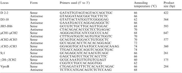
Table 2. Oligonucleotide primers used for DNA-PCR amplification
Locus Form Primers used (5’ to 3’) Annealing temperature (C)
Product size (bp)
D 2-2 Sense GATATTGTAGTAGTACCAGCTGC 66 422
Antisense GTAGGATAGCGGCTGCTTCTC 66
D3-10 Sense GTATTACTATGGTTCGGGGAG 62 564
Antisense GAAATGACCCAGGAGAGGCTC 66
JH5-JH6 Sense GCCGTCTGCTTGCAGTTGGAC 68 593
Antisense CTACAGACACCGCTCCTGAGAC 70
lCH1-lCH2 Sense AGGGAGTGCATCCGCCCCAAC 68 647
Antisense CTTTGATGGTCAGTGTGCTGGTC 70
dCH2-dCH3 Sense GCAGTGCAGGACCTGTGGCTC 70 577 Antisense GCCAGACACCTCACACAGGAGC 72
cCH2-cCH3 Sense GGAGGTGCATAATGCCAAGACAAAG 74 360 Antisense TTGACCAGGCAGGTCAGGCTGAC 74
Sc1 Sense GCAGAGCATCACAACGTCAGC 66 261
Antisense GAGCTAGTCCTGCTCACCTTC 66
c2H-c2CH2 Sense GCGCAAATGTTGTGTCGAGT 60 175 Antisense CGGTCCTGCCACAGGTGG 62
Using specific primers in the
IGHG1
switchregion (S
c
1)
or in the hinge region of the
IGHG2
gene (
c
2H and
c
2CH2), we finally demonstrated that the 3’ end of the
DNA deletion was located between the
IGHG1
and the
IGHG2
genes (Fig. 2). The size of the DNA deletion was
estimated to be between 165 to 265 kb [6].
Family studies
The PCR results obtained for the Ig
l
Dpatient suggested
the presence of a large homologous DNA deletion of
familial origin. A semi-quantitative PCR was performed
using VpreB and Ig
l
specific primers on the DNA
ex-tracted from peripheral blood mononuclear cells of the
father, mother, brother and a healthy donor (Fig. 3A).
Since
VPREB
is a unique gene in the human genome [4]
it was used to quantify Ig
l
PCR products. The results
clearly demonstrated that the DNA from both the father
and the mother contained only one copy of the
func-tional
IGHM
gene (Fig. 3B). In contrast, DNA of the
brother as well as control DNA contained two copies of
the
IGHM
allele (Fig. 3B). The patient’s brother was
therefore homozygous for the functional
IGHM
allele.
Discussion
Approximately 85% of patients withcongenital
hypo-gammaglobulinaemia and absent peripheral B cells are
males withXLA due to mutations in the
BTK
gene. The
remaining 15% of cases, clinically identical to XLA, are
still a heterogeneous group. Some mutations have been
already identified, especially in components of the
pre-BCR, suchas
IGHM
[12,15],
IGLL1
[9],
CD79A
[10], or
BLNK
[11] that result in impaired pre-B cell
differenti-ation. However, alteration in Ig
l
expression seems to be
relatively frequent. For example, Yel et al. [15] described
three different mutations in the
IGH
locus. For one
consanguineous family, the affected patients had a DNA
deletion covering 75–100 kb that encompassed the
di-versity genes (DH), the JH cluster and the
IGHM
gene.
Fig. 2. Molecular analysis of the patient’s gene defect. Schematic
representation (top) of the humanIGHlocus and localisation of the primers used for DNA amplification. Extent of the IglD patient
DNA deletion is shown. PCR amplification (bottom) of th e IglD
patient DNA using the indicated oligonucleotides (see Table 2). 0, C and P lanes correspond to PCR without DNA, control DNA and IglD patient DNA, respectively. Amplified DNA products were
For another consanguineous family, the alteration
cor-responded to a homozygous single G to A base pair
substitution at the –1 position of the alternative splice
site used to produce the membrane form of the Ig
l
transcript. This mutation would inhibit the production
of the membrane Ig
l
chain. It was also reported that a
boy, who did not suffer from a
BTK
alteration, had a
heterozygous alteration of the
IGHM
gene. In this case,
a single base pair substitution affecting an invariant
cysteine residue in the CH4 domain of the
IGHM
chain
for one allele, coupled to a large deletion of the
IGH
locus on the other allele, was detected. We have also
reported the case of a girl, referred to as the Ig
l
–/–pa-tient [7,12], in whom we identified a cytosine insertion at
the beginning of the CH1 exon of the
IGHM
gene,
re-sulting in a stop codon at position 48, and in the absence
of Ig
l
chain expression. The patient reported in this
paper, designated as the Ig
l
Dcase [8], witha large 165 to
265 kb homologous deletion of the
IGH
locus
encom-passing the
IGHM
gene, represents a new case of Ig
l
defect. Altogether we can conclude that Ig
l
alterations
account for most of the reported cases of autosomal
recessive agammaglobulinemia. These observations also
emphasise the requirement of signalling via a functional
pre-BCR to ensure normal early B cell differentiation.
Moreover, the absence of Ig
l
chains in Ig
l
–/–and Ig
l
Dpatients, has provided an opportunity to elucidate the
role of Ig
l
in selecting the antibody repertoire in humans
[8].
Along witha better understanding of B cell biology
and its developmental defects, identification of such
genetic alterations may open the way for new clinical
treatments, including somatic gene therapy. Moreover it
provides a basis for a more accurate diagnosis,
com-prising the identification of carrier status, prenatal
test-ing and counselltest-ing.
Acknowledgements This work was supported by CNRS (Centre
National de la Recherche Scientifique) and INSERM (Institut National de la Sante´ et de la Recherche Me´dicale).
References
1. Chomczynski P, Sacchi N (1987) Single-step method of RNA isolation by acid guanidium thiocyanate-phenol-chloroform extraction. Anal Biochem 162: 156–159
2. Conley ME (1992) Molecular approaches to analysis of X-linked immunodeficiencies. Annu Rev Immunol 10: 215–238 3. Conley ME, Rohrer J, Rapalus L, Boylin EC, Minegishi Y (2000) Defects in early B-cell development: comparing the consequences of abnormalities in pre-BCR signaling in the human and the mouse. Immunol Rev 178: 75–90
4. Frippiat JP, Williams SC, Tomlinson IM, Cook GP, Cherif D, Le Paslier D, Collins JE, Dunham I, Winter G, Lefranc MP (1995) Organization of the human immunoglobulin lambda light-chain locus on chromosome 22q11.2. Hum Mol Genet 4: 983–991
5. Guelpa-Fonlupt V, Bossy D, Alzari P, Fumoux F, Fougereau M, Schiff C (1994) The human pre-B cell receptor: structural constraints for a tentative model of the pseudo-light (psi L) chain. Mol Immunol 31: 1099–1108
6. Lefranc MP (2001) Nomenclature of the human immuno-globulin heavy (IGH) genes. Exp Clin Immunogenet 18: 100– 116
7. Meffre E, LeDeist F, de Saint-Basile G, Deville A, Fougereau M, Fischer A, Schiff C (1996) A human non-XLA immunodefi-ciency disease characterized by blockage of B cell development at an early proB cell stage. J Clin Invest 98: 1519–1526 8. Meffre E, Milili M, Blanco-Betancourt C, Antunes H,
Nussenzweig MC, Schiff C (2001) Immunoglobulin heavy chain expression shapes the B cell receptor repertoire in human B cell development. J Clin Invest 108: 879–886
9. Minegishi Y, Coustan-Smith E, Wang YH, Cooper MD, Campana D, Conley ME (1998) Mutations in the human lambda5/14.1 gene result in B cell deficiency and agamma-globulinemia. J Exp Med 187: 71–77
10. Minegishi Y, Coustan-Smith E, Rapalus L, Ersoy F, Campana D, Conley ME (1999a) Mutations in Igalpha (CD79a) result in a complete block in B-cell development. J Clin Invest 104: 1115–1121
11. Minegishi Y, Rohrer J, Coustan-Smith E, Lederman HM, Pappu R, Campana D, Chan AC, Conley ME (1999b) An es-sential role for BLNK in human B cell development. Science 286: 1954–1957
12. Schiff C, Lemmers B, Deville A, Fougereau M, Meffre E (2000) Autosomal primary immunodeficiencies affecting human bone marrow B cell differentiation. Immunol Rev 178: 91–98
Fig. 3. Molecular analysis
of family’s patient DNA at theIGHlocus.APCR was performed on DNA extracts from healthy donor’s pe-ripheral blood mononuclear cells (C), father (F), mother (M) brother (B) and patient (P) usinglCH1-lCH2 and
VpreB specific primers (see Table 2). 0 corresponds to PCR without DNA. Ampli-fied DNA products were separated on 2% agarose gels and revealed by BET staining.BFor eachDNA, Iglspecific amplification is
13. Tsukada S, Saffran DC, Rawlings DJ, Parolini O, Allen RC, Klisak I, Sparkes RS, Kubagawa H, Mohandas T, Quan S et al (1993) Deficient expression of a B cell cytoplasmic tyrosine kinase in human X-linked agammaglobulinemia. Cell 72: 279– 290
14. Vetrie D, Vorechovsky I, Sideras P, Holland J, Davies A, Flinter F, Hammarstrom L, Kinnon C, Levinsky R, Bobrow M et al (1993) The gene involved in X-linked
agammaglobulina-emia is a member of the src family of protein-tyrosine kinases. Nature 361: 226–233