Brazilian Journal of Microbiology (2012): 909-916 ISSN 1517-8382
OCCURRENCE OF PERIODONTAL PATHOGENS AMONG PATIENTS WITH CHRONIC PERIODONTITIS
B.C. Farias1*, P.R.E. Souza2, B. Ferreira1, R.S.A. Melo1, F.B. Machado1, E.S. Gusmão3, R. Cimões4
1
Universidade Federal de Pernambuco, Recife, Pernambuco, Brasil; 2Departamento de Genética, Universidade Federal Rural de
Pernambuco, Recife, Pernambuco, Brasil; 3Departamento de Medicina Oral da Universidade de Pernambuco, Recife, Pernambuco,
Brasil; 4Departamento de Prótese e Cirurgia Buco-Facial, da Faculdade de Odontologia da Universidade Federal de Pernambuco,
Recife, Pernambuco, Brasil.
Submitted: February 18, 2011; Returned to authors for corrections: November 29, 2011; Approved: June 07, 2012.
ABSTRACT
The aim of the present study was to evaluate the presence of the periodontal pathogens that form the red
complex (Tannerella forsythia, Porphyromonas gingivalis and Treponema denticola) and Aggregatibacter actinomycetemcomitans in patients with chronic periodontitis. The sample consisted of 29 patients with a clinical and radiographic diagnosis of chronic periodontitis based on the criteria of the American Academy of Periodontology (3). Samples for microbiological analysis were collected from the four sites of greatest
probing depth in each patient, totaling 116 samples. These samples were processed using conventional
polymerase chain reaction, which achieved the following positive results: 46.6% for P. gingivalis, 41.4% for T. forsythia, 33.6% for T. denticola and 27.6% for A. actinomycetemcomitans. P. gingivalis and T. forsythia were more prevalent (p < 0.05) in periodontal pockets ≥ 8 mm. The combinations T. forsythia + P. gingivalis (23.2%) and T. forsythia + P. gingivalis + T. denticola (20.0%) were more frequent in sites with a probing depth 8 mm. Associations with the simultaneous presence of A. actinomycetemcomitans + P.
gingivalis, A. actinomycetemcomitans + T. forsythia, P. gingivalis + T. forsythia and T. forsythia + T. denticola were statistically significant (p < 0.05). It was concluded that the red complex pathogens are related to chronic periodontitis, presenting a higher occurrence in deep periodontal pockets. Moreover, the
simultaneous presence of these bacteria in deep sites suggests a symbiotic relationship between these
virulent species, favoring, in this way, a further progression of periodontal disease.
Keywords: periodontitis; pathogens; Tannerella forsythia; Porphyromonas gingivalis; Treponema denticola;Aggregatibacter actinomycetemcomitans
INTRODUCTION
Periodontal disease affects a large number of individuals
around the world (2) and is a major oral health problem both in
developed and developing countries (12). Periodontitis is
defined as a chronic inflammatory disease that affects the tooth-supporting tissues and bacterial deposits play an essential
role in the pathogenesis of this condition 18, 22, 25).
Strong associations between periodontal disease and
certain microorganisms have been reported in the literature,
especially with regard to the presence of certain gram-negative anaerobic bacteria, such as A. actinomycetemcomitans, P. gingivalis, T. forsythia and T. denticola (6, 12, 17, 18, 19, 21, 24, 27). These organisms express potential virulence factors
and induce host inflammatory mediators, eventually leading to
the breakdown of connective tissue and alveolar bone
resorption (20, 23, 27).
The literature suggests that the distribution and prevalence of periodontal pathogens vary depending on geographic
locations as well as among different ethnic groups (9, 10, 12,
16, 24, 27). Such differences in global distribution place some
populations at greater risk for infection and periodontal disease
than others and this variation could be important to the
epidemiology and treatment of periodontal disease (9).
In Brazil, few studies have been conducted to investigate
the periodontal microbiota, most of these being conducted in the State of São Paulo (7, 8, 9, 13, 14, 15, 26). In these
investigations, the bacteria P. gingivalis had its prevalence ranging from 17.8% to 90%, T. forsythia 33.3% to 100%, A. actinomycetemcomitans 23% to 90% and T. denticola was only investigated by Avila-Campos and Velásquez-Meléndez (7)
with 60% of positive samples. In view of the importance of
knowledge on the microorganisms that make up the
subgingival microbiota and the limited number of studies involving South American populations, the aim of the present
study was to identify the occurrence of the periodontal
pathogens P. gingivalis, T. forsythia, T. denticola and A. actinomycetemcomitans in a convenience sample composed of adult Brazilian, nonsmokers with chronic periodontitis in the
city of Recife (northeastern Brazil).
MATERIALS AND METHODS
Participants and clinical examination
The present experimental, quantitative study was
conducted at the dental clinic of the Postgraduate Dentistry
Program, Universidade Federal de Pernambuco (UFPE), located in the city of Recife, state of Pernambuco, northeastern Brazil. A convenience sample comprised of 12 male and 17
female patients diagnosed with chronic periodontitis who
sought dental treatment between August 2008 and July 2009
participated in this study. After an explanation of the
objectives, the participants signed terms of informed consent.
The study received approval from the UFPE Ethics Committee
under process nº191/2008.
The following were the inclusion criteria: presence of
least 20 natural teeth; age at least 30 years; clinical and
radiographic diagnosis of chronic periodontitis (American
Academy of Periodontology, 1999) (3), where to be considered
as having chronic periodontitis patients should have at least
one site with probing depth (PD) and clinical attachment loss
(PIC) ≥ 4 mm; and voluntary agreement to participate. The
following were the exclusion criteria: smoking habits; any systemic condition that might affect the progression of
periodontal disease (diabetes, hypertension, autoimmune
disease, etc.); pregnancy or nursing; periodontal therapy or use
of antibiotics in previous six months; chronic use of
anti-inflammatory drug; HIV positivity; and use of orthodontic
appliance.
The following information on each patient was recorded:
visible plaque index (Ainamo and Bay) (1), bleeding index (Ainamo and Bay) (1), probing depth and clinical attachment
loss. The examination was performed by a single calibrated
examiner in an office with artificial light using a dental mirror
and Williams periodontal probe (Trinity ®, Brazil). The
diagnosis of chronic periodontitis was based on the criteria
established by the American Academy of Periodontology (4),
which state that the patient must have at least one site with a
probing depth and clinical attachment loss ≥ 4 mm. Subgingival plaque samples
The four sites of greatest probing depth in each patient
Farias, B.C. et al. Periodontal pathogens among patients with periodontitis
Sterilized nº 30 paper tips (Dentsply, Brazil) were inserted to
the depth of the pocket, left in place for 20 seconds, transferred
to microcentrifuge tubes and stored at -20 C for subsequent
DNA extraction and polymerase chain reaction (PCR) analysis
at the Molecular Biology Laboratory of the Postgraduate
Dentistry Program, UFPE.
DNA extraction
DNA from subgingival clinical samples was isolated
using the Geneclean kit (Qbiogene, Inc.), following the
manufacturer’s instructions.
PCR detection
The amplification reaction was performed with a total
volume of 25 μl, containing 1.3 μl of MgCl2 (50 mM) (LGC
Biotecnologia, Brazil), 2.5 μl of dNTP (2 mM) (LGC
Biotecnologia, Brazil), 1 μl of each primer (10 mM)
(Invitrogem ®, Brazil), 2.5 μl of 10X PCR buffer (LGC
Biotecnologia, Brazil), 0.2 μl of Taq DNA polymerase (5 U/μl)
(LGC Biotecnologia, Brazil), 13.5 μl of sterile Milli-Q water and 3 μl of DNA sample. At each run, an amplification reaction
without the DNA sample was used as the negative control to
check the possibility of contamination. The primers used for
PCR were species-specific for 16S rDNA. The sequence used
was based on Ashimoto et al. (5). The thermocycler (Biocycle) program and the annealing temperature was also based on
Avila-Campos and Velasques-Meléndez (7) (Table 1).
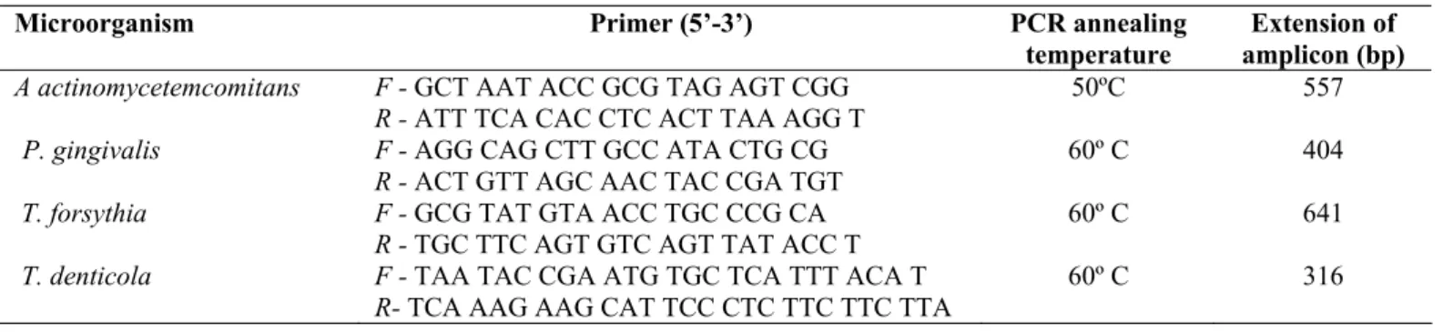
Table 1. Microorganisms and specific primers for PCR.
Microorganism Primer (5’-3’) PCR annealing
temperature
Extension of amplicon (bp) A actinomycetemcomitans F - GCT AAT ACC GCG TAG AGT CGG
R - ATT TCA CAC CTC ACT TAA AGG T
50ºC 557
P. gingivalis F - AGG CAG CTT GCC ATA CTG CG R - ACT GTT AGC AAC TAC CGA TGT
60º C 404
T. forsythia F - GCG TAT GTA ACC TGC CCG CA R - TGC TTC AGT GTC AGT TAT ACC T
60º C 641
T. denticola F - TAA TAC CGA ATG TGC TCA TTT ACA T R- TCA AAG AAG CAT TCC CTC TTC TTC TTA
60º C 316
The PCR products (9.5 μl) were added to 0.5 μl of blue
green fluorescence (LGC Biotecnologia, Brazil) and submitted
to electrophoresis in 1.5% agarose gel. The gels were then visualized and photographed on a UV transilluminator for later
analysis. A 100-bp DNA ladder (LGC Biotecnologia, Brazil)
was used as the molecular mass standard. All samples negative
for pathogens were repeated and confirmed.
Statistical Analysis
Analysis was performed using descriptive statistics
(frequency, mean, mode, median) and the chi-square test or Fisher’s exact test, when the conditions for using the chi-square
did not apply. A 5% level of significance and 95% confidence
interval were used for all statistical tests.
RESULTS
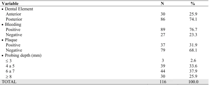
Patient age ranged from 30 to 59 years (mean: 41.8 years). More than half the patients (58.6%) were female. Regarding
the sites evaluated, 74.1% of samples were collected from
posterior teeth. Most (76.7%) exhibited bleeding upon probing,
37.9% had a probing depth of 6 to 7 mm and only 31.9% had
visible dental plaque (Table 2).
Among the 116 collection sites, 46.6% were positive for
P. gingivalis, 41.4% were positive for T. forsythia, 33.6% were positive for T. denticola and 27.6% were positive for A. actinomycetemcomitans (Figure 1). There were no significant associations (p > 0.05) between these bacteria and gender, age,
Table 2. Evaluation of the tooth, plaque, bleeding and probing depth
Variable N %
Dental Element
Anterior 30 25.9
Posterior 86 74.1
Bleeding
Positive 89 76.7
Negative 27 23.3
Plaque
Positive 37 31.9
Negative 79 68.1
Probing depth (mm)
3 3 2.6
4 a 5 39 33.6
6 a 7 44 37.9
8 30 25.9
TOTAL 116 100.0
Figure 1. Percentage of bacteria in sites
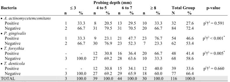
Significant associations were found between probing
depth and the bacteria P. gingivalis and T. forsythia (p < 0.05), demonstrating that these bacteria were more prevalent in deep pockets ( 8 mm), followed by sites with probing depths of 6
to 7 mm (Table 3). Table 4 displays the occurrence bacteria
either isolated or combined according to probing depth. At sites with a probing depth 8 mm, the two most frequent
Farias, B.C. et al. Periodontal pathogens among patients with periodontitis
forsythia + P. gingivalis + T. denticola (20.0%).
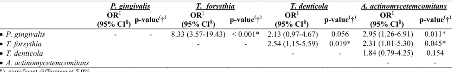
In the analysis of the relationship between periodontal
pathogens, there were significant associations for A.
actinomycetemcomitans + P. gingivalis and A.
actinomycetemcomitans + T. forsythia (p < 0.05), with odds ratios
of 2.95 and 2.31, respectively. The percentage of sites with T.
forsythia + T. denticola was higher when P. gingivalis was present
than when absent (66.7% vs. 19.4% and 42.6% vs. 25.8%,
respectively); however, only the only significant association was
for T. forsythia + P. gingivalis (p < 0.05), with an odds ratio 8.33.
There was also a significant association for T. forsythia + T.
denticola (p < 0.05), with an odds ratio of 2.54 (Table 5).
Table 3. Evaluation of bacteria at sites according to probing depth.
Probing depth (mm)
Bacteria 3 4 to 5 6 to 7 8 Total Group p-value
n % n % n % n % N %
A. actinomycetemcomitans
Positive 1 33.3 8 20.5 13 29.5 10 33.3 32 27.6 p(†) = 0.591
Negative 2 66.7 31 79.5 31 70.5 20 66.7 84 72.4
P. gingivalis
Positive 1 33.3 9 23.1 21 47.7 23 76.7 54 46.6 p(†) < 0.001*
Negative 2 66.7 30 76.9 23 52.3 7 23.3 62 53.4
T. forsythia
Positive - - 12 30.8 16 36.4 20 66.7 48 41.4 p(†) = 0.005*
Negative 3 100.0 27 69.2 28 63.6 10 33.3 68 58.6
T. denticola
Positive - - 12 30.8 15 34.1 12 40.0 39 33.6 p(†) = 0.660
Negative 3 100.0 27 69.2 29 65.9 18 60.0 77 66.4
TOTAL 3 100.0 39 100.0 44 100.0 30 100.0 116 100.0
(*): significant difference at 5.0% (†): Fisher’s exact test
Table 4. Assessment of isolated or combined bacteria in sites according to probing depth
Probing depth (mm)
Bacteria 3 4 to 5 6 to 7 8 Total Group
N % n % N % n % n %
None 2 66.7 18 46.2 15 34.1 2 6.7 37 31.9
A. actinomycetemcomitans - - 1 2.6 2 4.5 1 3.3 4 3.4
P. gingivalis - - - - 2 4.5 2 6.7 4 3.4
T. forsythia - - 1 2.6 1 2.3 1 3.3 3 2.6
T. denticola - - 5 12.8 3 6.8 - - 8 6.9
A. actinomycetemcomitans + P. gingivalis 1 33.3 - - 2 4.5 3 10.0 6 5.2
A. actinomycetemcomitans + T. forsythia - - 1 2.6 - - 1 3.3 2 1.7
A. actinomycetemcomitans + T. denticola - - 1 2.6 - - - - 1 0.9
P. gingivalis + T. forsythia - - 4 10.3 4 9.1 7 23.3 15 12.9
P. gingivalis + T. denticola - - 1 2.6 3 6.8 1 3.3 5 4.3
T. forsythia + T. denticola - - 1 2.6 1 2.3 1 3.3 3 2.6
A. actinomycetemcomitans + P. gingivalis + T.
forsythia - - 2 5.1 3 6.8 1 3.3 6 5.2
A. actinomycetemcomitans + P. gingivalis + T.
denticola - - 1 2.6 1 2.3 1 3.3 3 2.6
A. actinomycetemcomitans + T. forsythia + T.
denticola - - 2 5.1 1 2.3 1 3.3 4 3.4
P. gingivalis + T. forsythia + T. denticola - - 1 2.6 2 4.5 6 20.0 9 7.8
A. actinomycetemcomitans + P. gingivalis + T.
forsythia + T. denticola - - - - 4 9.1 2 6.7 6 5.2
Table 4. Associations between pathogens analyzed.
(*): significant difference at 5.0% (†): chi-square test
(‡): OR – odds ratio (§): CI – confidence interval
DISCUSSION
The present study assessed the occurrence of periodontal
pathogens in a convenience sample of adult Brazilian
nonsmokers with chronic periodontitis in the city of Recife
(northeastern Brazil). It takes on particular importance in light
of the small number of studies carried out on Brazilian
populations, particularly those in the northeastern region of the country.
Strict inclusion and exclusion criteria were employed in
the present study in order to obtain a homogeneous sample.
Previous other studies evaluating the subgingival microbiota
involved a considerable diversity of participants (8, 24, 25).
Diversity in samples leads to a greater number of biases, but
encourages the participation of a large number of individuals.
Factors such as the inclusion of both smokers and nonsmokers with no distinguishing between the two, patients in use of
antibiotics and anti-inflammatory agents, those with systemic
diseases and those with tooth loss are examples of selection
biases, as these aspects can influence the status of the
subgingival microbiota and, consequently, affect the results.
The eligibility criteria employed in the present study led to a
small number of patients, but with similar characteristics.
The pathogens investigated are known to be associated with the progression and severity of periodontal disease.
Epidemiological studies involving adults have demonstrated
significant variation in the prevalence of periodontal pathogens
depending on race, ethnicity, geographic location (24) and
methodology employed.
In Brazil, few studies have been conducted to investigate
the periodontal microbiota and most of these have been carried
out in the state of São Paulo (southeastern Brazil) (7, 8, 13, 14,
15, 26). Comparing the data to those of reported in other
studies involving Brazilian patients with periodontal disease,
the frequency of pathogens was lower in the present study (7,
8, 9, 13, 15, 26). However, differences in methodology and
distinct population characteristics (ethnic diversity) in the different regions investigated should be stressed.
Differences in the results reported in previous studies
carried out in Brazil and elsewhere may also be explained by
the different periodontal conditions assessed. The low presence
of bacteria in the sites evaluated may be explained by the
evaluation of a high number of shallow sites, as the patients in
the present study exhibited different degrees of chronic
periodontitis. A. actinomycetemcomitans
In sites with a probing depth 8 mm, a higher frequency
T. forsythia + P. gingivalis and P. gingivalis + T. denticola + T. forsythia combinations was found, confirming the strong association between chronic periodontitis and red complex
species, as also demonstrated by Socransky et al. (22) and Hamlet et al. (11). This fact can be explained by the deeper pockets offer a better medium for anaerobes and, consequently,
greater colonization of these pathogens (11, 22). In addition,
Zambon (28) also explains the strong association of T. forsythia and P. gingivalis pathogens in the pathogenesis of periodontitis, as these bacteria causes tissue loss and severe
alveolar bone resorption. Considering these facts, it is expected
that these pathogens be more present in deeper sites with P. gingivalis T. forsythia T. denticola A. actinomycetemcomitans OR‡
(95% CI§) p-value (
†) OR
‡
(95% CI§) p-value (
†) OR
‡
(95% CI§) p-value (
†) OR
‡
(95% CI§) p-value (
†)
P. gingivalis - - 8.33 (3.57-19.43) < 0.001* 2.13 (0.97-4.67) 0.056 2.95 (1.26-6.91) 0.011*
T. forsythia - - 2.54 (1.15-5.59) 0.019* 2.31 (1.01-5.30) 0.045*
T. denticola - - 1.84 (0.79-4.25) 0.154
Farias, B.C. et al. Periodontal pathogens among patients with periodontitis
greater signs of periodontal disease. Moreover, the
simultaneous presence of these bacteria in deep sites suggests a
symbiotic relationship between these virulent species, favoring,
in this way, a further progression of periodontal disease.
As found in the study carried out by Hamlet et al. (11), the bacterium A. actinomycetemcomitans exhibited a different behavior from the other pathogens, with a lower frequency at
deeper probing depths. This may be explained by the fact that
this pathogen is a facultative anaerobe.
It was concluded that the bacteria P. gingivalis, T. forsythia and T. denticola are related to chronic periodontitis, presenting a higher occurrence in deep periodontal pockets.
Moreover, the simultaneous presence of these bacteria in deep
sites suggests a symbiotic relationship between these virulent species, favoring, in this way, a further progression of
periodontal disease.
REFERENCES
1. Ainamo, J.; Bay, I. (1975). Problems and proposals for recording gingivitis and plaque. Int. Dent. J. 25, 229-35.
2. American Academy of Periodontology. (1996). Consensus report on periodontal disease: pathogenesis and microbial factors. Ann. Periodontol. 67, 926-932.
3. American Academy of Periodontology (1999). International workshop for classification of periodontal diseases and conditions. Ann. Periodontol. 4, 7–112.
4. American Academy of Periodontology. (2000). Parameter on chronic periodontitis with slight to moderate loss of periodontal support. J. Periodontol. 71, 853-855.
5. Ashimoto, A. ; Chen, C. ; Bakker, I. ; Slots, J. (1996). Polymerase chain reaction detection of 8 putative periodontal pathogens in subgingival plaque of gingivitis and advanced periodontal lesions. Oral Microbiol. Immunol. 11, 266-273.
6. Atieh, A. (2008). Accuracy of real-time polymerase chain reaction versus anaerobic culture in detection of Aggregatibacter actinomycetemcomitans and Porphyromonas gingivalis: a meta-analysis. J. Periodontol. 79, 1620-1629.
7. Ávila-Campos, M.J.; Velásquez-Meléndez, G. (2002). Prevalence of putative periodontopathogens from periodontal pacients and healthy subjects in São Paulo, SP, Brazil. Rev. Inst. Med. Trop. Sao Paulo. 44, 1-5.
8. Ávila-Campos, M.J. (2003). PCR detection of four periodontopathogens from subgingival clinical samples. Braz. J. Microbiol. 34, 81-84. 9. Cortelli, J.R.; Cortelli, S.C.; Jordan, S.; Haraszthy, V.I.; Zambon, J.J.
(2005). Prevalence of periodontal pathogens in Brazilians with aggressive or chronic periodontitis. J. Clin. Periodontol. 32, 860-866. 10. Dogan, B.; Antinhelmo, J.; Cetiner, D.; Bodur, A.; Emingil, G.;
Buduneli, E.; Uygur, C.; Firatli, E.; Lakio, L.; Asikainen, S. (2003). Subgingival microflora in Turkish patients with periodontitis. J. Periodontol. 74, 803-814.
11. Hamlet, S.M.; Cullinan, M.P.; Westerman, B.; Lindeman, M.; Bird, P.S.; Palmer, J.; Seymour, G.J. (2001). Distribution of Actinobacillus actinomycetemcomitans, Porphyromonas gingivalis and Prevotella intermedia in an Australian population. J. Clin. Periodontol. 28, 1163– 1171.
12. Herrera, D.; Contreras, A.; Gamonal, J.; Oteo, A.; Jaramillo, A.; Silva, N.; Sanz, M.; Botero, J.E; León, R. (2007). Subgingival microbial profiles in chronic periodontitis patients from Chile, Colombia and Spain. J. Clin. Periodontol. 35, 106-113.
13. Imbronito, A.V.; Okuda, O.S.; Freitas, N.M.; Lotufo, R.F.M; Nunes, F.D. (2008). Detection of herpes viruses and periodontal pathogens in subgingival plaque of patients with chronic periodontitis, generalized aggressive periodontitis, or gingivitis. J. Periodontol. 79, 2313-2321. 14. Kantorski, K.Z.; Rodrigues, A.S.; Zimmemann, G.S.; Lotufo, R.F.M.
(2006). Ocurrence of Porphyromonas gingivalis in patients with periodontitis in Brazil. Ciênc. Odontol. Bras. 9, 26-31.
15. Klein, M.I.; Gonçalves, R.B. (2003). Detection of Tannerella forsythensis (Bacteroides forsythus) and Porphyromonas gingivalis by polimerase chain reaction in subjects with different periodontal status. J. Periodontol. 74, 798-802.
16. Kim, T.S.; Kang, N.W.; Lee, S.B.; Eickholz, P.; Pretzl, B.; Kim, C.K. (2009). Differences in subgingival microflora of Korean and German periodontal patients. Arch. Oral Biol. 54, 223-229.
17. Mayanagi, G.; Sato, T.; Shimauchi, H.; Takahashi, N. (2004). Detection frequency of periodontitis-associated bacteria by polymerase chain reaction in subgingival and supragingival plaque of periodontitis and healthy subjects. Oral Microbiol. Immunol. 19, 379-385.
18. Mineoka, T.; Awano, S.; Rikimaru, T.; Kurata, H.; Yoshida, A.; Ansai, T.; Takehara, T. (2008). Site-specific development of periodontal disease is associated with increased levels of Porphyromonas gingivalis, Treponema denticola, and Tannerella forsythia in subgingival plaque. J. Periodontol. 79, 670-676.
19. Morikawa, M.; Chiba, T.; Tomil, N.; Sato, S.; Takahashi, Y.; Konishi, K.; Numabe, Y.; Iwata, K.; Imai, K. (2008). Comparative analysis of putatite periodontopathic bacteria by multiplex polymerase chain reaction. J. Periodont. Res. 43, 268-274.
21. Sakamoto, M.; Takeuchi, Y.; Umeda, M.; Ishikawa, I.; Benno, Y. (2001). Rapid detection and quantification of five periodontopathic bacteria by real-time PCR. Microbiol.Immunol. 45, 39-44.
22. Socransky, S.S.; Haffajee, A.D.; Cugni, M.A.; Smith, C.; Kent Jr., R.L. (1998). Microbial complexes in subgingival plaque. J. Clin. Periodontol. 25, 134-144.
23. Tatakis, D.N.; Kumar, P.S. (2005). Etiology and pathogenesis of periodontal diseases. Dent. Clin. North Am. 49, 491-516.
24. Torrungruang, K.; Bandhaya, P.; Likittanasombat, K.; Grittayaphong, C. (2009). Relationship between the presence of certain bacterial pathogens and periodontal status of urban Thai adults. J. Periodontol. 80, 122-129. 25. van Winkelhoff, A.J.; Loos, B.G.; van der Reijden, W.A.; van der
Velden, U. (2002). Porphyromonas gingivalis, Bacteroides forsythus and
other putative periodontal pathogens in subjects with and without periodontal destruction. J. Clin. Periodontol. 29, 1023–1028.
26. Victor, L.V.; Cortelli, S.H.; Aquino, D.R.; Carvalho Filho, J.; Cortelli, J.R. (2008). Periodontal profile and presence of periodontal pathogens in young African-Americans from Salvador, BA, Brazil. Braz. J.Microbiol 39, 226-232.
27. Wara-aswapati, N.; Pitiphat, W.; Chanchaimongkon, L.; Taweechaisupapong, S.; Boch, J.A.; Ishikawa, I. (2009). Red bacterial complex is associated with the severity of chronic periodontitis in a Thai population. Oral diseases. 15, 354-359.
28. Zambon, J.J. (1996). Periodontal disease: Microbial factors. Ann. Periodontol. 1, 879-925.