PLANKTONIC AND BIOFILM CELLS
Fernanda Gomes1, Bruna Leite1,2, Pilar Teixeira1, Joana Azeredo1, Rosário Oliveira1*
1
IBB-Institute for Biotechnology and Bioengineering, Centre of Biological Engineering, University of Minho, Campus de Gualtar,
4710-057, Braga, Portugal; 2Departamento de Biotecnologia, Universidade Federal de São Carlos, São Carlos, SP, Brasil.
Submitted: August 30, 2010; Approved: August 30, 2011.
ABSTRACT
Staphylococcus epidermidis is the most frequent cause of nosocomial sepsis and catheter-related infections,
in which biofilm formation is considered to be the main virulence mechanism. In biofilm environment,
microbes exhibit enhanced resistance to antimicrobial agents. This fact boosted the search of possible
alternatives to antibiotics. Farnesol and N-acetylcysteine (NAC) are non-antibiotic drugs that have
demonstrated antibacterial properties. In this study, the effect of farnesol and NAC isolated or in
combination (farnesol+NAC) was evaluated. NAC at 10 × MIC caused a total cell death in planktonic cells.
On the other hand, S. epidermidis biofilms exhibited 4 log reduction in viable cell number after a 24h
treatment with NAC at the former concentration. Our results demonstrated that there was a higher CFU log
reduction of S. epidermidis planktonic cells when farnesol was combined with NAC at 1 × MIC relatively to
each agent alone. However, these results were not relevant because NAC alone at 10 × MIC was always the
condition which gave the best results, having a very high killing effect on planktonic cells and a significant
bactericidal effect on biofilm cells. This study demonstrated that no synergy was observed between farnesol
and NAC. However, the pronounced antibacterial effect of NAC against S. epidermidis, on both lifestyles,
indicates the use of NAC as a potential therapeutic agent in alternative to antibiotics.
Key words: Nosocomial infection; biofilm; Staphylococci; farnesol; N- acetylcysteine.
INTRODUCTION
Staphylococcus epidermidis lives naturally on the skin and
mucous membrane as a commensal of the human skin flora (9)
and was primarely considered a natural human inhabitant
bacterium with a low pathogenic potential (22). However, in
recent decades, this bacterium was identified as a common
cause of numerous infections on indwelling medical devices
(22) and actually S. epidermidis is among the most leading
causes of nosocomial infections (16). These bacteria form
biofilms on implanted medical devices such as central venous
catheters (CVCs), urinary catheters, prosthetic heart valves,
orthopedic devices, contact lenses, etc, and cause persistent
infections (21) and diseases such as septicemia and
endocarditis (3). The biofilm-forming ability of Staphylococcus
epidermidis has been considered to be its main virulence
mechanism (5, 20) by which this organism is able to persist in
infections/diseases (11). Many implant infections sometimes
requires the implant removal, causing considerable suffering
for the patient, with pain and disability and even increased
mortality (8, 9). Moreover, the long-term systemic antibiotics,
surgical debridement, and prolonged hospitalization, greatly
increase the costs associated with implant replacement surgery.
The biofilm formation ability is a major clinical problem,
mainly due to the intrinsic tolerance/resistance of biofilm cells
to antibiotics (5). Antibiotic combination represents a
therapeutic option in the treatment of S. epidermidis infections
(14). However, increasing multiple resistance to antibiotics has
made the development of new treatment options for serious
infections a matter of urgent concern. In recent years, much
research has been devoted to investigating possible alternatives
to antibiotics, studying their mode of action and synergistic
effects with other antimicrobial compounds. Farnesol is a
sesquiterpene alcohol that has demonstrated to inhibit the
growth of some microorganisms, such as Staphylococcus
aureus and Streptococcus mutans, evidencing its potential use
as antimicrobial agent (6, 11). The mechanism of action of this
sesquiterpenoid probably involves cell membrane damages (6,
11, 13). N-acetylcysteine is another non-antibiotic drug that has
antibacterial properties (17). NAC is one of the smallest drug
molecules in use and it is generally used in the medical
treatment of chronic bronchitis, cancer and paracetamol
intoxication (15). The prevention of biofilm formation and
adherence to biomaterials devices is another possible role of
NAC (17).
Considering the results previously obtained with these
compounds, the purpose of this work was to investigate the
possible synergistic effect of farnesol with N-acetylcysteine
against S. epidermidis planktonic and biofilm cells.
MATERIALS AND METHODS
Bacterial strains and culture conditions
Two clinical isolates of S. epidermidis, known for their
ability to form biofilms, were used in this work: strain 1457
(isolated from an infected central venous catheter) and strain
9142, a known producer of the polysaccharide intracellular
adhesin (PIA). All strains were gently provided by Dr. G. B.
Pier, Channing Laboratory, Department of Medicine, Brigham
and Women’s Hospital, Harvard Medical School, Boston. Both
strains were grown for 18 ± 2 h, at 37ºC and 120 rpm in 30 mL
of Tryptic Soy Broth (TSB) (Merck, Darmstadt, Germany).
Then the cells were centrifuged (9500 ×g, 5 min, 4ºC), washed
twice with a saline solution [0.9% NaCl (Merck, Darmstadt,
Germany) in distilled water] and sonicated (Ultrasonic
Processor, Cole-Parmer Illinois, USA) (22% amplitude, 10s).
The cellular suspensions were adjusted to a final concentration
of approximately 1 × 10⁹ cells mL-1, determined by optical density at 640 nm, prior to be used in the subsequent assays.
Planktonic assays
Viability assays were performed in 100 mL Erlenmeyers
containing a S. epidermidis cell suspension(~ 2 × 10⁸ cells mL -1
) in the presence of farnesol (300 M) (Sigma, St Louis,
USA), NAC (NAC 1 × MIC = 4 mg mL-1 and 10 × MIC = 40 mg mL-1) (Sigma, St Louis, USA) and farnesol-NAC. It should be noted that 300 M farnesol was previously shown to
be highly effective against planktonic cells of S. epidermidis
(7). The suspensions were incubated for 24 hours, at 37ºC and
at 130 rpm. Afterwards, cellular viability was assessed by
colony forming units (CFU), while cell activity was determined
by the XTT
({2,3-bis(2-methoxy-4-nitro-5-sulfophenyl)-5-[(phenylamino)carbonyl]-2H-tetrazolium hydroxide}) (Sigma,
St Louis, USA) reduction assay (12).
CFU enumeration: CFU were obtained as follows: 1000
L of each cellular suspension after being washed with 0.9%
NaCl, were resuspended in 0.9% NaCl, followed by 20 s of
sonication at 22 W to homogenize the suspension. This
procedure disrupted the cell clumps without impairing cell
viability (4). Viable cells were determined by performing
10-fold serial dilutions in saline solution and plating in TSA
h incubation at 37ºC.
XTT assay: For XTT assay, aliquots of 200 L of each cell
suspension were collected. The cells were washed with 0.9% NaCl
by centrifugation for 10 min at 9500 ×g and 4ºC. The pellet was
resuspended in 200 L of 0.9% NaCl and dispensed in a well of a
microtiter plate. Then, 50 L of a solution containing 200 mg L-1 of XTT and 20 mg L-1 of phenazine methosulphate (PMS) (Sigma, St Louis, USA) were added. The microtiter plates were incubated
for 3 h at 37ºC in the dark. The absorbance was measured at 490
nm.
Controls included cells not exposed to farnesol or NAC
(positive control) and also cells exposed either to farnesol or to
NAC alone. All experiments were carried out in triplicate and
repeated three times.
Biofilm assays
Biofilm formation and treatment: Biofilms were formed in
96 well tissue culture plates containing 200 L of S. epidermidis
cell suspension (1 × 10⁶ cells mL-1) (1457 and 9142 strains) in TSB supplemented with 0.25% glucose (Merck, Darmstadt,
Germany) per well to promote biofilm formation. Plates were
incubated for 24 h at 37ºC on an orbital shaker (130 rpm). At the
end, planktonic cells were removed carefully, and the biofilm was
washed twice with 200 L of 0.9% NaCl. The biofilms were
incubated in fresh nutrient medium containing farnesol (300 M),
NAC (1 × MIC and 10 × MIC) and combination of both. XTT,
CFU and crystal violet (CV) assays were performed after 24 hours
of exposure to antimicrobial agents (alone and in combination)
tested. At time 0 (before exposure to antimicrobial agents) the
initial cellular concentration of biofilm (~ 2 × 10⁸ cells mL-1) was determined.
XTT assay: The quantification of biofilm cellular activity
was assessed through the XTT reduction assay. After exposure to
farnesol and NAC, biofilms were washed with 0.9% NaCl. Then,
250 L of a solution containing 200 mg L-1 of XTT and 20 mg L-1 of PMS were added to each well. The microtiter plates were
incubated for 3 h at 37ºC in the dark. The absorbance was
measured at 490 nm.
CFU enumeration: CFU were obtained as follows: the
planktonic cells were removed carefully and the biofilm was
washed twice with 200 L of 0.9% NaCl. The wells were
thoroughly scraped and ressuspended in 1 mL of 0.9% NaCl,
followed by centrifugation for 10 min at 9500 ×g. The pellet was
resuspended in 0.9% NaCl and washed twice, followed by 20 s of
sonication at 22 W to homogenize the suspension. Viable cells
were determined by performing 10-fold serial dilutions in saline
solution and plating in TSA. Colonies were counted after 24 h
incubation at 37ºC.
Crystal Violet assay: CV was used as indicator of total
biofilm biomass. For the measurement of this parameter, biofilms
were washed with 250 L of 0.9% NaCl, then 250 L of methanol
(Merck, Darmstadt, Germany) were added and left to act during 15
minutes. Afterwards, methanol was removed and 250 L of crystal
violet 1% (v/v) (Merck, Darmstadt, Germany) were added (5 min).
The wells were washed with distilled water and finally, acetic acid
33% (v/v) (Merck, Darmstadt, Germany) was added. The
absorbance was measured at 570 nm.
Controls were cells not exposed to farnesol or NAC (positive
control), and cells exposed either to farnesol or NAC alone. All
experiments were carried out in triplicate and repeated three times.
Scanning Electron Microscopy (SEM)
Biofilms were dehydrated by immersion in increasing ethanol
(Merck, Darmstadt, Germany) concentration solutions: 70 (10
min), 95 (10 min) and 100% (20 min) (v/v), having then been
placed in a sealed desiccator. Samples were mounted on
aluminium strubs with carbon tape, sputter coated with gold and
observed with a Field Emission Gun – Scanning Electron
Microscope (FEG/ESEM) - Nova Nano SEM 200 from FEI
Company.
Three fields were used for image analysis. All photographs
were taken at a magnification of × 40 000.
Statistical analysis
The results from all assays were compared by the one-way
analysis of variance by applying the Bonferroni and Tukey
multiple comparison tests, using the SPSS (Statistical Package for
the Social Sciences Inc, Chicago). All tests were performed with
RESULTS
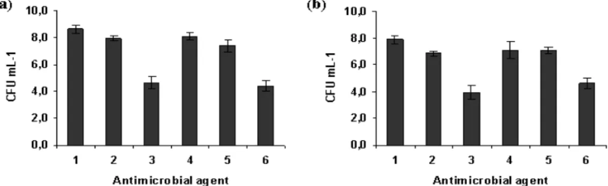
Figure 1 presents the effect of farnesol, NAC and the
association farnesol-NAC on Staphylococcus epidermidis
planktonic cells. NAC at 1 × MIC concentration is less
effective than farnesol at 300 M (p < 0.05) (Fig. 1). The
combination of farnesol at 300 M with NAC at 1 × MIC
caused a higher CFU log reduction when compared to each one
alone (p < 0.05). This combination resulted into an additional
log reduction of 0.5 and 1 for strains 1457 and 9142,
respectively (p < 0.05) and relatively to the most effective of
both antimicrobial agents tested, ie farnesol at 300 M.
However, NAC at 10 × MIC was more effective than farnesol
alone and farnesol and NAC 1 × MIC. After 24 hours, NAC 10
× MIC caused 8 log reduction resulting in total cell death (Fig.
1).
Figure 1. Effect of farnesol and/or NAC on planktonic cells of S. epidermidis 1457 (a) and 9142 (b), after 24 hours of contact with farnesol (300 M), NAC (4 mg mL-1 and 40 mg mL-1) and farnesol-NAC. Error bars represent standard deviation. Legend: 1- Positive control; 2- NAC 1 × MIC; 3- NAC 10 × MIC; 4- Farnesol 300 µM; 5- Farnesol 300 µM + NAC 1 × MIC; 6- Farnesol 300 µM + NAC 10 × MIC.
Relatively to biofilm cells, although NAC 10 × MIC did not
cause total cell death it was the most efficient against S.
epidermidis biofilm cells causing a reduction of approximately 4
log (Fig. 2). Conversely to planktonic cells, farnesol and NAC 1 ×
MIC had a similar effect in biofilms. For strain 1457, NAC 1 ×
MIC and farnesol worked better together than alone (p < 0.05)
(Fig. 2a). There was no synergistic or additional effect when NAC
10 × MIC was combined with farnesol at 300 M (p < 0.05).
The results of the XTT reduction assay, indicative of the
metabolic activity of cells within the biofilm and CV staining
assay, which allows the quantification of the total biofilm
biomass, after a 24h treatment with farnesol, NAC, and
farnesol+NAC are presented in table 1. These results
confirmed the absence of synergy between farnesol and NAC.
NAC at 10 × MIC was the antimicrobial agent treatment more
efficient against both S. epidermidis strains tested, causing a
significant decrease in the metabolic activity of biofilm cells
and total biofilm biomass (p < 0.05).
Table 1. Optical density (absorbance) and confidence interval obtained from XTT and Crystal Violet assays in biofilm cells of S.
epidermidis after exposure to farnesol, NAC and farnesol/NAC combination.
S. epidermidis 1457 S. epidermidis 9142
Condition XTT CV XTT CV
Positive control 3.124 ± 0.151 2.735 ± 0.280 2.314 ± 0.099 2.807 ± 0.279 NAC 1 × MIC 1.720 ± 0.149 2.649 ± 0.232 1.452 ± 0.078 2.577 ± 0.281
NAC 10 × MIC 1.277 ± 0.173 1.931 ± 0.117 1.124 ± 0.156 1.817 ± 0.204
Farnesol 300 µM 1.910 ± 0.185 2.763 ± 0.250 1.715 ± 0.097 2.260 ± 0.379 Farnesol 300 µM + NAC 1 × MIC 1.928 ± 0.148 2.707 ± 0.241 1.498 ± 0.161 2.132 ± 0.386
Farnesol 300 µM + NAC 10 × MIC 1.360 ± 0.106 1.868 ± 0.127 1.195 ± 0.169 1.681 ± 0.290
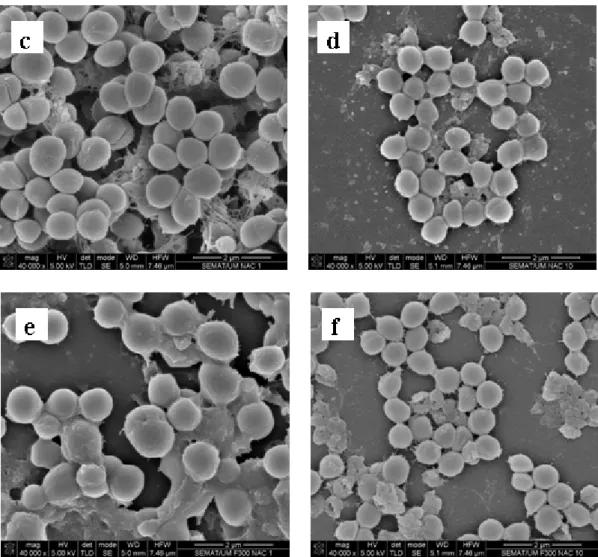
Representative scanning electron microscopy images of
1457 S. epidermidis biofilms after being exposed to farnesol,
NAC and farnesol-NAC are presented on figure 3. These
images specifically show the effect on the biofilm matrix and
biofilm cell viability, and are in agreement with the results
presented above. All biofilms treated with NAC revealed a
desintegration of the matrix which is more noticeable for NAC
Figure 3. Scanning electron micrographs of 24 h-biofilm of S. epidermidis 1457 after exposure to farnesol, NAC, and the
combination of both for 24 h. (a) Positive control; (b) 300 µM farnesol; (c) NAC 1 × MIC; (d) NAC 10 × MIC; (e) Farnesol 300
µM + NAC 1 × MIC; (f) Farnesol 300 µM + NAC 10 × MIC. Magnification × 40 000.
DISCUSSION
In this work, the effect of farnesol, NAC and
farnesol-NAC combination against S. epidermidis planktonic and
biofilm cells was studied. For that, two good biofilm-forming
strains were selected, strains 1457 and 9142 (19). Comparing
these two strains, 1457 produces slightly more biofilm than
9142 (19). The biofilm formation ability is due to the formation
of PIA homopolymer, which surrounds and connects S.
epidermidis cells in biofilm form (16). The extracellular matrix
is extremely important for intercellular connection during
surface colonization (10) and protection against the host
immune system and resistance to antibiotics (1). Figure 3a
represents a 48 hours biofilm of S. epidermidis 1457 and shows
the thickness of biofilm and the presence of a noticeable
amount of biofilm matrix.
N-acetylcysteine (NAC), a potent antioxidant that reduces
disulphide bonds linking mucin oligomers, has been widely
used as a mucolytic agent for inhalation therapy in patients
reduce adhesion but also to detach bacterial cells adhered to
surfaces and to inhibit bacterial growth in vitro (15). NAC
decreases biofilm formation by a variety of bacteria and
reduces the production of extracellular polysaccharide matrix,
while promoting the disruption of mature biofilm (2).
On the other hand, the principal interaction of farnesol
appears to be with the cytoplasmatic membrane (11). Farnesol
is a sesquiterpenoid that already demonstrated synergistic
effect with another antimicrobial agent (gentamicin) indicating
a potential application as an adjuvant therapeutic agent (11).
According to previous studies, where farnesol was tested at
concentrations ranging from 30 to 300 µM, the last
concentration demonstrated to have an antimicrobial effect
against S. epidermidis as well as against other bacteria (7, 11).
We hypothesized that the combination of NAC with
farnesol could be synergystic in the treatment of S. epidermidis
infections as they both act on different components of the
biofilm. Our results revealed that additionally to be bactericidal
NAC seems also to act against the matrix. In fact, NAC seems
to destroy the biofilm matrix resulting in the detachment of
cells and thus the biofilm cells become more exposed and
susceptible. This high effect against biofilm cells of S.
epidermidis must be due in part to the small molecular size of
NAC (Molecular Weight = 163.19), which easily penetrates
into the biofilm. NAC at 1 × MIC in combination with 300 µM
farnesol resulted in a higher antimicrobial effect against
planktonic cells of S. epidermidis 1457 and 9142 than both
antimicrobial agents alone. Nevertheless NAC alone at 10 x
MIC, similarly to biofilms, showed a very high bactericidal
effect. Although its very high effect on planktonic cells
promoting CFU reductions above 8 log, it is probably more
impressive its bactericidal effect on biofilms, which are always
very tolerant to the most common antibiotics (7). However,
unlike it was expected it did not work in synergy with farnesol
at 300 µM against biofilm cells.
Comparatively to planktonic cells, biofilm cells were
much more tolerant to the inhibitory effect of farnesol, NAC
and farnesol-NAC. As mentioned above, this fact must be due
to the protective effect of the matrix. The effect of NAC was
concentration dependent. While with NAC at 1 × MIC an
average reduction of 2.5 log was observed, NAC 10 × MIC
was enought to kill all planktonic cells. However, for biofilm
cells this concentration (10 × MIC) only promoted an
approximatly 4 log reduction in the number of viable cells
within the biofilm, while only 1 log was attained with 1 × MIC.
The peak serum concentration of NAC after a 600 mg oral
dose was estimated to be 0.465 mg mL-1 (18). The concentrations of NAC tested in our study (1 × MIC and 10 ×
MIC, 4 and 40 mg mL-1) are rather higher than those reached in serum when applied by the intravenous or oral route. However,
when applied locally it may be possible to obtain
concentrations that prevent the formation of biofilms and
consequently the adherence of S. epidermidis. (17).
In another study, a concentration of 80 mg mL-1 of NAC was tested in vitro based on preliminary data that showed a
dose-response relashionship on planktonic bacteria (2). Based
in these results it seems to be feasible the use of 40 mg mL-1in vivo.
In conclusion, NAC at 40 mg mL-1 was the only of the tested treatments that was bactericidal against S. epidermidis
cells both in planktonic or in biofilm form. Moreover, although
NAC and farnesol have different modes of action, the
combination of both has no significant synergistic effect.
ACKNOWLEDGEMENTS
Fernanda Gomes and Pilar Teixeira fully acknowledge the
financial support of Fundação para a Ciência e Tecnologia
(FCT) through the grants SFRH/BD/32126/2006 and
SFRH/BPD/26803/2006, respectively.
REFERENCES
2. Aslam, S.; Trautner, B.W.; Ramanathan, V.; Darouiche, R. (2007). Combination of tigecycline and N-acetylcysteine reduces biofilm-embedded bacteria on vascular catheters. Antimicrob. Agents Chemother. 51, 1556-1558.
3. Cargill, J.S.; Upton, M. (2009). Low concentration of vancomycin stimulate biofilm formation in some clinical isolates of Staphylococcus epidermidis. J. Clin. Pathol. 62, 1112-1116.
4. Cerca, N.; Martins, S.; Cerca, F.; Jefferson, K.K.; Pier, G.B.; Oliveira, R.; Azeredo, J. (2005a). Comparative assessment of antibiotic susceptibility of coagulase-negative staphylococci in biofilm versus planktonic culture as assessed by bacterial enumeration or rapid XTT colorimetry. J. Antimicrob. Chemother. 56, 331-336.
5. Cerca, N.; Martins, S.; Sillankorva, S.; Jefferson, K.K.; Pier, G.B.; Oliveira, R.; Azeredo, J. (2005b). Effects of growth in the presence of subinhibitory concentrations of dicloxacillin on Staphylococcus epidermidis and Staphylococcus haemolyticus biofilms. Appl. Environ.
Microbiol. 71, 8677-8682.
6. Derengowski, L.S.; De-Souza-Silva, C.; Braz, S.V.; Mello-De-Sousa, T.M.; Báo, S.N.; Kyaw, C.M.; Silva-Pereira, I. (2009). Antimicrobial effect of farnesol, a Candida albicans quorum sensing molecule, on Paracoccidioides brasiliensis growth and morphogenesis. Ann. Clin. Microbiol. Antimicrob. 8, 13.
7. Gomes, F.I.A.; Teixeira, P.; Azeredo, J.; Oliveira, R. (2009). Effect of farnesol on planktonic and biofilm cells of Staphylococcus epidermidis. Curr. Microbiol. 59, 118-122.
8. Hajdu, S.; Lassnigg, A.; Graninger, W.; Hirschl, A.M.; Presterl, E. (2009). Effects of vancomycin, daptomycin, fosfomycin, tigecycline, and ceftriaxone on Staphylococcus epidermidis biofilms. J. Orthop. Res. 27, 1361-1365.
9. Hellmark, B.; Unemo, M.; Nilsdotter-Augustinsson, A.; Söderquist, B. (2009). Antibiotic susceptibility among Staphylococcus epidermidis isolated from prosthetic joint infections with special focus on rifampicin and variability of the rpoB gene. Clin. Microbiol. Infect. 15, 238-244. 10. Hussain, M.; Hasting, J.G.M.; White, P.J. (1991). Isolation and
composition of the extracellular slime made by coagulase staphylococci in a chemically defined medium. J. Infect. Dis. 163, 534-541.
11. Jabra-Rizk, M.A.; Meiller, T.F.; James, C.E.; Shirtliff, M.E. (2006). Effect of farnesol on Staphylococcus aureus biofilm formation and antimicrobial susceptibility. Antimicrob. Agents Chemother. 50,
1463-1469.
12. Kuhn, D.M.; Balkis, M.; Chandra, J.; Mukherjee, P.K.; Ghannoum, M.A. (2003). Uses and limitations of the XTT assay in studies of Candida growth and metabolism. J. Clin. Microbiol. 41, 506-508.
13. Kuroda, M.; Nagasaki, S.; Ohta, T. (2007). Sesquiterpene farnesol inhibits recycling of the C55 lipid carrier of the murein monomer precursor contributing to increased susceptibility to β-lactams in methicillin-resistant Staphylococcus aureus. J. Antimicrob. Chemother. 59, 425-432.
14. Monzón, M.; Oteiza, C.; Leiva, J.; Amorena, B. (2001). Synergy of different antibiotic combinations in biofilms of Staphylococcus epidermidis. J. Antimicrob. Chemother. 48, 793-801.
15. Olofsson, A.; Hermansson, M.; Elwing, H. (2003). N-acetyl-L-Cysteine affects growth, extracellular polysaccharide production, and bacterial biofilm formation on solid surfaces. Appl. Environ. Microbiol. 69, 4814-4822.
16. Otto, M. (2009). Staphylococcus epidermidis- the “accidental” pathogen. Microbiology 7, 555-567.
17. Pérez-Giraldo, C.; Rodriguez-Benito, A.; Moran, F.J.; Hurtado, C.; Blanco, M.T.; Gomez-Garcia, A.C. (1997). Influence of N-acetylcysteine on the formation of biofilm by Staphylococcus epidermidis. J. Antimicrob. Chemother. 39, 643-646.
18. Rehman, T.; Fought, J.; Solomon, R. (2008). N-acetylcysteine effect on serum creatinine and cystatin C levels in CKD Patients. Clin. J. Am. Soc. Nephrol. 3, 1610-1614.
19. Sousa, C.; Teixeira, P.; Oliveira, R. (2009). The role of extracellular polymers on Staphylococcus epidermidis biofilm biomass and metabolic activity. J. Basic Microbiol. 49, 363-370.
20. Vuong, C.; Kocianova, S.; Yao, Y.; Carmody, A.B.; Otto, M. (2004). Increased colonization of indwelling medical devices by quorum-sensing mutants of Staphylococcus epidermidis in vivo. J. Infect. Dis. 2004; 190, 1498-1505.
21. Wang, C.; Li, M.; Dong, D.; Wang, J.; Ren, J.; Otto, M.; Gao, Q. (2007). Role of ClpP in biofilm formation and virulence of Staphylococcus epidermidis. Microbes Infect 9, 1376-1383.
22. Ziebuhr, W.; Hennig, S.; Eckart, M.; Kränzler, H.; Batzilla, C.; Kozitskaya, S. (2006). Nosocomial infections by Staphylococcus epidermidis: how a commensal bacterium turns into a pathogen. Int. J.
Antimicrob. Agents 28, 14-20.