A mutant cell line partially responsive
to both IFN-
αα
αα
α
and IFN-
γγγγγ
Laboratório de Vírus, Departamento de Microbiologia,
Universidade Federal de Minas Gerais, 31270-901 Belo Horizonte, MG, Brasil C.A. Bonjardim
Abstract
A recessive mutant cell line, B7, which is partially responsive to both interferon (IFN)-α and IFN-γ is described. B7 was FACS sorted from a cellular pool, which was obtained from the parental cell line 2C4, after several rounds of mutagenesis. The partial responsiveness to IFN was observed both in terms of expression of cell surface markers (CD2, class I and II HLAs) and mRNA expression of IFN-stimulated genes (9-27; 6-16; 2’-5’ OAS; GBP and HLA-DRα). A genetic cross with the U4 mutant (JAK1-, a member of the Janus family of nonre-ceptor tyrosine kinase) did not restore full IFN responsiveness to B7, and JAK1 cDNA transfection into B7 restored the wild phenotype of the cell line, defining B7 as a member of the U4 complementation group. Nevertheless, JAK1 mRNA was not detected in this mutant. Transcriptional regulator complexes such as IRF1/2 (IFN-regulatory factor) and ISGF3-γ (IFN-stimulated gene factor) were constitutively formed in the B7 mutant and co-migrated with the IFN-induced complexes expressed in the parental cell line 2C4. Thus, this cell line seems to be useful for understanding cis-acting elements governing JAK1 mRNA expression.
Correspondence
C.A. Bonjardim Laboratório de Vírus Departamento de Microbiologia Instituto de Ciências Biológicas Universidade Federal de Minas Gerais
Av. Antonio Carlos, 6627 Bloco F4, 258
31270-901 Belo Horizonte, MG Brasil
Fax: 55 (031) 498-1510 E-mail: [email protected]
The author was the recipient of a post-doctoral fellowship from CNPq.
Received December 28, 1995 Accepted October 16, 1996
Key words
•Interferons
•Transcriptional activation
•Molecular genetics
•JAK1
•FACS analysis
Introduction
Interferons (IFNs) constitute a family of multifunctional cytokines first identified as antiviral agents. However, other activities have been associated with these molecules such as the control of cell growth and differ-entiation, besides their modulatory regula-tion of the immune system (1).
After binding to the specific cell surface receptor, type I IFN (mainly α and ß) and type II IFN (γ) activate a number of IFN-stimulated genes (ISGs), which are believed to be responsible for the different IFN ac-tions (1,2). IFN-activated gene expression is mediated by latent cytoplasmic proteins which become phosphorylated at a tyrosine residue upon ligand-receptor binding,
mi-grate to the nucleus, bind to DNA and ini-tiate transcription (3-5).
re-sponse element (ISRE) located at the pro-moter of ISGs and initiate transcription (11-13). Due to the dual role of p91, p84 and p113 as signal transducers and activators of transcription, these molecules were renamed STAT1α, STAT1ß and STAT2, respectively. The nuclear signals resulting from the interaction of IFN-γ with its receptor occur in a fashion very similar to that described for IFN-α. After activation of STAT1α (the same component of ISGF3-α), and migra-tion to the nucleus, STAT1α activates tran-scription by binding as a dimer (14) to a regulatory sequence termed gamma-activated sequence (GAS). GAS was initially identi-fied at the promoter of the guanylate-bind-ing protein (GBP) gene (15,16), but has now been found in a variety of other genes (17-19), and STAT1α turned out to be the previ-ously described gamma-activated factor (GAF) (20).
It is known that the intracytoplasmic por-tions of both IFN-α/ß and IFN-γ receptors are devoid of an intrinsic tyrosine kinase activity (21,22), suggesting that a nonrecep-tor protein tyrosine kinase (NRPTK) is re-cruited to assemble the multisubunit recep-tor complexes in order to transduce the nuclear signals (23,24). By using a genetic approach, a number of IFN-unresponsive cell lines (U- and γ-mutants) have been gen-erated (25-28) and genetic complementa-tion of these mutants by cDNA transfeccomplementa-tion has proved valuable in dissecting both types of IFN signalling pathways (29-31).
The first evidence that a NRPTK is in-volved in these pathways came from the complementation of the U1 mutant which was transfected with the gene and cDNA encoding for TYK2 (29), a member of the Janus family of NRPTK (23,24). Comple-mentation of the U3 mutant, a STAT1α-/ STAT1ß- cell line, established the funda-mental role STAT1α plays in the IFN-α and IFN-γ transduction pathways (30), while complementation of the U4 mutant, a JAK1 -(a member of the Janus family of NRPTK)
cell line, clearly demonstrated the involve-ment of NRPTK-JAK1 in both signalling pathways (32). Another important finding was the restoration of the IFN-γ responsive-ness to the JAK2- cell line (γ1A) upon JAK2 cDNA transfection (28). Thus, the converg-ing biochemical and genetic approaches en-able to decipher the emerging picture of both IFN-α and IFN-γ signalling cascades (5), which can be summarized as follows: IFN-α: two JAK family members are re-quired: TYK2 and JAK1, plus the transcrip-tion factors p113 and/or STAT1α/STAT1ß, besides p48. IFN-γ: two JAK family mem-bers are required as well: JAK1 and JAK2 , plus the transcription activator STAT1α (dimer), probably associated with a DNA-binding protein.
In the present study, I describe the ge-netic characterization of a mutant cell line, B7, and its complementation by JAK1 cDNA. Although JAK1 mRNA expression was below detection levels in B7, a partial IFN-α and IFN-γ responsiveness was achieved. In addition, DNA-protein com-plexes are constitutively formed in this mu-tant. Thus, this cell line represents a useful model to analyze JAK1 structure-function relationships, concerning JAK1 expression.
Material and Methods
Cell culture, cell fusion and DNA transfection
(five rounds) with the frameshift agent ICR191 (25) and the cell population was subdivided into different pools. B7 was FACS sorted from one of these pools (a gift from D. Watling, ICRF, London), cloned and characterized. U3 and U4 mutants were characterized as described elsewere (27), and both are unresponsive to type I and type II IFN and correspond to STAT1α-/1ß- and JAK1- cell lines, respectively. For cell fu-sion experiments a puromycin- or hygromy-cin-dominant selectable marker was trans-fected into the cells and the resistant hybrid cell population was maintained in the pres-ence of the adequate drug selection. Cell fusion experiments employing PEG 4000 (Sigma) were performed as described else-where (27). DNA transfections using cal-cium phosphate were carried out essentially as described in Ref. 30.
FACS analysis
Fluorescence-activated cell sorter (FACSCAN-Becton Dickinson) analysis was performed as previously described (28). Briefly, 5 x 10⁵ cells were seeded in a 10-cm Petri dish and after an overnight incubation they were treated with 103 IU/ml of a highly purified mixture of IFNs-α (Wellferon, 1.5 x 10⁸ IU per mg protein) from Wellcome Research Laboratories, Beckenham, Kent, UK, or recombinant human IFN-γ (2 x 10⁷ IU per mg protein), a generous gift from Dr. G. Adolf, Ernst Boehringer Institut für Arzneimittelforschung, Vienna, Austria. Cells were treated with IFN for 48-72 h and then incubated with antibodies as described in Ref. 28 for 1 h at 0oC. Cells were pelleted, resuspended in PBS, fixed in 1% parafor-maldehyde and analyzed in a FACS using a Consort 30 program (3000 data points).
RNase protection assay
Cytoplasmic RNA was obtained from monolayer cells by NP40 lysis and phenol/
chloroform extraction. Probes were synthetized from SP6/T7 transcription vec-tors, pGEMs 3 and 4 (Promega), labelled with [32P]αUTP (Amersham, UK) to a spe-cific activity >108 cpm/µg DNA. Ten µg of RNA and 1-3 x 10⁵ cpm of each probe were employed in each assay. Single-stranded RNA was digested with ribonucleases and protected double-stranded RNA was re-solved on a 6% polyacrylamide-urea gel as described elsewhere (30). Details on the construction and length of the protected frag-ments of the specific RNase protection probes (2’-5’ OAS; 6-16; GBP; HLA class II DRα; 9-27 and γ-actin) used in this work have been described elsewhere (30). The 6-16 and GBP probes were gifts from S. Goodbourn and T. Decker, respectively.
Electrophoretic mobility shift assay (EMSA)
Northern blot analysis
mRNA poly A+ was obtained according to the manufacturer (Pharmacia). Five µg RNA per lane was electrophoresed on a denaturing 1.5% agarose-formaldehyde gel, transferred to Hybond N+ membranes (Amersham, UK) as previously described (36) and hybridized with an αdCTP [32P] human JAK1 cDNA probe, labelled to a
specific activity >108 cpm/µg DNA, by us-ing a multiprime DNA labellus-ing system from Amersham, UK. Hybridization was per-formed at 65oC and subsequent washes were carried out according to Church and Gilbert (37). After exposure for 7 days, the mem-brane was stripped of the JAK1 probe and then reprobed with GAPDH, labelled at the same specific activity as described above, and exposed for 16 h.
Cell number
A
2C4
B7
B7/JAK1
150 150
150
US
U α 1
U 2
U
3
6 U
5 U
4
U
US
7
U
US
U
8
U 9
γ
α γ
γ
γ α
γ
γ α
α γ
α
γ
γ
0
100 101 102 103 100 101 102 103 100 101 102 103
Fluorescence intensity (log)
Figure 1 - FACS analysis of the parental and mutant cell lines. A, 2C4, B7 and B7/JAK1 transfectants and B, genetic crosses B7/U3 and B7/U4. The expression of the cell surface markers CD2 (panels 2,5,8,11,14) and endogenous class I (panels 1,4,7,10,13) and class II HLAs (panels 3,6,9,12,15) upon IFN-α or IFN-γ stimulation (500 IU/ml for 48 h) was monitored by FACS analysis as described in Material and Methods. Transfectants were analyzed as a population. Panels 1-3: Parental cell line 2C4; panels 4-6: B7 cell line; panels 7-9: B7/JAK1 transfectants; panels 10-12: genetic cross between B7 and the U3 mutant; panels 13-15: genetic cross between B7 and the U4 mutant. Untreated cells (U), IFN-α (α) or IFN-γ (γ) treated cells, and unstained cells (US) as indicated. US cells were left untreated or IFN-α treated, but were not incubated with any antibody.
Class II CD2
B
150
US U α
10
U
11
U
12 γ
α
γ
γ
B7/U3
B7/U4
150
US
U
α
13
U
14
U
15
γ α
γ
γ Class II CD2
Class I
0
100 101 102 103 100 101 102 103 100 101 102 103
Fluorescence intensity (log) Results
FACS analysis of the parental cell line 2C4 and the B7 mutant
FACS analysis of the B7 mutant indi-cated that this cell line responded partially to IFN-α and IFN-γ when the cell surface marker (CD2) and the endogenous class I HLA were investigated, whereas no response to IFN-γ was observed when the expression of endogenous class II HLA was analyzed (Figure 1A, panels 4-6). The expression of these genes in response to both IFN-α and IFN-γ in the parental cell line 2C4 is shown in Figure 1A (panels 1-3). B7 is a recessive mutant since crossing with 2C4 and with the wild type 2fTGH (25) restores the full re-sponsiveness of the cells to both types of IFN (data not shown). The mutation(s) in B7 seems to be very stable since its
pheno-type remained the same after several pas-sages in culture for more than three months, as confirmed by FACS analysis and RNase protection experiments.
JAK1 complements B7 mutant
From the FACS analysis data (Figure 1A, panels 4-6), it was evident that the na-ture of the mutation(s) affecting both IFN-α and IFN-γ signalling pathways in this par-ticular cell line may reside in a common polypeptide(s) which serves simultaneously in both the IFN-α and IFN-γ transduction cascades. To address this question, the mu-tant cell lines U3 and U4, which are known to be unresponsive to both types of IFN (27), were fused to the B7 mutant and cell surface expression of CD2, and class I and II HLAs were monitored by FACS analysis as described elsewhere (28). Full
respon-Cell number
1 2 3 4 5 6 7 8 9 10 11 12
Actin GBP
9-27
2’-5’ OAS IFN
Cell line 2C4 B7 2C4
α γ α γ α γ α γ
DRα
6-16
GBP
Actin
U U U U
→ → → → → → → → → → → →→ → → → → → → →
→ → → → →
→ → → → →
→ → → → →
→ → → → →
Figure 2 - IFN-stimulated gene expression in the parental cell line 2C4 and in the B7 mutant. The expression of 2-5 oligo A synthetase (OAS), 9-27 and GBP (lanes 1-6), and class II HLA (DRα), 6-16 and GBP (lanes 7-12) mRNAs in the parental cell line 2C4 and in the B7 mutant was investigated upon IFN-α or IFN-γ treatment. Five hundred IU/ml of either IFN-α or IFN-γ was added to the cells for 6 h and 18 h, respectively, cytoplasmic RNA was isolated (10 µg per lane), hybridized with the specific probe and analyzed by the RNase protection assay as described in Material and Methods. γ-Actin was used as an internal standard for RNA loading.
siveness to both IFN-α and IFN-γ was ob-tained after fusing B7 with the U3 mutant (Figure 1B, panels 10-12). However, fusion with U4 just maintains the same original responsiveness of B7 to both types of inter-feron (Figure 1B, panels 13-15), indicating that B7 is a member of the U4 complemen-tation group. Human JAK1 cDNA was then transfected into the B7 cell line, and cell surface expression (the same markers as described above) was monitored by FACS analysis (Figure 1A, panels 7-9). Although the experiment was done with a cell popula-tion, an enhancement of responsiveness to both IFN-α and IFN-γ was achieved, con-firming B7 as a member of the U4 comple-mentation group.
IFN-inducible gene expression
I next asked whether the IFN-α- and
IFN-γ-partial responsiveness observed in this cell line by FACS analysis is correlated with the IFN-α- and IFN-γ-gene inducibility. Fig-ure 2 shows mRNA expression of some IFN-stimulated genes (2’-5’ OAS; 6-16; GBP; 9-27 and class II HLA-DRα) in response to either IFN-α or IFN-γ. Although the expres-sion of these genes varied to some extent in response to the specific inducer, as observed in the parental cell line, the reduction in the mRNA expression levels of these genes in mutant B7 was remarkable (compare Figure 2, lanes 1-3 to 4-6 and 7-9 to 10-12), which agrees with the phenomenon of IFN-partial responsiveness observed by FACS analysis.
IFN-activated transcriptional complex formation
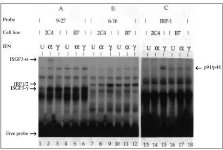
Upon IFN treatment a number of tran-scriptional activator complexes are formed depending on the type of ISG promoter (34,38,39). Thus, when the promoter of an ISG possessing a classical ISRE, such as the 9-27 gene, is used as a probe (Figure 3A), two complexes are observed after IFN stimu-lation in cell extracts prepared from a vari-ety of cell lines (34) including 2C4 cells (this work): i) ISGF3-α, which is activated/ formed upon IFN-α treatment (Figure 3A, lane 2); ii) ISGF3-γ, represented by p48, which is expressed at a low constitutive level in untreated cells, but whose level increases significantly after either IFN-α or IFN-γ treat-ment (Figure 3A, lanes 1-3). In contrast, the B7 mutant was completely inefficient in terms of ISGF3-α formation upon IFN-α treatment (Figure 3A, lane 5), whereas the expression level of ISGF3-γ in untreated cells was similar to that observed in IFN-treated cells (Figure 3A, lanes 4-6).
which comprises IRF1/IRF2 (IFN-regulator factor) proteins (34), and this was also ob-served for 2C4 cells (HT1080-derived cell line) (Figure 3B, lanes 7-9). On the other hand, the B7 mutant showed a constitutive level of IRF1/IRF2 complex formation re-gardless of the IFN treatment (Figure 3B, lanes 10-12).
When the promoter of the IRF1 gene was employed as a probe (Figure 3C), a tran-scriptional complex was detected in cell ex-tracts prepared from 2C4 cells after IFN-γ treatment (Figure 3C, lane 15), which was absent upon IFN-α treatment (Figure 3C, lane 14). Antibody characterization identi-fied p91 and p48 (or related proteins) as components of this complex (data not shown), which may act through the GAS element (17,20) or a different motif other than an ISRE (30,40), since the IRF1 pro-moter is devoid of an ISRE. In contrast, this complex formation was not observed in the B7 mutant. On the other hand, a slightly faster migrating complex was observed in the mutant, which is expressed at a low constitutive level, regardless of the IFN treat-ment (Figure 3C, lanes 16-18). The signifi-cance of this observation remains to be in-vestigated.
All the above DNA-protein complexes formed specifically competed for binding with the homologous oligonucleotide, at 50 molar excess, but did not compete with heterologous oligonucleotides (data not shown).
JAK1 mRNA expression in the B7 mutant
In order to investigate whether the par-tial responsiveness to both types of IFN in the B7 mutant (FACS analysis and ISGs mRNA expression) is correlated with the level of JAK1 mRNA expression in this cell line, poly A+ mRNA was hybridized with the human JAK1 probe. As shown in Figure 4, no detectable JAK1 message was identi-fied in the mutant cell line when compared to the parental cell line 2C4.
Figure 3 - IFN-activated transcriptional complex formation. EMSA of ISGF3-α, ISGF3-γ, IRF1/ 2 and p91/p48 in whole cell extracts from IFN-treated 2C4 and B7 cells. Cells were left untreated (U) or treated with 500 IU/ml of either IFN-α or IFN-γ for 6 h and 18 h, respectively. Cell extracts were incubated with an end-labelled oligodeoxynucleotide probe containing the promoter region of 9-27 (A, lanes 1-6); 6-16 (B, lanes 7-12) or IRF-1 (C, lanes 13-18) genes. DNA-protein complexes formed are indicated.
Discussion
By combining genetic and biochemical approaches, impressive progress has been made towards understanding the nuclear sig-nals triggered by IFN upon ligand-receptor binding (5). A converging approach employ-ing target gene disruption, which affects spe-cific genes such as IFN receptors (41), and regulatory proteins IRF1/IRF2 (42,43), has provided substantial evidence showing how the IFN system operates. On the other hand, structure-function analysis has also provid-ed important information on this emerging picture, for example i) a single amino acid substitution changing tyrosine 701 at the phosphorylation site in the STAT1 to phen-ylalanine (44) abrogates IFN-α and -γ re-sponsiveness, ii) the same substitution at position 690 in the STAT2 interferes with the phosphorylation of STAT1 (31,45), and iii) sequential deletions affecting different domains in the NRPTK-TYK2 restore the loss of kinase function (46).
In the present study I described a
reces-Probe Cell line IFN
ISGF3-α
IRF1/2
ISGF3-γ
Free probe
p91/p48
→ →→ →→
→ → → → → → → → → →
→ →→ →→
sive mutant cell line, B7, which is partially responsive to both IFN-α and IFN-γ. The defect in this mutant was associated with the NRPTK-JAK1, because this cell line is not complemented by fusion with the U4 mu-tant (JAK1-), and genetic transfer of human JAK1 cDNA into the cell line enhanced both IFN-α and IFN-γ responsiveness. Al-though the induction of ISGs, upon IFN-α and IFN-γ stimulation, was partially observed in this mutant, JAK1 mRNA expression was below detection levels. The nature of mutation(s) affecting JAK1 mRNA expres-sion remains to be further investigated, but one may speculate that it may map either at the promoter region of the gene or at the 3’ end, the latter contributing to the instability of the mRNA. However, the kinetics of ISG stimulation favors the former hypothesis. Whatever the mutation associated with the B7 phenotype, it seems that this mutant is particularly useful to understand cis-acting elements governing JAK1 expression. It is
tempting to speculate that even under these circumstances partial ISG stimulation occurs through the JAK/STAT signalling pathway (5).
Transcriptional complex formation, in-volving IRF-family members (IRF1, IRF2 and p48), which are inducible in the paren-tal cell line 2C4 upon IFN-α and IFN-γ treatment, becomes constitutively expressed in the B7 mutant. Whether this phenomenon is directly correlated with the nature of mutation(s) affecting JAK1 expression, or whether they are unrelated events remains to be determined. The possibility that addi-tional mutation(s) contributes to this par-ticular phenotype cannot be ruled out.
Thus, the mutant cell line B7 seems to be of particular importance for the understand-ing of cis-actunderstand-ing elements governunderstand-ing JAK1 mRNA expression and may represent regu-latory mechanisms associated with JAK1 expression and transcriptional activation.
Acknowledgments
I would like to thank Dr. I.M. Kerr, Im-perial Cancer Research Fund, London, for the opportunity to carry out this work in his laboratory. I also thank A. Wilks for provid-ing the human JAK1 cDNA, S. Goodbourn, T. Decker and M. Müller for kindly provid-ing the protection probes, R. Pine, D. Levy and C. Schindler for the antibodies, and Dima Guschin and L.F.L. Reis for a critical reading of the manuscript.
Figure 4 - Northern analysis of JAK1 mRNA. Five µg of poly A+ RNA from
the parental cell line 2C4 and the B7 mutant was hybridized with a human JAK1 cDNA probe as indicated. GAPDH was used as an internal standard for RNA loading.
2C4
JAK1
References
1. Pestka S, Langer JA, Zoon KC & Samuel C (1987). Interferons and their actions. An-nual Review of Biochemistry, 56: 727-777.
2. Uzé G, Lutfalla G & Mogensen KE (1995).
α and ß interferons and their receptor and their friends and relations. Journal of In-terferon and Cytokine Research, 15: 3-26.
3. Ihle JN (1995). Cytokine receptor signal-ling. Nature, 377: 591-594.
4. Pellegrini S & Schindler C (1993). Early events in signalling by interferons. Trends in Biochemical Sciences, 18: 338-342. 5. Darnell Jr JE, Kerr IM & Stark GR (1994).
JAK-STAT pathways and transcriptional activation in response to IFNs and other extracellular signaling proteins. Science, 264: 1415-1421.
6. Levy DE, Kessler DS, Pine R & Darnell Jr JE (1989). Cytoplasmic activation of ISGF3, the positive regulator of interfer-on-α-stimulated transcription, reconsti-tuted in vitro. Genes and Development, 3: 1362-1371.
7. Dale TC, Imam AMA, Kerr IM & Stark GR (1989). Rapid activation by interferon α of a latent DNA-binding protein present in the cytoplasm of untreated cells. Proceed-ings of the National Academy of Sciences, USA, 86: 1203-1207.
8. Fu XY, Kessler DS, Veals SA, Levy DE & Darnell Jr JE (1990). ISGF3, the transcrip-tional activator induced by interferon α, consists of multiple interacting polypep-tide chains. Proceedings of the National Academy of Sciences, USA, 87: 8555-8559.
9. Kessler DS, Veals SA, Fu XY & Levy DE (1990). Interferon-alpha regulates nuclear translocation and DNA binding affinity of ISGF3, a multimeric transcriptional activa-tor. Genes and Development, 4: 1753-1765.
10. Fu XY, Schindler C, Improta T, Aebersold R & Darnell Jr JE (1992). The proteins of ISGF-3, the interferon α-induced transcrip-tional activator, define a gene family in-volved in signal transduction. Proceedings of the National Academy of Sciences, USA, 89: 7840-7843.
11. Levy DE, Kessler DS, Pine R & Darnell Jr JE (1988). Interferon-induced nuclear fac-tors that bind a shared promoter element correlate with positive and negative tran-scription control. Genes and Develop-ment, 2: 383-393.
12. Dale TC, Rosen JM, Guille MJ, Lewin AR, Porter AC, Kerr IM & Stark GR (1989). Overlapping sites for constitutive and in-duced DNA binding factors involved in interferon-stimulated transcription.
EMBO Journal, 8: 831-839.
13. Schindler C, Shuai K, Prezioso V & Darnell Jr JE (1992). Interferon-dependent ty-rosine phosphorylation of a latent cyto-plasmic transcription factor. Science, 257: 809-812.
14. Shuai K, Horvath CM, Tsai Huang LH, Qureshi SA, Cowburn D & Darnell Jr JE (1994). Interferon activation of the tran-scription factor STAT91 involves dimer-ization through SH2-phosphotyrosyl pep-tide interactions. Cell, 76: 821-828. 15. Decker T, Lew DJ & Darnell Jr JE (1991).
Two distinct alpha-interferon-dependent signal transduction pathways may contri-bute to activation of transcription of the guanylate-binding protein gene. Molecu-lar and CelluMolecu-lar Biology, 11: 5147-5153. 16. Lew DJ, Decker T, Strehlow I & Darnell Jr
JE (1991). Overlapping elements in the guanylate-binding protein gene promoter mediate transcriptional induction by alpha and gamma interferons. Molecular and Cellular Biology, 11: 182-191.
17. Kanno Y, Kozak CA, Schindler C, Driggers PH, Ennist DL, Gleason SL, Darnell Jr JE & Ozato K (1993). The genomic structure of the murine ICSBP gene reveals the presence of the gamma interferon-re-sponsive element, to which an ISGF3-α subunit (or similar) molecule binds. Mo-lecular and Cellular Biology, 13: 3952-3963.
18. Khan KD, Shuai K, Lindwall G, Maher SE, Darnell Jr JE & Bothwell ALM (1993). In-duction of the LY-6A/E gene by interferon
α/ß and γ requires a DNA element to which a tyrosine-phosphorylated 91-kDa protein binds. Proceedings of the National Academy of Sciences, USA, 90: 6806-6809.
19. Pearse RN, Feinman RN, Shuai K, Darnell Jr JE & Ravetch JV (1993). Interferon gamma-induced transcription of the high affinity Fc receptor for IgG requires as-sembly of a complex that includes the 91-kDa subunit of the transcription factor ISGF3. Proceedings of the National Acad-emy of Sciences, USA, 90: 4314-4318. 20. Shuai K, Schindler C, Prezioso V & Darnell
Jr JE (1992). Activation of transcription by IFN-γ: tyrosine phosphorylation of a 91-kDa DNA binding protein. Science, 258: 1808-1812.
21. Aguet M, Dembic Z & Merli G (1988). Molecular cloning and expression of the human interferon-gamma receptor. Cell, 55: 273-280.
22. Uzé G, Lutfalla G & Gresser I (1990). Ge-netic transfer of a functional human inter-feron alpha receptor into mouse cells: cloning and expression of its cDNA. Cell, 60: 225-234.
23. Ziemiecki A, Harpur AG & Wilks AF (1994). JAK protein tyrosine kinases: their role in cytokine signalling. Trends in Cell Biol-ogy, 4: 207-212.
24. Ihle JN & Kerr IM (1995). JAKs and STATs in signaling by the cytokine receptor su-perfamily. Trends in Genetics, 11: 69-74. 25. Pellegrini S, John J, Shearer M, Kerr IM &
Stark GR (1989). Use of a selectable marker regulated by alpha interferon to obtain mutations in the signaling path-way. Molecular and Cellular Biology, 9: 4605-4612.
26. John J, McKendry R, Pellegrini S, Flavell D, Kerr IM & Stark GR (1991). Isolation and characterization of a new mutant cell line unresponsive to alpha and beta interferons. Molecular and Cellular Biol-ogy, 11: 4189-4195.
27. McKendry R, John J, Flavell D, Müller M, Kerr IM & Stark GR (1991). High-fre-quency mutagenesis of human cells and characterization of a mutant unresponsive to both α and γ interferons. Proceedings of the National Academy of Sciences, USA, 88: 11455-11459.
28. Watling D, Guschin D, Müller M, Silvennoinen O, Witthuhn BA, Quelle FW, Rodgers NC, Schindler C, Stark GR, Ihle JN & Kerr IM (1993). Complementation by the protein tyrosine kinase JAK2 of a mutant cell line defective in the interferon gamma signal transduction pathway. Na-ture, 366: 166-170.
29. Velazquez L, Fellous M, Stark GR & Pellegrini S (1992). A protein tyrosine ki-nase in the interferon α/ß signaling path-way. Cell, 70: 313-322.
30. Müller M, Laxton C, Briscoe J, Schindler C, Improta T, Darnell Jr JE, Stark GR & Kerr IM (1993). Complementation of a mutant cell line: central role of the 91 kDa polypeptide of ISGF3 in the interferon-alpha and gamma signal transduction pathways. EMBO Journal, 12: 4221-4228. 31. Leung S, Qureshi SA, Kerr IM, Darnell Jr JE & Stark GR (1995). Role of STAT2 in the alpha interferon signaling pathway.
32. Müller M, Briscoe J, Laxton C, Guschin D, Ziemiecki A, Silvennoinen O, Harpur AG, Barbieri G, Witthuhn BA, Schindler C, Pellegrini S, Wilks AF, Ihle JM, Stark GR & Kerr IM (1993). The protein tyrosine kinase JAK1 complements defects in in-terferon-α/ß and γ signal transduction.
Nature, 366: 129-135.
33. Zimarino W & Wu C (1987). Induction of sequence-specific binding of Drosophila
heat shock activator protein without pro-tein synthesis. Nature, 327: 727-730. 34. Parrington J, Rodgers NC, Gewert D, Pine
R, Veals SA, Levy DE, Stark GR & Kerr IM (1993). The interferon-stimulable re-sponse elements of two human genes detect overlapping sets of transcription factors. European Journal of Biochemis-try, 214: 617-626.
35. Pine R, Canova A & Schindler C (1994). Tyrosine phosphorylated p91 binds to a single element in the ISGF2/IRF-1 pro-moter to mediate induction by IFNα and IFNγ, and is likely to autoregulate the p91 gene. EMBO Journal, 13: 158-167. 36. Sambrook J, Fritsch EF & Maniatis T
(1987). Molecular Cloning. A Laboratory Manual. Vol. 1. 2ndedn. Cold Spring Har-bor LaHar-boratory Press, New York, 7.37-7.50.
37. Church GM & Gilbert W (1984). Genomic sequencing. Proceedings of the National Academy of Sciences, USA, 81: 1991-1995.
38. Stark GR & Kerr IM (1991). The control of interferon-inducible gene expression.
FEBS Letters, 285: 194-198.
39. Williams BRG (1991). Transcriptional regu-lation of interferon-stimulated genes. Eu-ropean Journal of Biochemistry, 200: 1-11.
40. Haque SJ & Williams BRG (1994). Identifi-cation and characterization of an interfer-on (IFN)-stimulated respinterfer-onse element-IFN-stimulated gene factor 3-independent signaling pathway for IFN-α. Journal of Biological Chemistry, 269: 19523-19529. 41. Müller U, Steiholff U, Reis LFL, Hemmi S,
Pavlovic J, Zinkernagel RM & Aguet M (1994). Functional role of type I and type II interferons in antiviral defense. Science, 264: 1918-1924.
42. Matsuyama T, Kimura T, Kitagawa M, Pfeffer K, Kawakami T, Watanabe N, Kündig TM, Amakawa R, Kishihara K, Wakeham A, Potter J, Furlonger CL, Narendran A, Suzuki H, Ohashi PS, Palge C, Taniguchi T & Mak TW (1993). Tar-geted disruption of IRF-1 or IRF-2 results in abnormal type I IFN gene induction and aberrant lymphocyte development. Cell, 75: 83-97.
43. Reis LFL, Ruffner H, Stark G, Aguet M & Weissmann C (1994). Mice devoid of in-terferon regulatory factor 1 (IRF-1) show normal expression of type I interferon genes. EMBO Journal, 13: 4798-4806. 44. Shuai K, Stark GR, Kerr IM & Darnell Jr JE
(1993). A single phosphotyrosine residue of Stat91 required for gene activation by interferon-γ. Science, 261: 1744-1746. 45. Improta T, Schindler C, Horvath CM, Kerr
IM, Stark GR & Darnell Jr JE (1994). Tran-scription factor ISGF-3 formation requires phosphorylated STAT91 protein, but Stat113 protein is phosphorylated inde-pendently of STAT91 protein. Proceed-ings of the National Academy of Sciences, USA, 91: 4776-4780.